

| Plane | Position | Flip |
| Show planes | Show edges |
0.0
M3#155
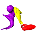
Left middle ear ossicles
Data citation:
Daisuke Koyabu , 2017. M3#155. doi: 10.18563/m3.sf.155
Tag legend:
Incus, Malleus, Stapes
Model solid/transparent
Flags:
Anterior crus, Anterior process, Footplate of stapes, Head of malleus, Head of stapes, Long process of incus, Manubrium, Orbicular apophysis, Posterior crus, Short process of incus, Transversal lamina

|
3D atlas and comparative osteology of the middle ear ossicles among Eulipotyphla (Mammalia, Placentalia).Daisuke KoyabuPublished online: 03/05/2017Keywords: aquatic adaptation; convergence; Eulipotyphla; fossorial adaptation; hearing https://doi.org/10.18563/m3.3.2.e3 Abstract Considerable morphological variations are found in the middle ear among mammals. Here I present a three-dimensional atlas of the middle ear ossicles of eulipotyphlan mammals. This group has radiated into various environments as terrestrial, aquatic, and subterranean habitats independently in multiple lineages. Therefore, eulipotyphlans are an ideal group to explore the form-function relationship of the middle ear ossicles. This comparative atlas of hedgehogs, true shrews, water shrews, mole shrews, true moles, and shrew moles encourages future studies of the middle ear morphology of this diverse group. M3 article infos Published in Volume 03, Issue 02 (2017) |
|