3D models of Cainotheriids Ossicular chain
Explodable 3D Dog Skull for Veterinary Education
3D models of Kalakocetus, the earliest Cetacea
3D GM dataset of bird skeletal variation
Skeletal embryonic development in the catshark
Bony connexions of the petrosal bone of extant hippos
bony labyrinth (14) , inner ear (11) , Eocene (11) , geometric morphometrics (10) , CT-scan (10) , Oligocene (9) , Micro-CT (9)
Lionel Hautier (25) , Maëva Judith Orliac (24) , Laurent Marivaux (18) , Renaud Lebrun (15) , Rodolphe Tabuce (15) , Pierre-Olivier Antoine (13) , Bastien Mennecart (13)
MorphoMuseuM Volume 05, issue 04
<< prev. article next article >>
|
3D dataset3D models related to the publication: Virtual reconstruction of cranial endocasts of traversodontid cynodonts (Eucynodontia: Gomphodontia) from the upper Triassic of Southern Brazil.Ane E. B. Pavanatto
Published online: 10/09/2019 |
|
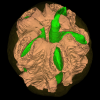
M3#4253D model of the brain endocast Type: "3D_surfaces"doi: 10.18563/m3.sf.425 state:published |
Download 3D surface file |
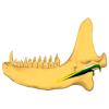
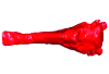
Exaeretodon riograndensis CAPPA/UFSM 0030 View specimen

|
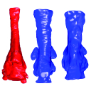
M3#4263D model of the brain endocast Type: "3D_surfaces"doi: 10.18563/m3.sf.426 state:published |
Download 3D surface file |
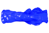
Exaeretodon riograndensis CAPPA/UFSM 0227 View specimen
|
M3#4273D model of the brain endocast Type: "3D_surfaces"doi: 10.18563/m3.sf.427 state:published |
Download 3D surface file |
Fedorov, A., Beichel, R., Kalphaty-Cramer, J., Finet, J., Fillion-Robbin, J.-C., Pujol, S., Bauer, C., Jennings, D., Fennessy, F., Sonka, M., Buatti, J., Aylward, S., Miller, J. V, Pieper, S., Kikinis, R., 2012. 3D Slicer as an Image Computing Platform for the Quantitative Imaging Network. Magnetic resonance imaging. 30, 1323–1341. https://doi.org/10.1016/j.mri.2012.05.001
Kammerer, C.F., Flynn, J.J., Ranivoharimanana, L., Wyss, A.R., 2008. New material of Menadon besairiei (Cynodontia: Traversodontidae) from the Triassic of Madagascar. Journal of Vertebrate Paleontology. 28, 445–462. https://doi.org/10.1671/0272-4634(2008)28[445:NMOMBC]2.0.CO;2
Liu, J., Abdala, F., 2014. Phylogeny and Taxonomy of the Traversodontidae. In: Kammerer, C.F., Angielczyk, K.D., Fröbisch, J. (Eds.), Early Evolutionary History of the Synapsida (Vertebrate Paleobiology and Paleoanthropology Series). Springer, Dordrecht, pp. 289–304. https://doi.org/10.1007/978-94-007-6841-3_15
Pavanatto, A.E.B., Kerber, L., Dias-da-Silva, S., 2019. Virtual reconstruction of cranial endocasts of traversodontid cynodonts (Eucynodontia: Gomphodontia) from the upper Triassic of Southern Brazil. Journal of Morphology. 280, 1267-1281. https://doi.org/10.1002/jmor.21029
Leonardo Kerber, Heloísa Moraes–Santos and Maria Inês Feijó Ramos (2023). Scanning holotypes from the Vertebrate Paleontology Collection at the Museu Paraense Emilio Goeldi (Brazil): Tools for research and science outreach. Curator: The Museum Journal. https://doi.org/10.1111/cura.12548