3D models of Cainotheriids Ossicular chain
3D models of Kalakocetus, the earliest Cetacea
The specimens of Speothos pacivorus
3D GM dataset of bird skeletal variation
Skeletal embryonic development in the catshark
Bony connexions of the petrosal bone of extant hippos
bony labyrinth (14) , inner ear (11) , Eocene (11) , geometric morphometrics (10) , CT-scan (10) , Oligocene (9) , Micro-CT (9)
Lionel Hautier (25) , Maëva Judith Orliac (24) , Laurent Marivaux (18) , Renaud Lebrun (15) , Rodolphe Tabuce (15) , Pierre-Olivier Antoine (13) , Bastien Mennecart (13)
Page 1 of 1, showing 6 record(s) out of 6 total
|
3D model related to the publication: Cranial morphology and phylogenetic relationships of Amynodontidae Scott & Osborn, 1883 (Perissodactyla, Rhinocerotoidea)Léa Veine-Tonizzo
Published online: 20/03/2023 |
|
M3#716Textured 3D surface model of the skull of the specimen UNISTRA.2015.0.1106 with right C1 and both rows of P2-M3. Type: "3D_surfaces"doi: 10.18563/m3.sf.716 state:published |
Download 3D surface file |
The present 3D Dataset contains the 3D model analyzed in Gaetano, L. C., Abdala, F., Seoane, F. D., Tartaglione, A., Schulz, M., Otero, A., Leardi, J. M., Apaldetti, C., Krapovickas, V., and Steinbach, E. 2021. A new cynodont from the Upper Triassic Los Colorados Formation (Argentina, South America) reveals a novel paleobiogeographic context for mammalian ancestors. Scientific Reports.
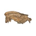
Tessellatia bonapartei PULR-V121 View specimen
|
M3#9603D surface model of PULR-V121 Type: "3D_surfaces"doi: 10.18563/m3.sf.960 state:published |
Download 3D surface file |
The present 3D Dataset contains the 3D model analyzed in the article : Dubied et al. (2021), Endocranium and ecology of Eurotherium theriodis, a European hyaenodont mammal from the Lutetian. Acta Palaeontologica Polonica 2021, https://doi.org/10.4202/app.00771.2020
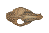
Eurotherium theriodis NMB.Em12 View specimen
|
M3#381NMB.Em12 unprepared specimen Type: "3D_surfaces"doi: 10.18563/m3.sf.381 state:published |
Download 3D surface file |
|
M3#382NMB.Em12 cranium Type: "3D_surfaces"doi: 10.18563/m3.sf.382 state:published |
Download 3D surface file |
|
M3#383NMB.Em12 endocast Type: "3D_surfaces"doi: 10.18563/m3.sf.383 state:published |
Download 3D surface file |
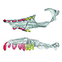
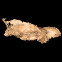
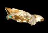
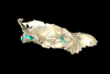
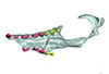
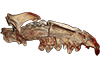
This contribution contains the 3D models described and figured in the following publication: Kassegne K. E., Mourlam M. J., Guinot G., Amoudji Y. Z., Martin J. E., Togbe K. A., Johnson A. K., Hautier L. 2021. First partial cranium of Togocetus from Kpogamé (Togo) and the protocetid diversity in the Togolese phosphate basin. Annales de Paléontologie, Issue 2, April–June 2021, 102488. https://doi.org/10.1016/j.annpal.2021.102488
Togocetus cf. traversei ULDG-KPO1 View specimen
|
M3#768The specimen consists of a partial cranium prepared out of a calcareous phosphate matrix. The partial cranium lacks the anterior part of the rostrum, the cranial roof, and most of the basicranium apart from the left zygomatic process of the squamosal. The maxilla, nasal, palatine, pterygoid, alisphenoid, and squamosal bones are preserved, as well as two incomplete dental rows described hereafter. Type: "3D_surfaces"doi: 10.18563/m3.sf.768 state:published |
Download 3D surface file |
|
M3#770µCT . Resolution: 0.3156mm. This scan can easily be opened with Fiji, MorphoDig, 3DSlicer, or any software that reads .MHD file format. Also, the .RAW file can be opened easily with other software such as Avizo/Amira when providing the correct dimensions (which are enclosed within the file name) Type: "3D_CT"doi: 10.18563/m3.sf.770 state:published |
Download CT data |
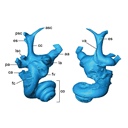
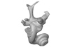
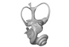
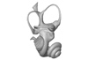
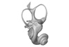
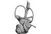
The present 3D Dataset contains the 3D models analyzed in Costeur L., Mennecart B., Müller B., Schulz G., 2016. Prenatal growth stages show the development of the ruminant bony labyrinth and petrosal bone. Journal of Anatomy. https://doi.org/10.1111/joa.12549
Bos taurus NMB3038 View specimen
|
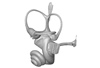
M3#124Right bony labyrinth of a Bos taurus foetus (gestational age 115 days) Type: "3D_surfaces"doi: 10.18563/m3.sf.124 state:published |
Download 3D surface file |
Bos taurus NMB3367 View specimen
|
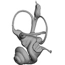
M3#125Right bony labyrinth of a Bos taurus foetus (gestational age 165 days) Type: "3D_surfaces"doi: 10.18563/m3.sf.125 state:published |
Download 3D surface file |
Bos taurus NMB3365 View specimen
|
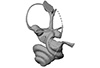
M3#126Right bony labyrinth of a Bos taurus foetus (gestational age 210 days) Type: "3D_surfaces"doi: 10.18563/m3.sf.126 state:published |
Download 3D surface file |
Bos taurus NMB2855 View specimen
|
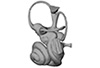
M3#127Right bony labyrinth of a Bos taurus foetus (gestational age 260 days) Type: "3D_surfaces"doi: 10.18563/m3.sf.127 state:published |
Download 3D surface file |
Bos taurus NMB1037 View specimen
|
M3#128Left bony labyrinth of an adult Bos taurus Type: "3D_surfaces"doi: 10.18563/m3.sf.128 state:published |
Download 3D surface file |
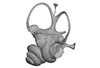
The present 3D Dataset contains the 3D models analyzed in the article Mennecart, B., and L. Costeur. 2016. A Dorcatherium (Mammalia, Ruminantia, Middle Miocene) petrosal bone and the tragulid ear region. Journal of Vertebrate Paleontology 36(6), 1211665(1)-1211665(7). DOI: 10.1080/02724634.2016.1211665.
Tragulus javanicus 10028 View specimen
|
M3#1193D surface of the left bony labyrinth of Tragulus javanicus NMB 10028 Type: "3D_surfaces"doi: 10.18563/m3.sf.119 state:published |
Download 3D surface file |
Moschiola meminna C.2453 View specimen
|
M3#1203D surface of the left bony labyrinth of Moschiola meminna NMB C.2453 Type: "3D_surfaces"doi: 10.18563/m3.sf.120 state:published |
Download 3D surface file |
Hyemoschus aquaticus C.1930 View specimen
|
M3#1223D surface of the right bony labyrinth of Hyemoschus aquaticus NMB C.1930 Type: "3D_surfaces"doi: 10.18563/m3.sf.122 state:published |
Download 3D surface file |
Dorcatherium crassum San.15053 View specimen
|
M3#1233D surface of the right bony labyrinth of Dorcatherium crassum NMB San.15053 Type: "3D_surfaces"doi: 10.18563/m3.sf.123 state:published |
Download 3D surface file |
Page 1 of 1, showing 6 record(s) out of 6 total


