3D model of a Late Oligocene madtsoiid snake
Allometric trajectories in Carboniferous unornamented Polygnathus
3D models of: The endocranial anatomy of Protocetus
3D GM dataset of bird skeletal variation
Skeletal embryonic development in the catshark
Bony connexions of the petrosal bone of extant hippos
bony labyrinth (11) , inner ear (10) , Eocene (8) , South America (8) , Paleobiogeography (7) , skull (7) , phylogeny (6)
Lionel Hautier (22) , Maëva Judith Orliac (21) , Laurent Marivaux (15) , Rodolphe Tabuce (13) , Pierre-Olivier Antoine (12) , Bastien Mennecart (12) , Renaud Lebrun (10)
MorphoMuseuM Volume 01, Issue 04
<< prev. article next article >>
|
Original article : anatomy atlasSkeletogenesis during the late embryonic development of the catshark Scyliorhinus canicula (Chondrichthyes; Neoselachii)Sébastien Enault, Sylvain Adnet
Published online: 25/04/2016 |

|
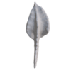
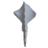
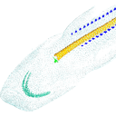
M3#50Mineralized skeleton of a 6,2 cm long embryo of Scyliorhinus canicula Type: "3D_surfaces"doi: 10.18563/m3.sf.50 state:published |
Download 3D surface file |
Scyliorhinus canicula SC6_7_2015_03_20 View specimen

|
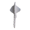
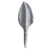
M3#51Mineralized skeleton of a 6,7 cm long embryo of Scyliorhinus canicula Type: "3D_surfaces"doi: 10.18563/m3.sf.51 state:published |
Download 3D surface file |
Scyliorhinus canicula SC7_1_2015_04_03 View specimen

|
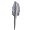
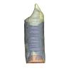
M3#52Mineralized skeleton of a 7,1 cm long embryo of Scyliorhinus canicula Type: "3D_surfaces"doi: 10.18563/m3.sf.52 state:published |
Download 3D surface file |
Scyliorhinus canicula SC7_5_2015_03_13 View specimen

|
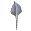
M3#53Mineralized skeleton of a 7,5 cm long embryo of Scyliorhinus canicula Type: "3D_surfaces"doi: 10.18563/m3.sf.53 state:published |
Download 3D surface file |
Scyliorhinus canicula SC8_2015_03_20 View specimen

|
M3#54Mineralized skeleton of a 8 cm long embryo of Scyliorhinus canicula Type: "3D_surfaces"doi: 10.18563/m3.sf.54 state:published |
Download 3D surface file |
Scyliorhinus canicula SC10_2015_02_27 View specimen

|
M3#55Mineralized skeleton of a 10 cm long embryo of Scyliorhinus canicula Type: "3D_surfaces"doi: 10.18563/m3.sf.55 state:published |
Download 3D surface file |
Applegate, S., 1967. A survey of shark hard parts. In: Gilbert, P., Mathewson, R., Rall, D. (Eds.), Shark, Skates and Rays. The Johns Hopkins Press, Baltimore, Maryland, pp. 37–67.
Ballard, W.W., Mellinger, J., Lechenault, H., 1993. A series of stages for development of Scyliorhinus canicula the lesser spotted dogfish (Chondrichthyes: Scyliorhinidae). Journal of Experimental Zoology. 267, 1–43. http://dx.doi.org/10.1002/jez.1402670309
Cappetta, H., 2012. Handbook of Paleoichthyology, Vol. 3E: Chondrichthyes · Mesozoic and Cenozoic Elasmobranchii: Teeth. Verlag Dr. Friedrich Pfeil.
Cignoni, P., Cignoni, P., Callieri, M., Callieri, M., Corsini, M., Corsini, M., Dellepiane, M., Dellepiane, M., Ganovelli, F., Ganovelli, F., Ranzuglia, G., Ranzuglia, G., 2008. MeshLab: an Open-Source Mesh Processing Tool. Sixth Eurographics Italian Chapter Conference. 129–136.
Compagno, L.J. V., 1988. Sharks of the order Carcharhiniformes. Princeton University Press, New Jersey.
Compagno, L.J. V., 1999. Endoskeleton. In: Hamlett, W.C. (Ed.), Sharks, Skates and Rays. The Biology of Elasmobranch Fishes.
Compagnucci, C., Debiais-Thibaud, M., Coolen, M., Fish, J., Griffin, J.N., Bertocchini, F., Minoux, M., Rijli, F.M., Borday-Birraux, V., Casane, D., Mazan, S., Depew, M.J., 2013. Pattern and polarity in the development and evolution of the gnathostome jaw: both conservation and heterotopy in the branchial arches of the shark, Scyliorhinus canicula. Developmental biology. 377, 428–48. http://dx.doi.org/10.1016/j.ydbio.2013.02.022
D’Souza, D.G., Rana, K., Milley, K.M., Maclean, H.E., Zajac, J.D., Bell, J., Brenner, S., Venkatesh, B., Richardson, S.J., Danks, J. a, 2013. Expression of Wnt signaling skeletal development genes in the cartilaginous fish, elephant shark (Callorhinchus milii). General and comparative endocrinology. 1–9. http://dx.doi.org/10.1016/j.ygcen.2013.06.021
Dahn, R.D., Davis, M.C., Pappano, W.N., Shubin, N.H., 2007. Sonic hedgehog function in chondrichthyan fins and the evolution of appendage patterning. Nature. 445, 311–314. http://dx.doi.org/10.1038/nature05436
de Beer, G.R., 1931. The development of the skull of Scyllium (Scyliorhinus) canicula L. Quarterly Journal of Microscopical Science. 74, 591–652. https://doi.org/10.1242/jcs.s2-74.296.591
Dean, M.N., Summers, A.P., 2006. Mineralized cartilage in the skeleton of chondrichthyan fishes. Zoology (Jena, Germany). 109, 164–8. http://dx.doi.org/10.1016/j.zool.2006.03.002
Debiais-Thibaud, M., Oulion, S., Bourrat, F., Laurenti, P., Casane, D., Borday-Birraux, V., 2011. The homology of odontodes in gnathostomes: insights from Dlx gene expression in the dogfish, Scyliorhinus canicula. BMC Evolutionary Biology. 11, 307. http://dx.doi.org/10.1186/1471-2148-11-307
Dingerkus, G., Seret, B., Guilbert, E., 1991. Multiple prismatic calcium phosphate layers in the jaws of present-day sharks (Chondrichthyes; Selachii). Experientia. 47, 38–40. http://dx.doi.org/10.1007/BF02041246
Eames, B.F., Allen, N., Young, J., Kaplan, A., Helms, J. a, Schneider, R. a, 2007. Skeletogenesis in the swell shark Cephaloscyllium ventriosum. Journal of anatomy. 210, 542–54. http://dx.doi.org/10.1111/j.1469-7580.2007.00723.x
Ellis, J.R., Shackley, S.E., 1997. The reproductive biology of Scyliorhinus canicula in the Bristol Channel, U.K. Journal of Fish Biology. 51, 361–372. http://dx.doi.org/10.1111/j.1095-8649.1997.tb01672.x
Enault, S., Muñoz, D.N., Silva, W.T.A.F., Borday-birraux, V., Bonade, M., Oulion, S., Ventéo, S., Marcellini, S., Debiais-Thibaud, M., 2015. Molecular footprinting of skeletal tissues in the catshark Scyliorhinus canicula and the clawed frog Xenopus tropicalis identifies conserved and derived features of vertebrate calcification. Frontiers in genetics. 6, 1–14. http://dx.doi.org/10.3389/fgene.2015.00283
Gegenbaur, C., 1872. Untersuchungen zur vergleichenden anatomie der wirbelthiere. Drittes heft. Das kopfskelet der selachier, ein beitrag Zur erkenntniss der genese des kopfskelete der wirbelthiere. Leipzig.
Godard, B.G., Mazan, S., 2012. Early patterning in a chondrichthyan model, the small spotted dogfish: towards the gnathostome ancestral state. Journal of anatomy. 222, 56–66. http://dx.doi.org/10.1111/j.1469-7580.2012.01552.x
Hasse, C., 1879. Das natürliche System der Elasmobranchier auf Grundlage des Baues und der Entwicklung ihrer Wirbelsäule. Eine morphologische und paläontologische Studie. I Allgemei, 1–76.
Hasse, C., 1882a. Das natürliche System der Elasmobranchier auf Grundlage des Baues und der Entwicklung ihrer Wirbelsäule. Eine morphologische und paläontologische Studie. II Besonde, 96–179.
Hasse, C., 1882b. Das natürliche System der Elasmobranchier auf Grundlage des Baues und der Entwicklung ihrer Wirbelsäule. Eine morphologische und paläontologische Studie. III, 181–285.
Hautier, L., Weisbecker, V., Sánchez-Villagra, M.R., Goswami, A., Asher, R.J., 2010. Skeletal development in sloths and the evolution of mammalian vertebral patterning. Proceedings of the National Academy of Sciences of the United States of America. 107, 18903–8. http://dx.doi.org/10.1073/pnas.1010335107
Holmgren, N., 1940. Studies of the head in fishes. Part 1. Development of the skull in sharks and rays. Acta Zoologica. 21, 51–257. https://doi.org/10.1111/j.1463-6395.1940.tb00339.x
Lebrun, R., 2014. ISE-MeshTools, a 3D interactive fossil reconstruction freeware. In: 12th Annual Meeting of EAVP, Torino, Italy.
Mellinger, J., Wrisez, F., 1993. Etude des écailles primaires de l’embryon de la roussette Scyliorhinus canicula (Chondrichthyes: Scyliorhinidae) au microscope électronique à balayage. Annales des Sciences Naturelles, Zoologie, Paris, 13e série. 14, 13–22.
O’Shaughnessy, K.L., Dahn, R.D., Cohn, M.J., 2015. Molecular development of chondrichthyan claspers and the evolution of copulatory organs. Nature Communications. 6, 6698. http://dx.doi.org/10.1038/ncomms7698
Peignoux-Deville, J., Lallier, F., Vidal, B., 1982. Evidence for the presence of osseous tissue in dogfish vertebrae. Cell and tissue research. 222, 605–614. http://dx.doi.org/10.1007/BF00213858
Reif, W.-E., 1980. Development of dentition and dermal skeleton in embryonic Scyliorhinus canicula. Journal of morphology. 166, 275–88. http://dx.doi.org/10.1002/jmor.1051660303
Reif, W.-E., 1985. Squamation and ecology of sharks. Cour Forsch Inst Senckenberg. 78, 1–255.
Ridewood, W.G., 1899. Some Observations on the Caudal Diplospondyly of Sharks. Journal of the Linnean Society of London, Zoology. 27, 46–59. https://doi.org/10.1111/j.1096-3642.1899.tb00248.x
Summers, A., 2000. Stiffening the stingray skeleton- an investigation of durophagy in myliobatid stingrays (Chondrichthyes, batoidea, myliobatidae). Journal of morphology. 243, 113–26. https://doi.org/10.1002/(SICI)1097-4687(200002)243:2<113::AID-JMOR1>3.0.CO;2-A
Raphaël Scherrer, Andrés Hurtado, Erik Garcia Machado and Mélanie Debiais-Thibaud (2017). MicroCT survey of larval skeletal mineralization in the Cuban gar Atractosteus tristoechus (Actinopterygii; Lepisosteiformes). MorphoMuseuM. https://doi.org/10.18563/m3.3.3.e3
Giuseppe Marramà, Kerin M. Claeson, Giorgio Carnevale and Jürgen Kriwet (2018). Revision of Eocene electric rays (Torpediniformes, Batomorphii) from the Bolca Konservat-Lagerstätte, Italy, reveals the first fossil embryoin situin marine batoids and provides new insights into the origin of trophic novelties in coral reef fishes. Journal of Systematic Palaeontology. https://doi.org/10.1080/14772019.2017.1371257
Jacob B. Pears, Zerina Johanson, Kate Trinajstic, Mason N. Dean and Catherine A. Boisvert (2020). Mineralization of the Callorhinchus Vertebral Column (Holocephali; Chondrichthyes). Frontiers in Genetics. https://doi.org/10.3389/fgene.2020.571694
Nicolas Leurs, Camille Martinand-Mari, Stéphanie Ventéo, Tatjana Haitina and Mélanie Debiais-Thibaud (2021). Evolution of Matrix Gla and Bone Gla Protein Genes in Jawed Vertebrates. Frontiers in Genetics. https://doi.org/10.3389/fgene.2021.620659
Fidji Berio, Morgane Broyon, Sébastien Enault, Nelly Pirot, Faviel A. López-Romero and Mélanie Debiais-Thibaud (2021). Diversity and Evolution of Mineralized Skeletal Tissues in Chondrichthyans. Frontiers in Ecology and Evolution. https://doi.org/10.3389/fevo.2021.660767
Rory L. Cooper, Ella F. Nicklin, Liam J. Rasch and Gareth J. Fraser (2023). Teeth outside the mouth: The evolution and development of shark denticles. Evolution & Development. https://doi.org/10.1111/ede.12427
Jamie L. Knaub, Michelle Passerotti, Lisa J. Natanson, Tricia Meredith and Marianne Porter (2024).
Vertebral morphology in the tail-whipping common thresher shark,
Alopias vulpinus
. Royal Society Open Science. https://doi.org/10.1098/rsos.231473