Explodable 3D Dog Skull for Veterinary Education
3D models of teeth and neurovascular system in the rostrum of Spinosauridae (theropod dinosaurs)
3D models of Ocnotherium skull
3D GM dataset of bird skeletal variation
Skeletal embryonic development in the catshark
Bony connexions of the petrosal bone of extant hippos
bony labyrinth (14) , inner ear (11) , Eocene (11) , geometric morphometrics (10) , CT-scan (10) , Oligocene (9) , Micro-CT (9)
Lionel Hautier (25) , Maëva Judith Orliac (24) , Laurent Marivaux (19) , Bastien Mennecart (16) , Renaud Lebrun (15) , Rodolphe Tabuce (15) , Pierre-Olivier Antoine (13)
Page 1 of 1, showing 4 record(s) out of 4 total
|
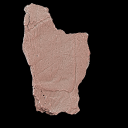
3D models related to the publication: Triassic turtles of France: the first European Rhaetian stem turtle taxon (clades Testudinata, Proterochersidae)Tomasz Szczygielski
Published online: 20/08/2026 |
|
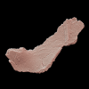
M3#1907Left axillary region and base of the anterior plastral lobe (left hyoplastron, small medial fragment of the right hyoplastron, part of the posterior process of the entoplastron) Type: "3D_surfaces"doi: 10.18563/m3.sf.1907 state:in_press |
Download 3D surface file |
Bartassochersis lopezi UM-PDE2-02 View specimen
|
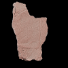
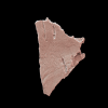
M3#1908Posterior plastral lobe Type: "3D_surfaces"doi: 10.18563/m3.sf.1908 state:in_press |
Download 3D surface file |
Bartassochersis lopezi UM-PDE2-03 View specimen
|
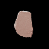
M3#1909First vertebral scute region, right side Type: "3D_surfaces"doi: 10.18563/m3.sf.1909 state:in_press |
Download 3D surface file |
Bartassochersis lopezi UM-PDE2-04 View specimen
|
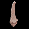
M3#1910?Carapace fragment Type: "3D_surfaces"doi: 10.18563/m3.sf.1910 state:in_press |
Download 3D surface file |
Bartassochersis lopezi UM-PDE2-05 View specimen
|
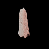
M3#1911Left scapula Type: "3D_surfaces"doi: 10.18563/m3.sf.1911 state:in_press |
Download 3D surface file |
Bartassochersis lopezi UM-PDE2-06 View specimen
|
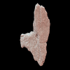
M3#1912Ungual Type: "3D_surfaces"doi: 10.18563/m3.sf.1912 state:in_press |
Download 3D surface file |
Bartassochersis lopezi UM-PDE2-08 View specimen
|
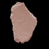
M3#1913Right hypoplastron Type: "3D_surfaces"doi: 10.18563/m3.sf.1913 state:in_press |
Download 3D surface file |
Bartassochersis lopezi UM-PDE2-09 View specimen
|
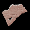
M3#1914Indeterminate Type: "3D_surfaces"doi: 10.18563/m3.sf.1914 state:in_press |
Download 3D surface file |
Bartassochersis lopezi UM-PDE2-10 View specimen
|
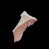
M3#1915?Mesoplastron/hypoplastron Type: "3D_surfaces"doi: 10.18563/m3.sf.1915 state:in_press |
Download 3D surface file |
Bartassochersis lopezi UM-PDE2-11 View specimen
|
M3#1916Left inguinal periphery (peripheral, hyoplastron) Type: "3D_surfaces"doi: 10.18563/m3.sf.1916 state:in_press |
Download 3D surface file |
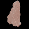
Bartassochersis lopezi UM-PDE2-12 View specimen
|
M3#1917?Left side of a vertebral scute area with two partial pleural scute areas Type: "3D_surfaces"doi: 10.18563/m3.sf.1917 state:in_press |
Download 3D surface file |
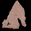
Bartassochersis lopezi UM-PDE2-13 View specimen
|
M3#1918?Posterior pleural and supramarginal of marginal areas Type: "3D_surfaces"doi: 10.18563/m3.sf.1918 state:in_press |
Download 3D surface file |
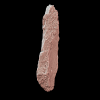
Bartassochersis lopezi UM-PDE2-14 View specimen
|
M3#1919Posterior peripherals Type: "3D_surfaces"doi: 10.18563/m3.sf.1919 state:in_press |
Download 3D surface file |
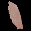
Bartassochersis lopezi UM-PDE2-15A View specimen
|
M3#1920?Left edge of the anterior plastral lobe Type: "3D_surfaces"doi: 10.18563/m3.sf.1920 state:in_press |
Download 3D surface file |
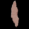
Bartassochersis lopezi UM-PDE2-15B View specimen
|
M3#1921Indeterminate Type: "3D_surfaces"doi: 10.18563/m3.sf.1921 state:in_press |
Download 3D surface file |
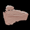
Bartassochersis lopezi UM-PDE2-16 View specimen
|
M3#1922Indeterminate Type: "3D_surfaces"doi: 10.18563/m3.sf.1922 state:in_press |
Download 3D surface file |
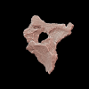
Bartassochersis lopezi UM-PDE2-17 View specimen
|
M3#1923?Bridge fragment Type: "3D_surfaces"doi: 10.18563/m3.sf.1923 state:in_press |
Download 3D surface file |
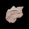
Bartassochersis lopezi UM-PDE2-19 View specimen
|
M3#1924Right bridge region Type: "3D_surfaces"doi: 10.18563/m3.sf.1924 state:in_press |
Download 3D surface file |
Bartassochersis lopezi UM-PDE2-20 View specimen
|
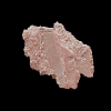
M3#1925Left femoral bulge Type: "3D_surfaces"doi: 10.18563/m3.sf.1925 state:in_press |
Download 3D surface file |
Bartassochersis lopezi UM-PDE2-21 View specimen
|
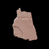
M3#1926Indeterminate Type: "3D_surfaces"doi: 10.18563/m3.sf.1926 state:in_press |
Download 3D surface file |
 , Raphaël Sarr , Dominique Chardon , Jean-Renaud Boisserie , Renaud Lebrun , Sylvain Adnet , Jeremy E. Martin , Bernard Sambou, Rodolphe Tabuce , Moustapha Thiam and Lionel Hautier
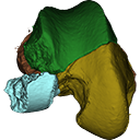
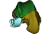
 The present contribution contains the 3D virtual restoration of a Pliocene Lutrine right femur of Tobène, Senegal, described and figured in Lihoreau et al. (2021) : "A fossil terrestrial fauna from Tobène (Senegal) provides a unique early Pliocene window in Western Africa ". https://doi.org/10.1016/j.gr.2021.06.013
Indet indet SN-Tob-12-02 View specimen
|
M3#441Virtual restoration of SN-Tob-12-02 Type: "3D_surfaces"doi: 10.18563/m3.sf.441 state:published |
Download 3D surface file |
 , Fabrice Lihoreau , Bernard Sambou, Moustapha Thiam, Jeremy E. Martin , Rodolphe Tabuce , Sylvain Adnet , Renaud Lebrun , Anne-Lise Charruault , Raphaël Sarr and Lionel Hautier
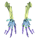
 The present 3D Dataset contains the 3D model analyzed in Vautrin et al. (2019), Palaeontology, From limb to fin: an Eocene protocetid forelimb from Senegal sheds new light on the early locomotor evolution of early cetaceans.
?Carolinacetus indet. SNTB 2011-01 View specimen
|
M3#3983D model of an articulated forelimb of a Carolinacetus-like protocetid from Senegal Type: "3D_surfaces"doi: 10.18563/m3.sf.398 state:published |
Download 3D surface file |
 and Mélanie Debiais-Thibaud
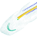
 Current knowledge on the skeletogenesis of Chondrichthyes is scarce compared with their extant sister group, the bony fishes. Most of the previously described developmental tables in Chondrichthyes have focused on embryonic external morphology only. Due to its small body size and relative simplicity to raise eggs in laboratory conditions, the small-spotted catshark Scyliorhinus canicula has emerged as a reference species to describe developmental mechanisms in the Chondrichthyes lineage. Here we investigate the dynamic of mineralization in a set of six embryonic specimens using X-ray microtomography and describe the developing units of both the dermal skeleton (teeth and dermal scales) and endoskeleton (vertebral axis). This preliminary data on skeletogenesis in the catshark sets the first bases to a more complete investigation of the skeletal developmental in Chondrichthyes. It should provide comparison points with data known in osteichthyans and could thus be used in the broader context of gnathostome skeletal evolution.
Scyliorhinus canicula SC6_2_2015_03_20 View specimen

|
M3#50Mineralized skeleton of a 6,2 cm long embryo of Scyliorhinus canicula Type: "3D_surfaces"doi: 10.18563/m3.sf.50 state:published |
Download 3D surface file |
Scyliorhinus canicula SC6_7_2015_03_20 View specimen

|
M3#51Mineralized skeleton of a 6,7 cm long embryo of Scyliorhinus canicula Type: "3D_surfaces"doi: 10.18563/m3.sf.51 state:published |
Download 3D surface file |
Scyliorhinus canicula SC7_1_2015_04_03 View specimen

|
M3#52Mineralized skeleton of a 7,1 cm long embryo of Scyliorhinus canicula Type: "3D_surfaces"doi: 10.18563/m3.sf.52 state:published |
Download 3D surface file |
Scyliorhinus canicula SC7_5_2015_03_13 View specimen

|
M3#53Mineralized skeleton of a 7,5 cm long embryo of Scyliorhinus canicula Type: "3D_surfaces"doi: 10.18563/m3.sf.53 state:published |
Download 3D surface file |
Scyliorhinus canicula SC8_2015_03_20 View specimen

|
M3#54Mineralized skeleton of a 8 cm long embryo of Scyliorhinus canicula Type: "3D_surfaces"doi: 10.18563/m3.sf.54 state:published |
Download 3D surface file |
Scyliorhinus canicula SC10_2015_02_27 View specimen

|
M3#55Mineralized skeleton of a 10 cm long embryo of Scyliorhinus canicula Type: "3D_surfaces"doi: 10.18563/m3.sf.55 state:published |
Download 3D surface file |
Page 1 of 1, showing 4 record(s) out of 4 total