3D models of early strepsirrhine primate teeth from North Africa
3D models of Protosilvestria sculpta and Coloboderes roqueprunetherion
3D models of Miocene vertebrates from Tavers
3D GM dataset of bird skeletal variation
Skeletal embryonic development in the catshark
Bony connexions of the petrosal bone of extant hippos
bony labyrinth (11) , inner ear (10) , Eocene (8) , South America (8) , Paleobiogeography (7) , skull (7) , phylogeny (6)
Lionel Hautier (22) , Maëva Judith Orliac (21) , Laurent Marivaux (16) , Rodolphe Tabuce (14) , Bastien Mennecart (13) , Pierre-Olivier Antoine (12) , Renaud Lebrun (11)

|
3D models related to the publication: Prenatal growth stages show the development of the ruminant bony labyrinth and petrosal bone.Loïc Costeur
Published online: 19/10/2016 |
|

|
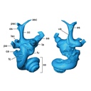
M3#125Right bony labyrinth of a Bos taurus foetus (gestational age 165 days) Type: "3D_surfaces"doi: 10.18563/m3.sf.125 Data citation: Loïc Costeur and Bastien Mennecart, 2016. M3#125. doi: 10.18563/m3.sf.125 state:published |
Download 3D surface file |