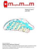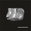
Explodable 3D Dog Skull for Veterinary Education
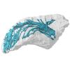
3D models of teeth and neurovascular system in the rostrum of Spinosauridae (theropod dinosaurs)
3D models of Cainotheriids Ossicular chain
3D GM dataset of bird skeletal variation
Skeletal embryonic development in the catshark
Bony connexions of the petrosal bone of extant hippos
bony labyrinth (14) , inner ear (11) , Eocene (11) , geometric morphometrics (10) , CT-scan (10) , Oligocene (9) , Micro-CT (9)
Lionel Hautier (25) , Maëva Judith Orliac (24) , Laurent Marivaux (19) , Bastien Mennecart (16) , Renaud Lebrun (15) , Rodolphe Tabuce (15) , Pierre-Olivier Antoine (13)
Page 1 of 1, showing 5 record(s) out of 5 total
|
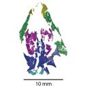
3D model related to the publication: Earliest-diverging rhynchocephalian from Gondwana: a new sphenodontian from the Upper Triassic Los Colorados Formation (northwestern Argentina).Leandro C. Gaetano
Published online: 31/08/2026 |
|
M3#1855fragmentary skull and lower jaws Type: "3D_surfaces"doi: 10.18563/m3.sf.1855 state:in_press |
Download 3D surface file |
 , Ruihan R. Liu, Alexis M. Slack , Emma R. Schachner , William G. Ryerson and Brandon P. Hedrick
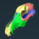
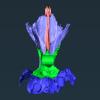
 Veterinary education often relies on cadaveric specimens, but there is increasing demand for alternatives due to limited resources and ethical considerations. To address this, we developed a 3D printed ‘explodable’ model of a dog cranium with detachable, magnetized cranial components for teaching anatomy to students. This model was generated from a computed tomographic scan of a juvenile dog cranium for which cranial sutures were still partially open and segmented such that major cranial bones were isolated. All bones are printed at actual size and retain openings for cranial nerves and major vessels. This interactive model enhances anatomical education by supplying a hands-on tool that can be used either in the classroom setting or for independent learning and can be incorporated at the high school, college, or veterinary school level. It is currently being integrated into the first-year anatomy foundation course at Cornell University’s College of Veterinary Medicine. The model can be printed using any hobbyist or specialist 3D printer and we outline assembly instructions on how to attach magnets at prefabricated attachment points. Using both digital and 3D printed resources, we hope to help to address current shortages of anatomical resources and also inspire future generations of practicing veterinarians by making anatomy more accessible and engaging.
Canis lupus familiaris CUHL 9 View specimen
|
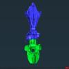
M3#1858PLYs of the segmented cranial bones with pre-fabricated magnetic casings and shelves for assembly following 3D printing Type: "3D_surfaces"doi: 10.18563/m3.sf.1858 state:published |
Download 3D surface file |
|
M3#1859PLYs of the segmented cranial bones of the "BOTTOM" cranial component. Downloadable for additional learning opportunities for students Type: "3D_surfaces"doi: 10.18563/m3.sf.1859 state:published |
Download 3D surface file |
 , Fernando Abdala, Adriana Mancuso and Nahuel Vega
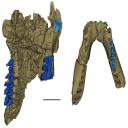
This contribution contains the 3D models described and figured in the following publication: Gaetano, L. C., Abdala, F., Mancuso, C, and Vega N.2025. New traversodontid cynodont from the Late Triassic Chañares Formation. Publicación Electrónica de la Asociación Paleontológica Argentina.
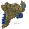
Pontognathus ignotus PULR-V 287 View specimen
|
M3#1647partial snout preserving the lateralmost incisor, the base of the canine, and several postcanines Type: "3D_surfaces"doi: 10.18563/m3.sf.1647 state:published |
Download 3D surface file |
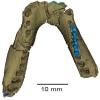
Massetognathus pascuali PULR-V 289 View specimen
|
M3#1646partial lower jaw Type: "3D_surfaces"doi: 10.18563/m3.sf.1646 state:published |
Download 3D surface file |
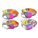 , Pierre-Henri Fabre and Lionel Hautier
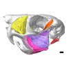
 This contribution contains the 3D model(s) described and figured in the following publication: Da Cunha, L., Fabre, P.-H. & Hautier, L. (2024) Springhares, flying and flightless scaly-tailed squirrels (Anomaluromorpha, Rodentia) are the squirrely mouse: comparative anatomy of the masticatory musculature and its implications on the evolution of hystricomorphy in rodents. Journal of Anatomy, 244, 900–928.
Anomalurus derbianus 21804 View specimen
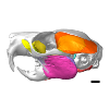
|
M3#1493Masticatory apparatus of Anomalurus Type: "3D_surfaces"doi: 10.18563/m3.sf.1493 state:published |
Download 3D surface file |
Idiurus macrotis 29335 View specimen
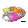
|
M3#1492Masticatory apparatus of Idiurus Type: "3D_surfaces"doi: 10.18563/m3.sf.1492 state:published |
Download 3D surface file |
Zenkerella insignis 5.5.23.27 View specimen
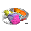
|
M3#1490Masticatory apparatus of Zenkerella Type: "3D_surfaces"doi: 10.18563/m3.sf.1490 state:published |
Download 3D surface file |
Pedetes capensis NA View specimen
|
M3#1491Masticatory apparatus of Pedetes Type: "3D_surfaces"doi: 10.18563/m3.sf.1491 state:published |
Download 3D surface file |
 , Morgane Dubied , Kévin Le Verger and Bastien Mennecart
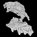
 The present 3D Dataset contains the 3D model analyzed in the following publication: Solé et al. (2018), Niche partitioning of the European carnivorous mammals during the paleogene. Palaios. https://doi.org/10.2110/palo.2018.022
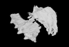
Hyaenodon leptorhynchus FSL848325 View specimen
|
M3#336The specimen FSL848325 is separated in two fragments: the anterior part bears the incisors, the deciduous and permanent canines, while the posterior part bears the right P3, P4, M1 and M2. The P2 is isolated. When combined, the cranium length is approximatively 10.5 cm long. The anterior part is 6.9 cm long and 2.15 cm wide (taken at the level of the P1). The posterior part is 4.8 cm long. The anterior part of the cranium is very narrow. Type: "3D_surfaces"doi: 10.18563/m3.sf.336 state:published |
Download 3D surface file |
Page 1 of 1, showing 5 record(s) out of 5 total