|
3D Printing an Explodable Dog Skull for Veterinary Education
Published online: 17/12/2025
Keywords:
anatomy; cranium; Education; Outreach; Teaching
https://doi.org/10.18563/journal.m3.276
Cite this article:
William C. Hooker, Ruihan R. Liu, Alexis M. Slack, Emma R. Schachner, William G. Ryerson and Brandon P. Hedrick, 2025. 3D Printing an Explodable Dog Skull for Veterinary Education. MorphoMuseuM e276. doi: 10.18563/journal.m3.276
Export citation
Abstract
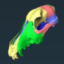
Veterinary education often relies on cadaveric specimens, but there is increasing demand for alternatives due to limited resources and ethical considerations. To address this, we developed a 3D printed ‘explodable’ model of a dog cranium with detachable, magnetized cranial components for teaching anatomy to students. This model was generated from a computed tomographic scan of a juvenile dog cranium for which cranial sutures were still partially open and segmented such that major cranial bones were isolated. All bones are printed at actual size and retain openings for cranial nerves and major vessels. This interactive model enhances anatomical education by supplying a hands-on tool that can be used either in the classroom setting or for independent learning and can be incorporated at the high school, college, or veterinary school level. It is currently being integrated into the first-year anatomy foundation course at Cornell University’s College of Veterinary Medicine. The model can be printed using any hobbyist or specialist 3D printer and we outline assembly instructions on how to attach magnets at prefabricated attachment points. Using both digital and 3D printed resources, we hope to help to address current shortages of anatomical resources and also inspire future generations of practicing veterinarians by making anatomy more accessible and engaging.
M3 article infos
Published in Volume 11, issue 04 (2025)
|
PDF
S.I. Data
|


