3D models of teeth and neurovascular system in the rostrum of Spinosauridae (theropod dinosaurs)
Explodable 3D Dog Skull for Veterinary Education
3D models related to the publication : Nouvelles données géologiques et biostratigraphiques du gisement paléontologique à vertébrés de Mauvières, à Marcilly-sur-Maulne (Miocène Inférieur et Moyen ; Indre-et-Loire, France)
3D GM dataset of bird skeletal variation
Skeletal embryonic development in the catshark
Bony connexions of the petrosal bone of extant hippos
bony labyrinth (14) , Eocene (11) , inner ear (11) , CT-scan (10) , geometric morphometrics (10) , Miocene (9) , Oligocene (9)
Lionel Hautier (25) , Maëva Judith Orliac (24) , Laurent Marivaux (20) , Bastien Mennecart (16) , Renaud Lebrun (15) , Rodolphe Tabuce (15) , Pierre-Olivier Antoine (13)
MorphoMuseuM Volume 05, issue 04
<< prev. article next article >>
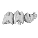
|
3D dataset3D reconstructions of dental epithelium during Oryctolagus cuniculus embryonic development related to the publication ”Morphological features of tooth development and replacement in the rabbit Oryctolagus cuniculus”Ludivine Bertonnier-Brouty
Published online: 30/09/2019 |
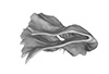
|
M3#390Right cheek teeth, Left and right incisors at 14 dpf Type: "3D_surfaces"doi: 10.18563/m3.sf.390 state:published |
Download 3D surface file |
Oryctogalus cuniculus E16 View specimen
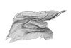
|
M3#391Left cheek teeth, Left and right incisors at 16 dpf Type: "3D_surfaces"doi: 10.18563/m3.sf.391 state:published |
Download 3D surface file |
Oryctogalus cuniculus E18 View specimen
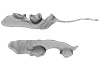
|
M3#392Left cheek teeth and incisors at 18 dpf Type: "3D_surfaces"doi: 10.18563/m3.sf.392 state:published |
Download 3D surface file |
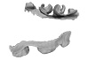
Oryctogalus cuniculus E20 View specimen
|
M3#393Left cheek teeth and incisors at 20 dpf Type: "3D_surfaces"doi: 10.18563/m3.sf.393 state:published |
Download 3D surface file |
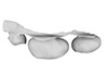
Oryctogalus cuniculus E22 View specimen
|
M3#394Left lower cheek teeth and incisors, right upper cheek teeth and incisors at 22 dpf Type: "3D_surfaces"doi: 10.18563/m3.sf.394 state:published |
Download 3D surface file |
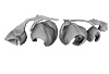
Oryctogalus cuniculus E24 View specimen
|
M3#395Left cheek teeth and incisors at 24 dpf Type: "3D_surfaces"doi: 10.18563/m3.sf.395 state:published |
Download 3D surface file |
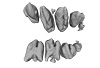
Oryctogalus cuniculus E28 View specimen
|
M3#396Right cheek teeth and incisors at 28 dpf Type: "3D_surfaces"doi: 10.18563/m3.sf.396 state:published |
Download 3D surface file |
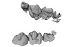
Oryctogalus cuniculus E26 View specimen
|
M3#397Right cheek teeth and incisors at 26 dpf Type: "3D_surfaces"doi: 10.18563/m3.sf.397 state:published |
Download 3D surface file |
Bertonnier Brouty L., Viriot L., Joly T., Charles C. 2019, 3D reconstructions of dental epithelium during Oryctolagus cuniculus embryonic development. Archives of Oral Biology, https://doi.org/10.1016/j.archoralbio.2019.104576
Ludivine Bertonnier-Brouty, Laurent Viriot, Thierry Joly and Cyril Charles (2020). Morphological features of tooth development and replacement in the rabbit Oryctolagus cuniculus. Archives of Oral Biology. https://doi.org/10.1016/j.archoralbio.2019.104576
Ludivine Bertonnier‐Brouty, Laurent Viriot, Thierry Joly and Cyril Charles (2021). Gene expression patterns associated with dental replacement in the rabbit, a new model for the mammalian dental replacement mechanisms. Developmental Dynamics. https://doi.org/10.1002/dvdy.335