3D models of Cainotheriids Ossicular chain
Explodable 3D Dog Skull for Veterinary Education
3D models of Kalakocetus, the earliest Cetacea
3D GM dataset of bird skeletal variation
Skeletal embryonic development in the catshark
Bony connexions of the petrosal bone of extant hippos
bony labyrinth (14) , inner ear (11) , Eocene (11) , geometric morphometrics (10) , CT-scan (10) , Oligocene (9) , Micro-CT (9)
Lionel Hautier (25) , Maëva Judith Orliac (24) , Laurent Marivaux (18) , Renaud Lebrun (15) , Rodolphe Tabuce (15) , Pierre-Olivier Antoine (13) , Bastien Mennecart (13)
Page 1 of 1, showing 4 record(s) out of 4 total

|
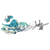
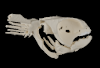
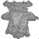
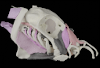
3D models related to the publication: The pharynx of the iconic stem-group chondrichthyan Acanthodes Agassiz, 1833 revisited with micro computed tomography.Richard Dearden
Published online: 25/06/2024 |
|
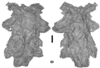
M3#14703D surfaces representing the three-dimensionally fossilised head of Acanthodes confusus Type: "3D_surfaces"doi: 10.18563/m3.sf.1470 state:published |
Download 3D surface file |
Acanthodes confusus MNHN-F-SAA21 View specimen
|
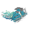
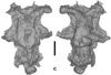
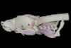
M3#14713D surfaces representing the three-dimensionally fossilised head of Acanthodes confusus Type: "3D_surfaces"doi: 10.18563/m3.sf.1471 state:published |
Download 3D surface file |
Acanthodes confusus MNHN-F-SAA24 View specimen
|
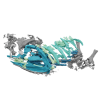
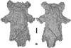
M3#14723D surfaces representing the three-dimensionally fossilised head of Acanthodes confusus Type: "3D_surfaces"doi: 10.18563/m3.sf.1472 state:published |
Download 3D surface file |
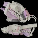
The present 3D Dataset contains 3D models of the cranial, visceral, and pectoral endoskeleton of Iniopera, an iniopterygian stem-group holocephalan from the Pennsylvanian of the USA. These data formed the basis for the analyses carried out in Dearden et al. (2023) “Evidence for high-performance suction feeding in the Pennsylvanian stem-group holocephalan Iniopera” PNAS.
Iniopera sp. KUNHM 22060, 158289 View specimen
|
M3#1034plys of the head endoskeleton of Iniopera sp. Type: "3D_surfaces"doi: 10.18563/m3.sf.1034 state:published |
Download 3D surface file |
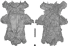
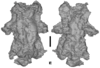
The present 3D Dataset contains the 3D models of Carboniferous-Permian chondrichthyan neurocrania analyzed in “Phylogenetic implications of the systematic reassessment of Xenacanthiformes and ‘Ctenacanthiformes’ (Chondrichthyes) neurocrania from the Carboniferous-Permian Autun Basin (France)”.
cf. Triodus sp MNHN.F.AUT811 View specimen
|
M3#834MHNH.F.AUT811 (isolated neurocranium) in dorsal view. Type: "3D_surfaces"doi: 10.18563/m3.sf.834 state:published |
Download 3D surface file |
indet indet MNHN.F.AUT812 View specimen
|
M3#835MHNH.F.AUT812 (isolated neurocranium) in dorsal view. Type: "3D_surfaces"doi: 10.18563/m3.sf.835 state:published |
Download 3D surface file |
indet indet MNHN.F.AUT813 View specimen
|
M3#836MHNH.F.AUT813 (isolated neurocranium) in dorsal view. Type: "3D_surfaces"doi: 10.18563/m3.sf.836 state:published |
Download 3D surface file |
cf. Triodus sp MNHN.F.AUT814 View specimen
|
M3#837MHNH.F.AUT814 (isolated neurocranium) in dorsal view. Type: "3D_surfaces"doi: 10.18563/m3.sf.837 state:published |
Download 3D surface file |
cf. Triodus sp MHNE.2021.9.1 View specimen
|
M3#838MHNE.2021.9.1 (isolated neurocranium) in dorsal view. Type: "3D_surfaces"doi: 10.18563/m3.sf.838 state:published |
Download 3D surface file |
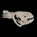
This contribution contains 3D models of the cranial skeleton and muscles in an elephantfish (Callorhinchus milii) and a catshark (Scyliorhinus canicula), based on synchrotron tomographic scans. These datasets were analyzed and described in Dearden et al. (2021) “The morphology and evolution of chondrichthyan cranial muscles: a digital dissection of the elephantfish Callorhinchus milii and the catshark Scyliorhinus canicula.” Journal of Anatomy.
Callorhinchus milii 001 View specimen
|
M3#7083D models of the cranial skeleton and muscles of Callorhinchus milii, created using Mimics. Type: "3D_surfaces"doi: 10.18563/m3.sf.708 state:published |
Download 3D surface file |
Scyliorhinus canicula 002 View specimen
|
M3#7093D models of the cranial skeleton and muscles of Scyliorhinus canicula, created using Mimics. Type: "3D_surfaces"doi: 10.18563/m3.sf.709 state:published |
Download 3D surface file |
Page 1 of 1, showing 4 record(s) out of 4 total


