|
3D models related to the publication: The morphology and evolution of chondrichthyan cranial muscles: a digital dissection of the elephantfish Callorhinchus milii and the catshark Scyliorhinus canicula
Published online: 11/01/2021
Keywords:
chondrichthyan; cranial muscles; digital dissection; elasmobranch; holocephalan
https://doi.org/10.18563/journal.m3.133
Cite this article:
Richard Dearden, Rohan Mansuit, Anthony Herrel, Antoine Cuckovic, Dominique Didier, Paul Tafforeau and Alan Pradel, 2021.
3D models related to the publication: The morphology and evolution of chondrichthyan cranial muscles: a digital dissection of the elephantfish Callorhinchus milii and the catshark Scyliorhinus canicula. MorphoMuseuM 6:e133. doi: 10.18563/journal.m3.133
Export citation
Abstract
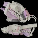
This contribution contains 3D models of the cranial skeleton and muscles in an elephantfish (Callorhinchus milii) and a catshark (Scyliorhinus canicula), based on synchrotron tomographic scans. These datasets were analyzed and described in Dearden et al. (2021) “The morphology and evolution of chondrichthyan cranial muscles: a digital dissection of the elephantfish Callorhinchus milii and the catshark Scyliorhinus canicula.” Journal of Anatomy.
M3 article infos
Published in Volume 07, issue 01 (2021)
|
PDF
|


