3D models of Cainotheriids Ossicular chain
Explodable 3D Dog Skull for Veterinary Education
3D models of Kalakocetus, the earliest Cetacea
3D GM dataset of bird skeletal variation
Skeletal embryonic development in the catshark
Bony connexions of the petrosal bone of extant hippos
bony labyrinth (14) , inner ear (11) , Eocene (11) , geometric morphometrics (10) , CT-scan (10) , Oligocene (9) , Micro-CT (9)
Lionel Hautier (25) , Maëva Judith Orliac (24) , Laurent Marivaux (18) , Renaud Lebrun (15) , Rodolphe Tabuce (15) , Pierre-Olivier Antoine (13) , Bastien Mennecart (13)
MorphoMuseuM Volume 03, Issue 03
<< prev. article next article >>
|
Original article : anatomy atlasMicroCT survey of larval skeletal mineralization in the Cuban gar Atractosteus tristoechus (Actinopterygii; Lepisosteiformes)Raphaël Scherrer
Published online: 17/05/2017 |
|
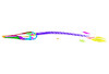
M3#94At1-13dph : 13 dph larvae, 21 mm TL Type: "3D_surfaces"doi: 10.18563/m3.sf.94 state:published |
Download 3D surface file |
Atractosteus tristoechus At2-16dph View specimen
|
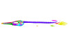
M3#95Atractosteus tristoechus larva, 16 dph, 26mm SL. Type: "3D_surfaces"doi: 10.18563/m3.sf.95 state:published |
Download 3D surface file |
Atractosteus tristoechus At3-19dph View specimen
|
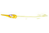
M3#96Atractosteus tristoechus larva, 19 dph, 27mm SL. Type: "3D_surfaces"doi: 10.18563/m3.sf.96 state:published |
Download 3D surface file |
Atractosteus tristoechus At4-22dph View specimen
|
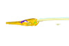
M3#97Atractosteus tristoechus larva, 22dph, 30mm SL. Type: "3D_surfaces"doi: 10.18563/m3.sf.97 state:published |
Download 3D surface file |
Atractosteus tristoechus At5-26dph View specimen
|
M3#98Atractosteus tristoechus larva, 26 dph, 32mm SL. Type: "3D_surfaces"doi: 10.18563/m3.sf.98 state:published |
Download 3D surface file |
Atractosteus tristoechus At6-31dph View specimen
|
M3#99Atractosteus tristoechus larva, 31 dph, 39mm SL. Type: "3D_surfaces"doi: 10.18563/m3.sf.99 state:published |
Download 3D surface file |
Atractosteus tristoechus At7-37dph View specimen
|
M3#100Atractosteus tristoechus larva, 37 dph, 43mm SL. Type: "3D_surfaces"doi: 10.18563/m3.sf.100 state:published |
Download 3D surface file |
Atractosteus tristoechus At8-52dph View specimen
|
M3#101Atractosteus tristoechus larva, 52 dph, 46mm SL. Type: "3D_surfaces"doi: 10.18563/m3.sf.101 state:published |
Download 3D surface file |
Atractosteus tristoechus At9-74dph View specimen
|
M3#102Atractosteus tristoechus larva, 74 dph, 61mm SL. Not all structures are colored, only newly ossified ones. Type: "3D_surfaces"doi: 10.18563/m3.sf.102 state:published |
Download 3D surface file |
Atractosteus tristoechus At10-89dph View specimen
|
M3#103Atractosteus tristoechus larva, 89 dph, 63mm SL. Not all structures are colored, only newly ossified ones. You may find the tag file in the At1-13dph reconstruction data. Type: "3D_surfaces"doi: 10.18563/m3.sf.103 state:published |
Download 3D surface file |
Atractosteus tristoechus At11-104dph View specimen
|
M3#104Atractosteus tristoechus larva, 104 dph, 70mm SL. Not all structures are colored, only newly ossified ones. Type: "3D_surfaces"doi: 10.18563/m3.sf.104 state:published |
Download 3D surface file |
Atractosteus tristoechus At12-118dph View specimen
|
M3#105Atractosteus tristoechus larva, 118 dph, 87mm SL. Type: "3D_surfaces"doi: 10.18563/m3.sf.105 state:published |
Download 3D surface file |
Arratia, G., 2009. Identifying patterns of diversity of the actinopterygian fulcra. Acta Zoologica. 90, 220–235. https://doi.org/10.1111/j.1463-6395.2008.00375.x
Arratia, G., Schultze, H.-P., 1991. Palatoquadrate and Its Ossifications: Development and Homology Within Osteichthyans. Journal of Morphology. 208, 1–81. https://doi.org/10.1002/jmor.1052080102
Arratia, G., Schultze, H.-P., 1992. Reevaluation of the caudal skeleton of certain actinopterygian fishes: III. Salmonidae. Homologization of caudal skeletal structures. Journal of Morphology. 214, 187–249. https://doi.org/10.1002/jmor.1052140209
Betancur-R, R., Broughton, R.E., Wiley, E.O., Carpenter, K., López, J.A., Li, C., Holcroft, N.I., Arcila, D., Sanciangco, M., Ii, J.C.C., Zhang, F., Campbell, M.A., Ballesteros, J.A., Roa-varon, A., Willis, S., Borden, W.C., Hough, D.J., Lu, G., 2013. The Tree of Life and a New Classification of Bony Fishes. PLOS Currents Tree of Life. 732988, 1–45. https://doi.org/10.1371/currents.tol.53ba26640df0ccaee75bb165c8c26288
Boughner, J.C., Buchtová, M., Fu, K., Diewert, V., Hallgrímsson, B., Richman, J.M., 2007. Embryonic development of Python sebae - I: Staging criteria and macroscopic skeletal morphogenesis of the head and limbs. Zoology. 110, 212–230. https://doi.org/10.1016/j.zool.2007.01.005
Britz, R., Johnson, G.D., 2010. Occipito-vertebral fusion in actinopterygians: conjecture, myth and reality. Part 1: Non-teleosts. In: Mesozoic Fishes 4-Homology and Phylogeny. J. S. Nelson, H.-P. Schultze, and M. V. H. Wilson (Eds.). Verlag Dr. Freidrich Pfeil, München. pp. 77–93.
Broughton, R.E., Betancur-R., R., Li, C., Arratia, G., Ortí, G., 2013. Multi-locus phylogenetic analysis reveals the pattern and tempo of bony fish evolution, PLoS Currents Tree of Life. https://doi.org/10.1371/currents.tol.2ca8041495ffafd0c92756e75247483e
Cignoni, P., Cignoni, P., Callieri, M., Callieri, M., Corsini, M., Corsini, M., Dellepiane, M., Dellepiane, M., Ganovelli, F., Ganovelli, F., Ranzuglia, G., Ranzuglia, G., 2008. MeshLab: an Open-Source Mesh Processing Tool. Sixth Eurographics Italian Chapter Conference. 129–136.
Dean, M.N., Mull, C.G., Gorb, S.N., Summers, A.P., 2009. Ontogeny of the tessellated skeleton: Insight from the skeletal growth of the round stingray Urobatis halleri. Journal of Anatomy. 215, 227–239. https://doi.org/10.1111/j.1469-7580.2009.01116.x
Dufeau, D.L., Witmer, L.M., 2015. Ontogeny of the Middle-Ear Air-Sinus System in Alligator mississippiensis (Archosauria: Crocodylia). PLoS ONE. 10, 1–25. https://doi.org/10.1371/journal.pone.0137060
Enault S., Adnet S., Debiais-Thibaud M., 2016. Skeletogenesis during the late embryonic development of the catshark Scyliorhinus canicula (Chondrichthyes; Neoselachii). MorphoMuseuM 1 (4)-e2. https://doi.org/10.18563/m3.1.4.e2
Friedman, M., 2015. The early evolution of ray-finned fishes. Palaeontology. 58, 213–228. https://doi.org/10.1111/pala.12150
Gardiner, B.G., Littlewood, D.T.G., Maisey, J.G., 1996. Interrelationships of basal neopterygians. In: Stiassny, M.L.J., Parenti, L.R., Johnson, G.D. (Eds.), Interrelationships of Fishes. Academic Press, San Diego, California, pp. 117–146. https://doi.org/10.1016/B978-012670950-6/50007-2
Gardiner, B.G., Schaeffer, B., 1989. Interrelationships of lower actinopterygian fishes. Zoological Journal of the Linnean Society. 1989, 135–187. https://doi.org/10.1111/j.1096-3642.1989.tb00550.x
Gardiner, B.G., Schaeffer, B., Masserie, J.A., 2005. A review of the lower actinopterygian phylogeny. Zoological Journal of the Linnean Society. 144, 511–525. https://doi.org/10.1111/j.1096-3642.2005.00181.x
Gegenbauer, C., 1887. Ueber die Occipitalregion und die ihr benachbarten Wirbel der Fische. In: Festschrift Albert von Kölliker Zur Feier Seines Siebenzigsten Geburtstages Gewidmet von Seinen Schülern. W. Engelmann, Leipzig.
Grande, L., 2010. An empirical synthetic pattern study of gars (Lepisosteiformes) and closely related species, based mostly on skeletal anatomy. The resurrection of Holostei., American Society of Ichthyologists and Herpetologists Special Publication 6:i–x, 1–871; supplementary issue of Copeia 10 (2A).
Grande, L., Bemis, W.E., 1991. Osteology and phylogenetic relationships of fossil and recent paddlefishes (Polyodontidae) with comments on the interrelationships of Acipenseriformes. Journal of Vertebrate Paleontology. 11, 1-121. https://doi.org/10.1080/02724634.1991.10011424
Grande, L., Bemis, W.E., 1998. A Comprehensive Phylogenetic Study of Amiid Fishes (Amiidae) Based on Comparative Skeletal Anatomy. an Empirical Search for Interconnected Patterns of Natural History. Journal of Vertebrate Paleontology. 18, 1–696. https://doi.org/10.1080/02724634.1998.10011114
Gregory, W.K.K., 1959. Fish skulls - A study of the evolution of natural mechanisms, Transactions of the American Philosophical Society. Eric Lundberg, Laurel, Florida.
Hammarberg, F., 1937. Zur Kenntnis der Ontogenetischen Entwicklung des Schädels von Lepisosteus platystomus. Acta Zoologica. 18, 210–336. https://doi.org/10.1111/j.1463-6395.1937.tb00680.x
Helfman, G., Collette, B., Facey, D., 2009. The diversity of fishes—biology, evolution, and ecology, 2nd edition. Wiley-Blackwell, Oxford, UK.
Hilton, E.J., Konstantinidis, P., Schnell, N.K., Dillman, C.B., 2014. Identity of a unique cartilage in the buccal cavity of gars (Neopterygii: Lepisosteiformes: Lepisosteidae). Copeia. 2014, 50–55. https://doi.org/10.1643/CI-12-173
Hilton, E.J., Schnell, N.K., Konstantinidis, P., 2015. When Tradition Meets Technology: Systematic Morphology of Fishes in the Early 21st Century. Copeia. 103, 858–873. https://doi.org/10.1643/CI-14-178
Ho, T.V., Iwata, J., Ho, H.A., Grimes, W.C., Park, S., Sanchez-Lara, P.A., Chai, Y., 2015. Integration of comprehensive 3D microCT and signaling analysis reveals differential regulatory mechanisms of craniofacial bone development. Developmental Biology. 400, 180–190. https://doi.org/10.1016/j.ydbio.2015.02.010
Hurley, I.A., Mueller, R.L., Dunn, K.A., Schmidt, E.J., Friedman, M., Ho, R.K., Prince, V.E., Yang, Z., Thomas, M.G., Coates, M.I., 2007. A new time-scale for ray-finned fish evolution. Proceedings. Biological sciences / The Royal Society. 274, 489–498. https://doi.org/10.1098/rspb.2006.3749
Huysseune, A., Sire, J.-Y., 1998. Evolution of patterns and processes in teeth and tooth-related tissues in non-mammalian vertebrates. European journal of oral sciences. 106, 437–481. https://doi.org/10.1111/j.1600-0722.1998.tb02211.x
Jessen, H., 1972. Schultergürtel und Pectoralflosse bei Actinopterygiern. Fossil Strata. 1, 1–101.
Jollie, M., 1984. Development of Cranial and Pectoral Girdle Bones of Lepisosteus with a Note on Scales. Copeia. 2, 476–502. https://doi.org/10.2307/1445204
Kammerer, C.F.F., Grande, L., Westneat, M.W.W., 2006. Comparative and Developmental Functional Morphology of the Jaws of Living and Fossil Gars (Actinopterygii : Lepisosteidae). Journal of morphology. 267, 1017–31. https://doi.org/10.1002/jmor.10293
Konstantinidis, P., Warth, P., Naumann, B., Metscher, B., Hilton, E.J., Olsson, L., 2015. The Developmental Pattern of the Musculature Associated with the Mandibular and Hyoid Arches in the Longnose Gar, Lepisosteus osseus (Actinopterygii, Ginglymodi, Lepisosteiformes). Copeia. 103, 920–932. https://doi.org/10.1643/OT-14-195
Kryzanovsky, S., 1927. Die Entwicklung der paarigen Flossen bei Acipenser, Amia und Lepidosteus. Acta Zoologica. 8, 278–352. https://doi.org/10.1111/j.1463-6395.1927.tb00654.x
Lauder, G. V., 1980. Evolution of the feeding mechanism in primitive actionopterygian fishes: A functional anatomical analysis of Polypterus, Lepisosteus, and Amia. Journal of Morphology. 163, 283–317. https://doi.org/10.1002/jmor.1051630305
Lebrun, R., 2014. ISE-MeshTools, a 3D interactive fossil reconstruction freeware. In: 12th Annual Meeting of EAVP, Torino, Italy.
Lecointre, G., Philippe, H., Van Le, H.L., Le Guyader, H., 1994. How many nucleotides are required to resolve a phylogenetic problem? The use of a new statistical method applicable to available sequences. Molecular Phylogenetics and Evolution. https://doi.org/10.1006/mpev.1994.1037
Long, J.M., Snow, R.A., 2016. Ontogenetic development of otoliths in alligator gar. Transactions of the American Fisheries Society. 145, 537–544. https://doi.org/10.1080/00028487.2015.1135189
Long, W.L., Ballard, W.W., 2001. Normal embryonic stages of the longnose gar, Lepisosteus osseus. BMC developmental biology. 1, 6. https://doi.org/10.1186/1471-213X-1-6
López-Arbarello, A., 2012. Phylogenetic Interrelationships of Gynglymodian Fishes (Actinopterygii: Neopterygii). PLoS ONE. 7, 1–44. https://doi.org/10.1371/journal.pone.0039370
Luther, A., 1913. Ueber die vom N. trigeminus versorgte Muskulatur der Ganoiden und Dipneusten. Helsingfors Acta Societas Scientiarum Fennica. 41, 1–72.
Maxwell, E.E., Wilson, L.A.B., 2013. Regionalization of the axial skeleton in the “ambush predator” guild -- are there developmental rules underlying body shape evolution in ray-finned fishes? BMC Evolutionary Biology. 13, 265. https://doi.org/10.1186/1471-2148-13-265
Mayhew, R.L., 1924. The skull of Lepidosteus platostomus. Journal of Morphology. 38, 315–346. https://doi.org/10.1002/jmor.1050380303
Metscher, B.D., 2009. MicroCT for developmental biology: A versatile tool for high-contrast 3D imaging at histological resolutions. Developmental Dynamics. 238, 632–640. https://doi.org/10.1002/dvdy.21857
Near, T.J., Eytan, R.I., Dornburg, A., Kuhn, K.L., Moore, J.A., Davis, M.P., Wainwright, P.C., Friedman, M., Smith, W.L., 2012. Resolution of ray-finned fish phylogeny and timing of diversification. Proceedings of the National Academy of Sciences. 109, 13698–13703. https://doi.org/10.1073/pnas.1206625109
Nelson, J.S., Grande, T.C., M. V. H. W, 2016. Order LEPISOSTEIFORMES - gars. In: Fishes of the World. John Wiley & Sons, New York.
Normark, B.B., McCune, a R., Harrison, R.G., 1991. Phylogenetic relationships of neopterygian fishes, inferred from mitochondrial DNA sequences. Molecular biology and evolution. 8, 819–834.
Palci, A., Lee, M.S.Y., Hutchinson, M.N., 2016. Patterns of postnatal ontogeny of the skull and lower jaw of snakes as revealed by micro-CT scan data and three-dimensional geometric morphometrics. Journal of Anatomy. https://doi.org/10.1111/joa.12509
Perry, S.F., Wilson, R.J. a, Straus, C., Harris, M.B., Remmers, J.E., 2001. Which came first, the lung or the breath? Comparative Biochemistry and Physiology - A Molecular and Integrative Physiology. 129, 37–47. https://doi.org/10.1016/S1095-6433(01)00304-X
Rice, R., Kallonen, A., Cebra-Thomas, J., Gilbert, S.F., 2016. Development of the turtle plastron, the order-defining skeletal structure. Proceedings of the National Academy of Sciences of the United States of America. 113, 5317–5322. https://doi.org/10.1073/pnas.1600958113
Rücklin, M., Donoghue, P.C.J., Cunningham, J.A., Marone, F., Stampanoni, M., 2014. Developmental Paleobiology of the Vertebrate Skeleton. Journal of Paleontology. 88, 676–683. https://doi.org/10.1666/13-107
Schindelin, J., Arganda-Carreras, I., Frise, E., Kaynig, V., Longair, M., Pietzsch, T., Preibisch, S., Rueden, C., Saalfeld, S., Schmid, B., Tinevez, J.-Y., White, D.J., Hartenstein, V., Eliceiri, K., Tomancak, P., Cardona, A., 2012. Fiji: an open-source platform for biological-image analysis. Nature Methods. 9, 676–682. https://doi.org/10.1038/nmeth.2019
Schmalhausen, J.J., 1913. Zur Morphologie der unpaaren Flossen. II. Bau und Phylogenese der unpaaren Flossen und insbesonders der Schwanzflosse der Fische. Zeitschrift für wissenschaftliche Zoologie. 104, 1–80.
Schreiner, K.E., 1902. Einige Ergebnisse über den Bau und der Occipitalregion von Amia und Lepidosteus. Zetischriften. wiss. Zool. 72, 467–524.
Schultze, H.-P., Cloutier, R., 1991. Computed Tomography and Magnetic Resonance Imaging studies of Latimeria chalumnae. Environmental Biology of Fishes. 32, 159–181. https://doi.org/10.1007/BF00007451
Sewertzoff, A.N., 1895. Die Entwicklung der Occipitalregion der niederen Vertebraten im Zusammenhang mit der Frage nach der Metamerie des Kopfes. Bulletin de la société impériale des naturalistes de Moscou. 9, 186–284.
Shen, L., Ai, H., Liang, Y., Ren, X., Anthony, C.B., Goodlett, C.R., Ward, R., Zhou, F.C., 2013. Effect of prenatal alcohol exposure on bony craniofacial development: a mouse MicroCT study. Alcohol (Fayetteville, N.Y.). 47, 405–15. https://doi.org/10.1016/j.alcohol.2013.04.005
Sire, J.-Y., 1989. Scales in Young Polypterus senegalus Are Elasmoid : New Phylogenetic ImpIications. The American Journal of Anatomy. 186, 315–323. https://doi.org/10.1002/aja.1001860308
Sire, J.-Y., 1990. From ganoid to elasmoid scales in the actinopterygian fishes. Netherlands journal of zoology. 40, 75–92. https://doi.org/10.1163/156854289X00192
Sire, J.-Y., Donoghue, P.C.J., Vickaryous, M.K., 2009. Origin and evolution of the integumentary skeleton in non-tetrapod vertebrates. Journal of Anatomy. 214, 409–440. https://doi.org/10.1111/j.1469-7580.2009.01046.x
Sire, J.-Y., Géraudie, J., Meunier, F.J., Zylberberg, L., 1987. On the Origin of Ganoine: Histological and Ultrastructural Data on the Experimental Regeneration of the Scales of Calamoichthys calabaricus (Osteichthyes, Brachyopterygii, Polypteridae). The American Journal of Anatomy. 180, 391–402. https://doi.org/10.1002/aja.1001800409
Slater, B.J., Liu, K.J., Kwan, M.D., Quarto, N., Longaker, M.T., 2009. Cranial osteogenesis and suture morphology in Xenopus laevis: A unique model system for studying craniofacial development. PLoS ONE. 4. https://doi.org/10.1371/journal.pone.0003914
Veit, O., 1907. Über einige Besonderheiten am Primordialcranium von Lepidosteus osseus. Anatomische Hefte. 99, 155–204. https://doi.org/10.1007/BF02214184
Vickaryous, M.K., Sire, J.Y., 2009. The integumentary skeleton of tetrapods: Origin, evolution, and development. Journal of Anatomy. 214, 441–464. https://doi.org/10.1111/j.1469-7580.2008.01043.x
Wiley, E.O., 1976. The phylogeny and biogeography of fossil and recent gars (Actinopterygii: Lepisosteidae). University of Kansas Museum of Natural History Miscellaneous Publications. 64, 1–111.
James M. Long and Richard A. Snow (2018). Posthatch Development of Otoliths and Daily Ring Genesis in Age‐0 Spotted Gars. Transactions of the American Fisheries Society. https://doi.org/10.1002/tafs.10108
N. G. Smith, D. J. Daugherty, E. L. Brinkman, M. G. Wegener, B. R. Kreiser, A. M. Ferrara, K. D. Kimmel and S. R. David (2020). Advances in Conservation and Management of the Alligator Gar: A Synthesis of Current Knowledge and Introduction to a Special Section. North American Journal of Fisheries Management. https://doi.org/10.1002/nafm.10369
Alyssa Enny, Anusha Shanabag, Andrew W. Thompson, Brett Racicot, Ingo Braasch and Tetsuya Nakamura (2021). Cellular mechanisms of frontal bone development in spotted gar (Lepisosteus oculatus). Developmental Dynamics. https://doi.org/10.1002/dvdy.356
Simrith E. Cordova-de la Cruz, Marta F. Riesco, Gil Martínez-Bautista, Daniel Calzada-Ruiz, Talhia Martínez-Burguete, Emyr S. Peña-Marín, Carlos Alfonso Álvarez-Gonzalez and Ignacio Fernández (2022). Larval Development in Tropical Gar (Atractosteus tropicus) Is Dependent on the Embryonic Thermal Regime: Ecological Implications under a Climate Change Context. Fishes. https://doi.org/10.3390/fishes7010016