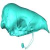
3D models of Ocnotherium skull
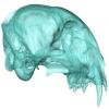
3D models of Kalakocetus, the earliest Cetacea
Explodable 3D Dog Skull for Veterinary Education
3D GM dataset of bird skeletal variation
Skeletal embryonic development in the catshark
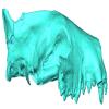
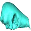
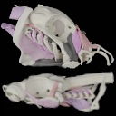
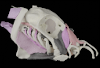
Bony connexions of the petrosal bone of extant hippos
bony labyrinth (14) , inner ear (11) , Eocene (11) , geometric morphometrics (10) , CT-scan (10) , Oligocene (9) , Micro-CT (9)
Maëva Judith Orliac (24) , Lionel Hautier (24) , Laurent Marivaux (18) , Renaud Lebrun (15) , Rodolphe Tabuce (14) , Pierre-Olivier Antoine (13) , Bastien Mennecart (13)
MorphoMuseuM Volume 07, issue 01
<< prev. article next article >>
|
3D dataset
|
|
M3#7083D models of the cranial skeleton and muscles of Callorhinchus milii, created using Mimics. Type: "3D_surfaces"doi: 10.18563/m3.sf.708 state:published |
Download 3D surface file |
Scyliorhinus canicula 002 View specimen
|
M3#7093D models of the cranial skeleton and muscles of Scyliorhinus canicula, created using Mimics. Type: "3D_surfaces"doi: 10.18563/m3.sf.709 state:published |
Download 3D surface file |
Dearden, R.P., Mansuit, R., Cuckovic, A., Herrel, A., Didier, D., Tafforeau, P., Pradel, A. 2021. The morphology and evolution of chondrichthyan cranial muscles: a digital dissection of the elephantfish Callorhinchus milii and the catshark Scyliorhinus canicula. Journal of Anatomy. https://doi.org/10.1111/joa.13362
Richard P. Dearden, Rohan Mansuit, Antoine Cuckovic, Anthony Herrel, Dominique Didier, Paul Tafforeau and Alan Pradel (2021). The morphology and evolution of chondrichthyan cranial muscles: A digital dissection of the elephantfish Callorhinchus milii and the catshark Scyliorhinus canicula. Journal of Anatomy. https://doi.org/10.1111/joa.13362
Faviel A. López-Romero, Sebastian Stumpf, Pepijn Kamminga, Christine Böhmer, Alan Pradel, Martin D. Brazeau and Jürgen Kriwet (2023). Shark mandible evolution reveals patterns of trophic and habitat-mediated diversification. Communications Biology. https://doi.org/10.1038/s42003-023-04882-3
Richard P. Dearden, Anthony Herrel and Alan Pradel (2023).
Evidence for high-performance suction feeding in the Pennsylvanian stem-group holocephalan
Iniopera
. Proceedings of the National Academy of Sciences. https://doi.org/10.1073/pnas.2207854119