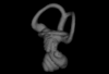
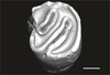
3D models of Cainotheriids Ossicular chain
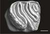
3D models of Kalakocetus, the earliest Cetacea
The specimens of Speothos pacivorus
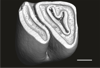
3D GM dataset of bird skeletal variation
Skeletal embryonic development in the catshark
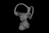
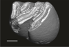
Bony connexions of the petrosal bone of extant hippos
bony labyrinth (14) , inner ear (11) , Eocene (11) , geometric morphometrics (10) , CT-scan (10) , Oligocene (9) , Micro-CT (9)
Lionel Hautier (25) , Maëva Judith Orliac (24) , Laurent Marivaux (18) , Renaud Lebrun (15) , Rodolphe Tabuce (15) , Pierre-Olivier Antoine (13) , Bastien Mennecart (13)
Page 1 of 1, showing 5 record(s) out of 5 total
|
3D models related to the publication: Hidden diversity of Palaeogene metatherians: a new family of polydolopimorphian marsupials from Peruvian AmazoniaNarla Stutz
Published online: 17/04/2026 |

|
M3#1874Pozodolops manuelorum, fragmentary left dentary with p3–m1 Type: "3D_surfaces"doi: 10.18563/m3.sf.1874 state:published |
Download 3D surface file |
Wamradolops telloi MUSM 4032 View specimen

|
M3#1875Wamradolops telloi, fragmentary right dentary with m1–m2 Type: "3D_surfaces"doi: 10.18563/m3.sf.1875 state:published |
Download 3D surface file |
Pozodolops manuelorum MUSM 4036 View specimen
|
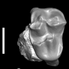
M3#1876Pozodolops manuelorum, holotype, fragmentary right maxilla with M1 Type: "3D_surfaces"doi: 10.18563/m3.sf.1876 state:published |
Download 3D surface file |
Pozodolops manuelorum MUSM 4041 View specimen

|
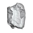
M3#1877Pozodolops manuelorum, fragmentary right dentary with m1 Type: "3D_surfaces"doi: 10.18563/m3.sf.1877 state:published |
Download 3D surface file |
Pozodolops manuelorum MUSM 4058 View specimen

|
M3#1878Pozodolops manuelorum, fragmentary left P3 Type: "3D_surfaces"doi: 10.18563/m3.sf.1878 state:published |
Download 3D surface file |
Wamradolops telloi MUSM 4144 View specimen

|
M3#1879Wamradolops telloi, fragmentary left dentary with p2–m1 Type: "3D_surfaces"doi: 10.18563/m3.sf.1879 state:published |
Download 3D surface file |
Wamradolops telloi MUSM 4179 View specimen

|
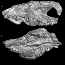
M3#1880Wamradolops telloi, holotype, partial skull, with right P2–M4 and left I4–M3, plus boneless lower teeth below upper teeth, belonging to the same individual Type: "3D_surfaces"doi: 10.18563/m3.sf.1880 state:published |
Download 3D surface file |
Wamradolops telloi MUSM 4221 View specimen

|
M3#1881Wamradolops telloi, fragmentary of right dentary with p3–m2 Type: "3D_surfaces"doi: 10.18563/m3.sf.1881 state:published |
Download 3D surface file |
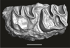
This contribution contains the 3D models described and figured in the following publication: Pujos F., Hautier L., Antoine P-O., Boivin M., Moison B, Salas-Gismondi R, Tejada J.V. , Varas-Malca R.M., Yans J., Marivaux L. (2025). Unexpected pampatheriid from the early Oligocene of Peruvian Amazonia: insights into the tropical differentiation of cingulate xenarthrans. Historical Biology.
Bradypus tridactylus UM-ZOOL-V69 View specimen
|
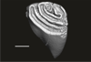
M3#1600Molariform and associated dentinal microstructure Type: "3D_surfaces"doi: 10.18563/m3.sf.1600 state:published |
Download 3D surface file |
Choloepus didactylus UM-ZOOL-V12 View specimen
|
M3#1601Molariform and associated dentinal microstructure Type: "3D_surfaces"doi: 10.18563/m3.sf.1601 state:published |
Download 3D surface file |
Dasypus mexicanus UM-ZOOL-2787 View specimen

|
M3#1602Molariform and associated dentinal microstructure Type: "3D_surfaces"doi: 10.18563/m3.sf.1602 state:published |
Download 3D surface file |
Tolypeutes matacus UM-ZOOL-2789 View specimen
|
M3#1603Molariform and associated dentinal microstructure Type: "3D_surfaces"doi: 10.18563/m3.sf.1603 state:published |
Download 3D surface file |
Euphractus sexcinctus UM-ZOOL-2790 View specimen
|
M3#1604Molariform and associated dentinal microstructure Type: "3D_surfaces"doi: 10.18563/m3.sf.1604 state:published |
Download 3D surface file |
Holmesina septrionalis UM-FLD-1 View specimen
|
M3#1605Molariform and associated dentinal microstructure Type: "3D_surfaces"doi: 10.18563/m3.sf.1605 state:published |
Download 3D surface file |
Megatherium sp. UM-TAR-1 View specimen
|
M3#1607Molariform and associated dentinal microstructure Type: "3D_surfaces"doi: 10.18563/m3.sf.1607 state:published |
Download 3D surface file |
Indet indet MUSM-3965 View specimen
|
M3#1606Molariform and associated dentinal microstructure Type: "3D_surfaces"doi: 10.18563/m3.sf.1606 state:published |
Download 3D surface file |
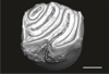
This contribution contains the three-dimensional models of the inner ear of the hetaxodontid rodents Amblyrhiza, Clidomys and Elasmodontomys from the West Indies. These specimens were analyzed and discussed in : The inner ear of caviomorph rodents: phylogenetic implications and application to extinct West Indian taxa.
Amblyrhiza inundata 11842 View specimen
|
M3#11543D surface of the left-oriented inner ear of Amblyrhiza. Type: "3D_surfaces"doi: 10.18563/m3.sf.1154 state:published |
Download 3D surface file |
Clidomys sp NA View specimen
|
M3#11553D surface of the left-oriented inner ear of Clidomys sp. Type: "3D_surfaces"doi: 10.18563/m3.sf.1155 state:published |
Download 3D surface file |
Elasmodontomys obliquus 17127 View specimen
|
M3#11563D surface of the left-oriented inner ear of Elasmodontomys obliquus. Type: "3D_surfaces"doi: 10.18563/m3.sf.1156 state:published |
Download 3D surface file |
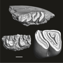
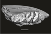
This contribution contains the three-dimensional digital models of a part of the dental fossil material (the large specimens) of caviomorph rodents, discovered in late middle Miocene detrital deposits of the TAR-31 locality in Peruvian Amazonia (San Martín, Peru). These fossils were described, figured and discussed in the following publication: Boivin, Marivaux et al. (2021), Late middle Miocene caviomorph rodents from Tarapoto, Peruvian Amazonia. PLoS ONE 16(11): e0258455. https://doi.org/10.1371/journal.pone.0258455
Microscleromys paradoxalis MUSM 4643 View specimen
|
M3#1115Fragment of left mandibule preserving dp4, m1 and a portion of incisor Type: "3D_surfaces"doi: 10.18563/m3.sf.1115 state:published |
Download 3D surface file |
Ricardomys longidens MUSM 4375 View specimen
|
M3#1116Fragment of left maxillary preserving DP4 and M1 (or M1 and M2) Type: "3D_surfaces"doi: 10.18563/m3.sf.1116 state:published |
Download 3D surface file |
"Scleromys" sp. MUSM 4272 View specimen
|
M3#1117Isolated left upper molar Type: "3D_surfaces"doi: 10.18563/m3.sf.1117 state:published |
Download 3D surface file |
"Scleromys" sp. MUSM 4275 View specimen
|
M3#1118Isolated right upper molar Type: "3D_surfaces"doi: 10.18563/m3.sf.1118 state:published |
Download 3D surface file |
"Scleromys" sp. MUSM 4273 View specimen
|
M3#1119Isolated left upper molar Type: "3D_surfaces"doi: 10.18563/m3.sf.1119 state:published |
Download 3D surface file |
"Scleromys" sp. MUSM 4276 View specimen
|
M3#1120Isolated right upper molar Type: "3D_surfaces"doi: 10.18563/m3.sf.1120 state:published |
Download 3D surface file |
"Scleromys" sp. MUSM 4282 View specimen
|
M3#1121Isolated right lower molar Type: "3D_surfaces"doi: 10.18563/m3.sf.1121 state:published |
Download 3D surface file |
"Scleromys" sp. MUSM 4281 View specimen
|
M3#1122Isolated right lower molar Type: "3D_surfaces"doi: 10.18563/m3.sf.1122 state:published |
Download 3D surface file |
"Scleromys" sp. MUSM 4280 View specimen
|
M3#1123Isolated left p4 Type: "3D_surfaces"doi: 10.18563/m3.sf.1123 state:published |
Download 3D surface file |
"Scleromys" sp. MUSM 4277 View specimen
|
M3#1124Isolated left lower dp4 Type: "3D_surfaces"doi: 10.18563/m3.sf.1124 state:published |
Download 3D surface file |
"Scleromys" sp. MUSM 4279 View specimen
|
M3#1125Isolated right lower dp4 (mesial fragment) Type: "3D_surfaces"doi: 10.18563/m3.sf.1125 state:published |
Download 3D surface file |
gen.indet sp. indet MUSM 4283 View specimen
|
M3#1126Isolated right lower p4 Type: "3D_surfaces"doi: 10.18563/m3.sf.1126 state:published |
Download 3D surface file |
Microscleromys sp. MUSM 4658 View specimen
|
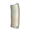
M3#1127Isolated left tarsal bone (astragalus) Type: "3D_surfaces"doi: 10.18563/m3.sf.1127 state:published |
Download 3D surface file |
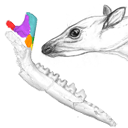
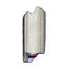
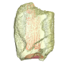
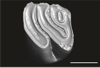
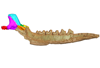
This note presents the 3D model of the hemi-mandible UM-PAT 159 of the MP7 Diacodexis species D. cf. gigasei and 3D models corresponding to the restoration of the ascending ramus, broken on the original specimen, and to a restoration of a complete mandible based on the preserved left hemi-mandible.
Diacodexis cf. gigasei UMPAT159 View specimen
|
M3#3153D models of UM PAT 159 after the restoration of the ascending ramus Type: "3D_surfaces"doi: 10.18563/m3.sf.315 state:published |
Download 3D surface file |
|
M3#316restoration of a complete mandible based on the preserved left hemi-mandible UM PAT 159 Type: "3D_surfaces"doi: 10.18563/m3.sf.316 state:published |
Download 3D surface file |
|
M3#3173D model of the hemi-mandible UM PAT 159 Type: "3D_surfaces"doi: 10.18563/m3.sf.317 state:published |
Download 3D surface file |
Page 1 of 1, showing 5 record(s) out of 5 total


