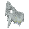
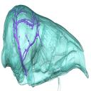
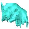
Explodable 3D Dog Skull for Veterinary Education
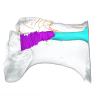
3D models of the paratympanic sinus system, the endocast and the neurovascular bony canal of the maxilla, premaxilla and the jugal of Leidyosuchus canadensis and Stangerochampsa mccabei
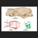
3D Topography and food processing in Palmatolepis
3D GM dataset of bird skeletal variation
Skeletal embryonic development in the catshark
Bony connexions of the petrosal bone of extant hippos
bony labyrinth (14) , inner ear (11) , geometric morphometrics (10) , Eocene (10) , CT-scan (9) , Micro-CT (9) , Miocene (8)
Lionel Hautier (24) , Maëva Judith Orliac (21) , Laurent Marivaux (17) , Rodolphe Tabuce (14) , Pierre-Olivier Antoine (13) , Bastien Mennecart (13) , Renaud Lebrun (12)
|
3D models related to the publication: The neotropical giant ground sloth Ocnotherium giganteum (Xenarthra, Mylodontinae) from the Late Pleistocene of Brazil: anatomy, paleoneurology, and phylogenetic relationshipsFrançois Pujos
Published online: 26/03/2026 |
|
M3#1870skull, endocast, inner ear Type: "3D_surfaces"doi: 10.18563/m3.sf.1870 state:in_press |
Download 3D surface file |
The present 3D Dataset contains a selection of 3D models analyzed in Billet G, Hautier L, Gaudin TJ, Flynn JJ, Ruf I, Carrillo JD, Ladevèze S, Lehmann T, Nicolas V, Orliac MJ, Tornero C, Wible JR, Wong N, Gaubert P. Submitted. Brain drain: Exceptional pattern of calvarial venation in pangolins and its phylogenetic significance for Ferae. Zoological Journal of the Linnean Society.
Phataginus tricuspis NHM-UK 48.13.26 View specimen
|
M3#1847cranium & intradiploic canals (sinuses & diploic veins) Type: "3D_surfaces"doi: 10.18563/m3.sf.1847 state:in_press |
Download 3D surface file |
Manis javanica NHM-UK 9.1.5.858 View specimen
|
M3#1848cranium & intradiploic canals (sinuses & diploic veins) Type: "3D_surfaces"doi: 10.18563/m3.sf.1848 state:in_press |
Download 3D surface file |
Felis silvestris UM-ZOOL-149N View specimen
|
M3#1849cranium & intradiploic canals (sinuses & diploic veins) Type: "3D_surfaces"doi: 10.18563/m3.sf.1849 state:in_press |
Download 3D surface file |
Erinaceus europaeus SMNS40759 View specimen

|
M3#1850cranium & intradiploic canals (sinuses & diploic veins) Type: "3D_surfaces"doi: 10.18563/m3.sf.1850 state:in_press |
Download 3D surface file |
Pterodon dasyuroides MNHN.F.Qu8301 View specimen
|
M3#1851cranium & intradiploic canals (sinuses & diploic veins) Type: "3D_surfaces"doi: 10.18563/m3.sf.1851 state:in_press |
Download 3D surface file |
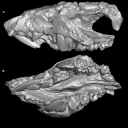
This contribution contains the 3D digital models of some fossil specimens of Wamradolops telloi Stutz and Pozodolops manuelorum Stutz (Metatheria: Polydolopimorphia), from several Palaeogene locations of Peruvian Amazonia. These taxa were described and analyzed in detail in the following publication: Stutz et al. (2026), Hidden diversity of Palaeogene metatherians: a new family of polydolopimorphian marsupials from Peruvian Amazonia. Zoological Journal of the Linnean Society. https://doi.org/10.1093/zoolinnean/zlag006.
Pozodolops manuelorum MUSM 4029 View specimen

|
M3#1874Pozodolops manuelorum, fragmentary left dentary with p3–m1 Type: "3D_surfaces"doi: 10.18563/m3.sf.1874 state:in_press |
Download 3D surface file |
Wamradolops telloi MUSM 4032 View specimen

|
M3#1875Wamradolops telloi, fragmentary right dentary with m1–m2 Type: "3D_surfaces"doi: 10.18563/m3.sf.1875 state:in_press |
Download 3D surface file |
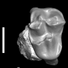
Pozodolops manuelorum MUSM 4036 View specimen
|
M3#1876Pozodolops manuelorum, holotype, fragmentary right maxilla with M1 Type: "3D_surfaces"doi: 10.18563/m3.sf.1876 state:in_press |
Download 3D surface file |
Pozodolops manuelorum MUSM 4041 View specimen

|
M3#1877Pozodolops manuelorum, fragmentary right dentary with m1 Type: "3D_surfaces"doi: 10.18563/m3.sf.1877 state:in_press |
Download 3D surface file |
Pozodolops manuelorum MUSM 4058 View specimen

|
M3#1878Pozodolops manuelorum, fragmentary left P3 Type: "3D_surfaces"doi: 10.18563/m3.sf.1878 state:in_press |
Download 3D surface file |
Wamradolops telloi MUSM 4144 View specimen

|
M3#1879Wamradolops telloi, fragmentary left dentary with p2–m1 Type: "3D_surfaces"doi: 10.18563/m3.sf.1879 state:in_press |
Download 3D surface file |
Wamradolops telloi MUSM 4179 View specimen

|
M3#1880Wamradolops telloi, holotype, partial skull, with right P2–M4 and left I4–M3, plus boneless lower teeth below upper teeth, belonging to the same individual Type: "3D_surfaces"doi: 10.18563/m3.sf.1880 state:in_press |
Download 3D surface file |
Wamradolops telloi MUSM 4221 View specimen

|
M3#1881Wamradolops telloi, fragmentary of right dentary with p3–m2 Type: "3D_surfaces"doi: 10.18563/m3.sf.1881 state:in_press |
Download 3D surface file |