3D models of Kalakocetus, the earliest Cetacea
The specimens of Speothos pacivorus
3D models related to the publication: Hidden diversity of Palaeogene metatherians: a new family of polydolopimorphian marsupials from Peruvian Amazonia
3D GM dataset of bird skeletal variation
Skeletal embryonic development in the catshark
Bony connexions of the petrosal bone of extant hippos
bony labyrinth (14) , inner ear (11) , Eocene (11) , geometric morphometrics (10) , CT-scan (10) , Oligocene (9) , Micro-CT (9)
Maëva Judith Orliac (24) , Lionel Hautier (24) , Laurent Marivaux (18) , Renaud Lebrun (15) , Rodolphe Tabuce (14) , Pierre-Olivier Antoine (13) , Bastien Mennecart (13)
|
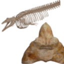
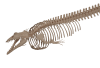
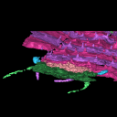
3D models related to the publication: A heavyweight early whale pushes the boundaries of vertebrate morphologyGiovanni Bianucci, Olivier Lambert
Published online: 04/08/2023 |
|
M3#1131Thirteen vertebrae, rib, and innominate of Perucetus colossus (holotype, MUSM NNNN). Type: "3D_surfaces"doi: 10.18563/m3.sf.1131 state:published |
Download 3D surface file |
Cynthiacetus peruvianus MNHN.F.PRU10 View specimen
|
M3#1130Articulated skeleton of the holotype of Cynthiacetus peruvianus MNHN.F.PRU10 Type: "3D_surfaces"doi: 10.18563/m3.sf.1130 state:published |
Download 3D surface file |
The present dataset contains the 3D models of the cheek teeth of eight raccoons analyzed in Koomen, S. E., Lang, A. J. & Martin, T. (2026). Tooth Function of the Northern Raccoon (Procyon lotor) and Adaptations to Omnivory in the Order Carnivora. Journal of Morphology.
Procyon lotor ZFMK-MAM-2013.0341 View specimen
|
M3#1838Cheek teeth of P. lotor specimen ZFMK-MAM-2013.0341 Type: "3D_surfaces"doi: 10.18563/m3.sf.1838 state:in_press |
Download 3D surface file |
Procyon lotor ZFMK-MAM-1993.0289 View specimen
|
M3#1839Cheek teeth of P. lotor specimen ZFMK-MAM-1993.0289 Type: "3D_surfaces"doi: 10.18563/m3.sf.1839 state:in_press |
Download 3D surface file |
Procyon lotor ZFMK-MAM-2016.0912 View specimen
|
M3#1840Cheek teeth of P. lotor specimen ZFMK-MAM-2016.0912 Type: "3D_surfaces"doi: 10.18563/m3.sf.1840 state:in_press |
Download 3D surface file |
Procyon lotor ZFMK-MAM-2016.0914 View specimen
|
M3#1841Cheek teeth of P. lotor specimen ZFMK-MAM-2016.0914 Type: "3D_surfaces"doi: 10.18563/m3.sf.1841 state:in_press |
Download 3D surface file |
Procyon lotor ZFMK-MAM-2016.0923 View specimen
|
M3#1842Cheek teeth of P. lotor specimen ZFMK-MAM-2016.0923 Type: "3D_surfaces"doi: 10.18563/m3.sf.1842 state:in_press |
Download 3D surface file |
Procyon lotor ZFMK-MAM-2016.0937 View specimen
|
M3#1843Cheek teeth of P. lotor specimen ZFMK-MAM-2016.0937 Type: "3D_surfaces"doi: 10.18563/m3.sf.1843 state:in_press |
Download 3D surface file |
Procyon lotor ZFMK-MAM-2016.0938 View specimen
|
M3#1844Cheek teeth of P. lotor specimen ZFMK-MAM-2016.0938 Type: "3D_surfaces"doi: 10.18563/m3.sf.1844 state:in_press |
Download 3D surface file |
Procyon lotor ZFMK-MAM-2016.0967 View specimen
|
M3#1845Cheek teeth of P. lotor specimen ZFMK-MAM-2016.0967 Type: "3D_surfaces"doi: 10.18563/m3.sf.1845 state:in_press |
Download 3D surface file |
The present 3D Dataset contains the 3D models analyzed in the publication: Head anatomy and phylogenomics show the Carboniferous giant Arthropleura was a relative to both millipedes and centipedes. Lhéritier Mickaël, Edgecombe Gregory D., Garwodd Russell J., Buisson Adrien, Gerbe Alexis, Mongiardino Koch Nicolás, Vannier Jean, Escarguel Gilles, Adrien Jérome, Fernandez Vincent, Bergeret-Medina Aude, Giupponi Alexandra and Perrier Vincent. Sciences Advances. https://www.science.org/doi/10.1126/sciadv.adp6362
Arthropleura sp. MNHN.F.SOT002123 View specimen
|
M3#1481Reconstitution of MNH.F.SOT002123 made from Phoenix X-ray Phoenix V|tome|x CT-scan Type: "3D_surfaces"doi: 10.18563/m3.sf.1481 state:published |
Download 3D surface file |
|
M3#1482Ct-scan (X-ray Phoenix V|tome|x) of MNHN.F.SOT002123 Type: "3D_CT"doi: 10.18563/m3.sf.1482 state:published |
Download CT data |
|
M3#1484Reconstitution of MNH.F.SOT002123 made from synchrotron X-ray micro-Computed tomography Type: "3D_surfaces"doi: 10.18563/m3.sf.1484 state:published |
Download 3D surface file |
|
M3#1485Synchrotron data of MNHN.F.SOT002123 (bin4) Type: "3D_CT"doi: 10.18563/m3.sf.1485 state:published |
Download CT data |
Arthropleura sp. MNHN.F.SOT002118 View specimen
|
M3#1480Reconstitution of MNH.F.SOT002118 made from Phoenix X-ray Phoenix V|tome|x CT-scan Type: "3D_surfaces"doi: 10.18563/m3.sf.1480 state:published |
Download 3D surface file |
|
M3#1483Ct-scan (X-ray Phoenix V|tome|x) of MNHN.F.SOT002118 Type: "3D_CT"doi: 10.18563/m3.sf.1483 state:published |
Download CT data |
Arthropleura sp. MNHN.F.SOT002123 (synchrotron data) View specimen
The present 3D Dataset contains the 3D models of three specimens of Spinosauridae: two Cristatusaurus lapparenti Taquet & Russell, 1998 (MNHN.F.GDF365 and MNHN.F.GDF366) and one Spinosaurus maroccanus Russell, 1996 (MNHN.F.SAM124). These specimens are analyzed and discussed in Pittet F. 2026. Neurovascular system and dental renewal in the rostrum of Spinosauridae: new descriptions and implications on non-olfactive snout sensitivity of dinosaurs, Geodiversitas.
Cristatusaurus lapparenti MNHN.F.GDF365 View specimen
|
M3#1755Right and left premaxillae of MNHN.F.GDF365 (mature specimen of Cristatusaurus lapparenti): bony surface, teeth and neurovascular system Type: "3D_surfaces"doi: 10.18563/m3.sf.1755 state:in_press |
Download 3D surface file |
Spinosaurus maroccanus MNHN.F.SAM124 View specimen
|
M3#1756Premaxillae and maxillae of MNHN.F.SAM124 (Spinosaurus maroccanus): bony surface, teeth and neurovascular system Type: "3D_surfaces"doi: 10.18563/m3.sf.1756 state:in_press |
Download 3D surface file |
Cristatusaurus lapparenti MNHN.F.GDF366 View specimen
|
M3#1757Right and left premaxillae of MNHN.F.GDF366 (young specimen of Cristatusaurus lapparenti): bony surface, teeth and neurovascular system Type: "3D_surfaces"doi: 10.18563/m3.sf.1757 state:in_press |
Download 3D surface file |
|
M3#1758Right maxilla portion of MNHN.F.GDF366 (young specimen of Cristatusaurus lapparenti): bony surface, teeth and neurovascular system Type: "3D_surfaces"doi: 10.18563/m3.sf.1758 state:in_press |
Download 3D surface file |
This contribution contains the three-dimensional models of the turbinal complex of 10 myrmecophagous and 10 non-myrmecophagous placental species. These specimens were analyzed and discussed in: Wright et. al (2024), Sniffing out morphological convergence in the turbinal complex of myrmecophagous placentals. https://doi.org/10.1002/ar.25603
Priodontes maximus NHMUK 732-a View specimen
|
M3#1536Turbinals of Priodontes maximus Type: "3D_surfaces"doi: 10.18563/m3.sf.1536 state:published |
Download 3D surface file |
Dasypus pilosus NHMUK 94-10-1-13 View specimen
|
M3#1537Turbinals of Dasypus pilosus Type: "3D_surfaces"doi: 10.18563/m3.sf.1537 state:published |
Download 3D surface file |
Dasypus novemcinctus AMNH 263287 View specimen
|
M3#1538Turbinals of Dasypus novemcinctus Type: "3D_surfaces"doi: 10.18563/m3.sf.1538 state:published |
Download 3D surface file |
Bradypus tridactylus UM 789N View specimen
|
M3#1539Turbinals of Bradypus tridactylus Type: "3D_surfaces"doi: 10.18563/m3.sf.1539 state:published |
Download 3D surface file |
Choloepus didactylus UM 767V View specimen
|
M3#1540Turbinals of Choloepus didactylus Type: "3D_surfaces"doi: 10.18563/m3.sf.1540 state:published |
Download 3D surface file |
Cyclopes didactylus NHMUK 88-8-8-14 View specimen
|
M3#1541Turbinals of Cyclopes didactylus Type: "3D_surfaces"doi: 10.18563/m3.sf.1541 state:published |
Download 3D surface file |
Myrmecophaga tridactyla UM 065V View specimen
|
M3#1542Turbinals of Myrmecophaga tridactyla Type: "3D_surfaces"doi: 10.18563/m3.sf.1542 state:published |
Download 3D surface file |
Tamandua tetradactyla NHMUK 3-7-7-135 View specimen
|
M3#1543Turbinals of Tamandua tetradactyla Type: "3D_surfaces"doi: 10.18563/m3.sf.1543 state:published |
Download 3D surface file |
Tamandua mexicana NHMUK 79-1-6-1 View specimen
|
M3#1544Turbinals of Tamandua mexicana Type: "3D_surfaces"doi: 10.18563/m3.sf.1544 state:published |
Download 3D surface file |
Orycteropus afer NHMUK 2-9-9-58 View specimen
|
M3#1545Turbinals of Orycteropus afer Type: "3D_surfaces"doi: 10.18563/m3.sf.1545 state:published |
Download 3D surface file |
Tenrec eucaudatus UM N439 View specimen
|
M3#1546Turbinals of Tenrec eucaudatus Type: "3D_surfaces"doi: 10.18563/m3.sf.1546 state:published |
Download 3D surface file |
Elephantulus rozeti UM N227 View specimen
|
M3#1547Turbinals of Elephantulus rozeti Type: "3D_surfaces"doi: 10.18563/m3.sf.1547 state:published |
Download 3D surface file |
Phataginus tetradactyla NHMUK 1-11-21-35 View specimen
|
M3#1548Turbinals of Phataginus tetradactyla Type: "3D_surfaces"doi: 10.18563/m3.sf.1548 state:published |
Download 3D surface file |
Smutsia gigantea KMMA 25479 View specimen
|
M3#1549Turbinals of Smutsia gigantea Type: "3D_surfaces"doi: 10.18563/m3.sf.1549 state:published |
Download 3D surface file |
Manis culionensis MNHN ZM-MO 1884-1822 View specimen
|
M3#1550Turbinals of Manis culionensis Type: "3D_surfaces"doi: 10.18563/m3.sf.1550 state:published |
Download 3D surface file |
Vulpes vulpes UM N140 View specimen
|
M3#1551Turbinals of Vulpes vulpes Type: "3D_surfaces"doi: 10.18563/m3.sf.1551 state:published |
Download 3D surface file |
Alopex lagopus UM N110 View specimen
|
M3#1552Turbinals of Alopex lagopus Type: "3D_surfaces"doi: 10.18563/m3.sf.1552 state:published |
Download 3D surface file |
Felis silvestris UM N149 View specimen
|
M3#1553Turbinals of Felis sylvestris Type: "3D_surfaces"doi: 10.18563/m3.sf.1553 state:published |
Download 3D surface file |
Hyaena hyaena UM N109 View specimen
|
M3#1554Turbinals of Hyaena hyaena Type: "3D_surfaces"doi: 10.18563/m3.sf.1554 state:published |
Download 3D surface file |
Proteles cristata NHMUK 4-3-1-58 View specimen
|
M3#1555Turbinals of Proteles cristata Type: "3D_surfaces"doi: 10.18563/m3.sf.1555 state:published |
Download 3D surface file |
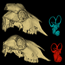
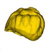
This contribution contains the 3D models described and figured in the following publication:Skull and Inner Ear Morphometrics in Sheep and Goats: Species and Breed Differentiation with Bioarchaeological Applications (Hemelsdael et al. submitted). The models include the external surface of a complete skull and inner ear of both a sheep (Ovis aries) and a goat (Capra hircus), generated from micro-CT scans. In the associated paper, we used 3D geometric morphometric data to assess inter and intra (i.e. between breeds) discrimination based on complete skulls, skull fragments and the semi-circular canals of the inner ear.
Capra hircus Amp_1 View specimen
|
M3#1806Skull of the goat Amp_1 Type: "3D_surfaces"doi: 10.18563/m3.sf.1806 state:published |
Download 3D surface file |
|
M3#1807Inner ear of the goat Amp_1 Type: "3D_surfaces"doi: 10.18563/m3.sf.1807 state:published |
Download 3D surface file |
Ovis aries UM_RR_2331 View specimen
|
M3#1808Skull of the sheep UM_RR_2331 Type: "3D_surfaces"doi: 10.18563/m3.sf.1808 state:published |
Download 3D surface file |
|
M3#1809Inner ear of the sheep UM_RR_2331 Type: "3D_surfaces"doi: 10.18563/m3.sf.1809 state:published |
Download 3D surface file |
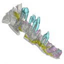
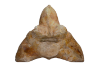
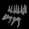
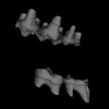
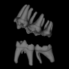
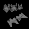
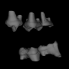
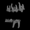
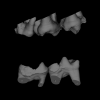
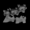
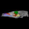
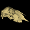
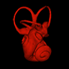
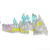
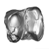
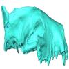
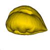
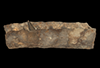
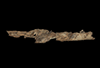
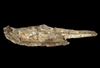
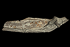
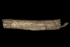
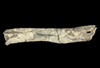
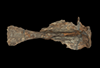
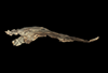
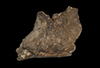
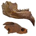
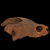
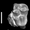
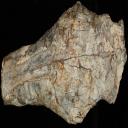
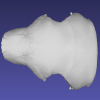
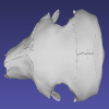
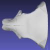
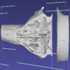
The present 3D Dataset contains the 3D models of the holotype and only specimen of Kalakocetus aurorae, a new cetacean retrieved from the Kalakot area in northwestern India. This specimen consists in a left hemimandible preserving the root of i3, p2, p4, m1 and m3 in situ. Its primitive morphology, with a tricuspid m3 morphologically intermediate between Raoellidae and Pakicetidae, makes it the first offshoot of Cetacea and provides crucial new elements to understand the setting up of the peculiar dental morphology of early cetaceans.
Kalakocetus aurorae GU/RJ/07 View specimen
|
M3#1803left hemi mandible with p2, p4, m1, m3 Type: "3D_surfaces"doi: 10.18563/m3.sf.1803 state:in_press |
Download 3D surface file |
|
M3#1804digitaly restored m1 Type: "3D_surfaces"doi: 10.18563/m3.sf.1804 state:in_press |
Download 3D surface file |
|
M3#1805digital restoration of complete mandible Type: "3D_surfaces"doi: 10.18563/m3.sf.1805 state:in_press |
Download 3D surface file |

|
M3#1810Scan Type: "3D_CT"doi: 10.18563/m3.sf.1810 state:in_press |
Download CT data |
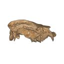
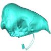
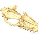
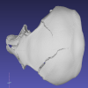
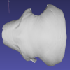
The present 3D Dataset contains the 3D model of a specimen of Metamynodon planifrons (UNISTRA.2015.0.1106) described and figured in: Veine-Tonizzo, L., Tissier, J., Bukhsianidze, M., Vasilyan, D., Becker, D., 2023, Cranial morphology and phylogenetic relationships of Amynodontidae Scott & Osborn, 1883 (Perissodactyla, Rhinocerotoidea).
Metamynodon planifrons UNISTRA.2015.0.1106 View specimen
|
M3#716Textured 3D surface model of the skull of the specimen UNISTRA.2015.0.1106 with right C1 and both rows of P2-M3. Type: "3D_surfaces"doi: 10.18563/m3.sf.716 state:published |
Download 3D surface file |
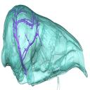
The present 3D Dataset contains a selection of 3D models analyzed in Billet G, Hautier L, Gaudin TJ, Flynn JJ, Ruf I, Carrillo JD, Ladevèze S, Lehmann T, Nicolas V, Orliac MJ, Tornero C, Wible JR, Wong N, Gaubert P. Submitted. Brain drain: Exceptional pattern of calvarial venation in pangolins and its phylogenetic significance for Ferae. Zoological Journal of the Linnean Society.
Phataginus tricuspis NHM-UK 48.13.26 View specimen
|
M3#1847cranium & intradiploic canals (sinuses & diploic veins) Type: "3D_surfaces"doi: 10.18563/m3.sf.1847 state:in_press |
Download 3D surface file |
Manis javanica NHM-UK 9.1.5.858 View specimen
|
M3#1848cranium & intradiploic canals (sinuses & diploic veins) Type: "3D_surfaces"doi: 10.18563/m3.sf.1848 state:in_press |
Download 3D surface file |
Felis silvestris UM-ZOOL-149N View specimen
|
M3#1849cranium & intradiploic canals (sinuses & diploic veins) Type: "3D_surfaces"doi: 10.18563/m3.sf.1849 state:in_press |
Download 3D surface file |
Erinaceus europaeus SMNS40759 View specimen

|
M3#1850cranium & intradiploic canals (sinuses & diploic veins) Type: "3D_surfaces"doi: 10.18563/m3.sf.1850 state:in_press |
Download 3D surface file |
Pterodon dasyuroides MNHN.F.Qu8301 View specimen
|
M3#1851cranium & intradiploic canals (sinuses & diploic veins) Type: "3D_surfaces"doi: 10.18563/m3.sf.1851 state:in_press |
Download 3D surface file |
The present 3D Dataset contains the 3D models analyzed in the following publication: Paulina-Carabajal, A., and Porfiri, J.D. 2026. Novel information on the braincase of Megaraptor namunhuaiquii (Dinosauria: Theropoda) using X-ray tomography: pneumaticity, paleoneurology and their paleobiological implications. Ameghiniana 63(1), 16-32
Megaraptor namunhuaiquii MUC-PV 595 View specimen
|
M3#18733D models of the braincase, lateral wall of the braincase, brain, right and left inner ears Type: "3D_surfaces"doi: 10.18563/m3.sf.1873 state:in_press |
Download 3D surface file |
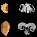
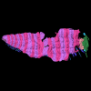
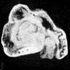
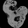
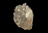
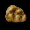
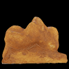
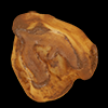
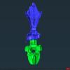
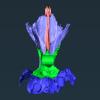
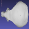
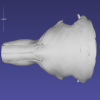
The present 3D dataset contains the 3D models of two fossil seeds of Vitaceae from the Quercy fossiliferous area, southwestern France. One seed comes from the Eocene locality of Fontoffre 2, and the other from the Oligocene locality of Baraval. These seeds document new fossil species within the Vitaceae family and illustrate the morphological diversity of this family during the Palaeogene. The CT scan data were processed with ImageJ and Mimics Innovation Suite version 1.13 to reconstruct the specimens. Here we provide .stl files that can be easily opened with the software MeshLab.
Vitis quercyensis UM-FTF2-167 View specimen
|
M3#1930Surface of the seed Type: "3D_surfaces"doi: 10.18563/m3.sf.1930 state:in_press |
Download 3D surface file |
|
M3#1933Seed : 8.43 μm µCT scan Type: "3D_CT"doi: 10.18563/m3.sf.1933 state:in_press |
Download CT data |
Vitis praerotundifolia UM-BAV-660 View specimen
|
M3#1931Seed : 8.43 μm µCT scan Type: "3D_CT"doi: 10.18563/m3.sf.1931 state:in_press |
Download CT data |
|
M3#1932Surface of the seed Type: "3D_surfaces"doi: 10.18563/m3.sf.1932 state:in_press |
Download 3D surface file |
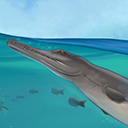
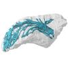
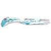
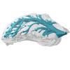
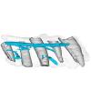
The democratization of 3D techniques in recent years provides exciting new opportunities for the study of complex fossils. In the present contribution, we provide a virtual reconstruction of a partial, disarticulated metriorhynchid (Metriorhynchidae, Thalattosuchia, Crocodylomorpha) skull from the Late Jurassic of northwestern Switzerland. This virtual reconstruction was used to produce high quality scientific illustrations of the whole skull for descriptive purposes. The reconstructed skull also served for the estimation of the total body length of the specimen and to propose a life reconstruction of the animal in its paleoenvironment. In an effort for transparency, we review the sources that were consulted for the life reconstruction and explain the choices that we had to make.
Torvoneustes jurensis BSY008-465 View specimen
|
M3#1037Left dentary (3 meshes) Type: "3D_surfaces"doi: 10.18563/m3.sf.1037 state:published |
Download 3D surface file |
|
M3#1038Right dentary (3 meshes) Type: "3D_surfaces"doi: 10.18563/m3.sf.1038 state:published |
Download 3D surface file |
|
M3#1039Left ramus (2 meshes) Type: "3D_surfaces"doi: 10.18563/m3.sf.1039 state:published |
Download 3D surface file |
|
M3#1040Right ramus (3 meshes) Type: "3D_surfaces"doi: 10.18563/m3.sf.1040 state:published |
Download 3D surface file |
|
M3#1041Left splenial (2 meshes) Type: "3D_surfaces"doi: 10.18563/m3.sf.1041 state:published |
Download 3D surface file |
|
M3#1042Right splenial (2 meshes) Type: "3D_surfaces"doi: 10.18563/m3.sf.1042 state:published |
Download 3D surface file |
|
M3#1043Frontal and left prefrontal Type: "3D_surfaces"doi: 10.18563/m3.sf.1043 state:published |
Download 3D surface file |
|
M3#1044Left maxilla (4 meshes) Type: "3D_surfaces"doi: 10.18563/m3.sf.1044 state:published |
Download 3D surface file |
|
M3#1045Right maxilla Type: "3D_surfaces"doi: 10.18563/m3.sf.1045 state:published |
Download 3D surface file |
|
M3#1046Left nasal Type: "3D_surfaces"doi: 10.18563/m3.sf.1046 state:published |
Download 3D surface file |
|
M3#1047Right nasal Type: "3D_surfaces"doi: 10.18563/m3.sf.1047 state:published |
Download 3D surface file |
|
M3#1048Parietal Type: "3D_surfaces"doi: 10.18563/m3.sf.1048 state:published |
Download 3D surface file |
|
M3#1049Right postorbital Type: "3D_surfaces"doi: 10.18563/m3.sf.1049 state:published |
Download 3D surface file |
|
M3#1050Right prefrontal Type: "3D_surfaces"doi: 10.18563/m3.sf.1050 state:published |
Download 3D surface file |
|
M3#1051Right premaxilla Type: "3D_surfaces"doi: 10.18563/m3.sf.1051 state:published |
Download 3D surface file |
|
M3#1052Left squamosal Type: "3D_surfaces"doi: 10.18563/m3.sf.1052 state:published |
Download 3D surface file |

|
M3#1053Right squamosal Type: "3D_surfaces"doi: 10.18563/m3.sf.1053 state:published |
Download 3D surface file |

|
M3#1054Reconstruction of the mandible Type: "3D_surfaces"doi: 10.18563/m3.sf.1054 state:published |
Download 3D surface file |

|
M3#1055Reconstruction of the cranium Type: "3D_surfaces"doi: 10.18563/m3.sf.1055 state:published |
Download 3D surface file |
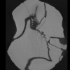
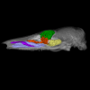
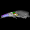
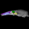
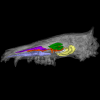
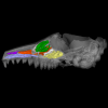
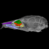
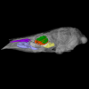
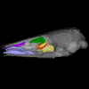
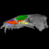
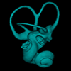
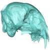
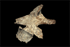
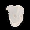
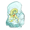
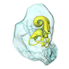
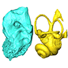
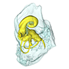
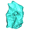
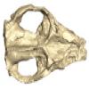
The present 3D dataset contains 3D models of the endocranial cast of the raoellid Khirtharia inflata retrieved from the middle Eocene of the Upper Subathu Formation in the Kalakot area (India). Raoellidae are closely related to stem cetaceans and bring crucial information to understand the earliest phase of land to water transition in Cetacea.
Khirtharia inflata GU/RJ/197 View specimen
|
M3#1608labeled cast of the endocranial cavity Type: "3D_surfaces"doi: 10.18563/m3.sf.1608 state:published |
Download 3D surface file |
|
M3#1609endocast and associated sinuses Type: "3D_surfaces"doi: 10.18563/m3.sf.1609 state:published |
Download 3D surface file |
The present 3D Dataset contains the 3D models produced in the frame of the article Perthuis, A. de, Mennecart, B., Barrier, P., Chenot, É., Falconnet, J., Gagnaison, J.-C., Georgalis, G. L., Gilbert, C., Guevel, B., Langevin, D., Lapparent de Broin, F. de, Lemierre, A., Maubert, F., Ossó, À., Potel, S., Thivaiou, D., Tissier, J., Toullec, R., Xerri, S., Gagnaison, C. 2025. Révision des données sédimentologiques et biostratigraphiques des gisements à vertébrés des sables de l’Orléanais, à Beaugency, Tavers et Le Bardon (Miocène Moyen ; Loiret, France). Geodiversitas 47 (12): 2-76. https://doi.org/10.5252/geodiversitas2025v47a12
Bunolistriodon lockharti ULB-TAV-21 View specimen
|
M3#1837Left upper M3 Type: "3D_surfaces"doi: 10.18563/m3.sf.1837 state:published |
Download 3D surface file |
Megamphicyon giganteus ULB-TAV-13 View specimen
|
M3#1531Left first lower molar Type: "3D_surfaces"doi: 10.18563/m3.sf.1531 state:published |
Download 3D surface file |
Hispanotherium matritense ULB-TAV-17 View specimen
|
M3#1532Left first lower molar Type: "3D_surfaces"doi: 10.18563/m3.sf.1532 state:published |
Download 3D surface file |
Plesiaceratherium lumiarense ULB-TAV-18 View specimen
|
M3#1533Left third upper molar Type: "3D_surfaces"doi: 10.18563/m3.sf.1533 state:published |
Download 3D surface file |
Chelydropsis aff. sansaniensis ULB-TAV-23 View specimen
|
M3#1535Cast of a skull Type: "3D_surfaces"doi: 10.18563/m3.sf.1535 state:published |
Download 3D surface file |
Ronzotherium romani ULB-TAV-4 View specimen
|
M3#1556Right fourth upper premolar Type: "3D_surfaces"doi: 10.18563/m3.sf.1556 state:published |
Download 3D surface file |
Prodeinotherium bavaricum ULB-TAV-24 View specimen
|
M3#1557left hemimandibule Type: "3D_surfaces"doi: 10.18563/m3.sf.1557 state:published |
Download 3D surface file |
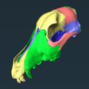
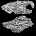
Veterinary education often relies on cadaveric specimens, but there is increasing demand for alternatives due to limited resources and ethical considerations. To address this, we developed a 3D printed ‘explodable’ model of a dog cranium with detachable, magnetized cranial components for teaching anatomy to students. This model was generated from a computed tomographic scan of a juvenile dog cranium for which cranial sutures were still partially open and segmented such that major cranial bones were isolated. All bones are printed at actual size and retain openings for cranial nerves and major vessels. This interactive model enhances anatomical education by supplying a hands-on tool that can be used either in the classroom setting or for independent learning and can be incorporated at the high school, college, or veterinary school level. It is currently being integrated into the first-year anatomy foundation course at Cornell University’s College of Veterinary Medicine. The model can be printed using any hobbyist or specialist 3D printer and we outline assembly instructions on how to attach magnets at prefabricated attachment points. Using both digital and 3D printed resources, we hope to help to address current shortages of anatomical resources and also inspire future generations of practicing veterinarians by making anatomy more accessible and engaging.
Canis lupus familiaris CUHL 9 View specimen
|
M3#1858PLYs of the segmented cranial bones with pre-fabricated magnetic casings and shelves for assembly following 3D printing Type: "3D_surfaces"doi: 10.18563/m3.sf.1858 state:published |
Download 3D surface file |
|
M3#1859PLYs of the segmented cranial bones of the "BOTTOM" cranial component. Downloadable for additional learning opportunities for students Type: "3D_surfaces"doi: 10.18563/m3.sf.1859 state:published |
Download 3D surface file |
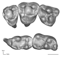
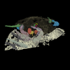
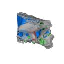
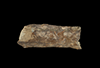
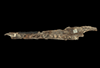
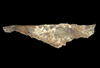
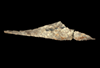
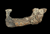
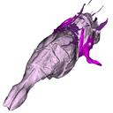
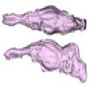
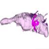
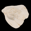
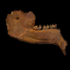
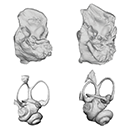
This contribution contains the 3D digital models of some fossil specimens of Wamradolops telloi Stutz and Pozodolops manuelorum Stutz (Metatheria: Polydolopimorphia), from several Palaeogene locations of Peruvian Amazonia. These taxa were described and analyzed in detail in the following publication: Stutz et al. (2026), Hidden diversity of Palaeogene metatherians: a new family of polydolopimorphian marsupials from Peruvian Amazonia. Zoological Journal of the Linnean Society. https://doi.org/10.1093/zoolinnean/zlag006.
Pozodolops manuelorum MUSM 4029 View specimen

|
M3#1874Pozodolops manuelorum, fragmentary left dentary with p3–m1 Type: "3D_surfaces"doi: 10.18563/m3.sf.1874 state:in_press |
Download 3D surface file |
Wamradolops telloi MUSM 4032 View specimen

|
M3#1875Wamradolops telloi, fragmentary right dentary with m1–m2 Type: "3D_surfaces"doi: 10.18563/m3.sf.1875 state:in_press |
Download 3D surface file |
Pozodolops manuelorum MUSM 4036 View specimen
|
M3#1876Pozodolops manuelorum, holotype, fragmentary right maxilla with M1 Type: "3D_surfaces"doi: 10.18563/m3.sf.1876 state:in_press |
Download 3D surface file |
Pozodolops manuelorum MUSM 4041 View specimen

|
M3#1877Pozodolops manuelorum, fragmentary right dentary with m1 Type: "3D_surfaces"doi: 10.18563/m3.sf.1877 state:in_press |
Download 3D surface file |
Pozodolops manuelorum MUSM 4058 View specimen

|
M3#1878Pozodolops manuelorum, fragmentary left P3 Type: "3D_surfaces"doi: 10.18563/m3.sf.1878 state:in_press |
Download 3D surface file |
Wamradolops telloi MUSM 4144 View specimen

|
M3#1879Wamradolops telloi, fragmentary left dentary with p2–m1 Type: "3D_surfaces"doi: 10.18563/m3.sf.1879 state:in_press |
Download 3D surface file |
Wamradolops telloi MUSM 4179 View specimen

|
M3#1880Wamradolops telloi, holotype, partial skull, with right P2–M4 and left I4–M3, plus boneless lower teeth below upper teeth, belonging to the same individual Type: "3D_surfaces"doi: 10.18563/m3.sf.1880 state:in_press |
Download 3D surface file |
Wamradolops telloi MUSM 4221 View specimen

|
M3#1881Wamradolops telloi, fragmentary of right dentary with p3–m2 Type: "3D_surfaces"doi: 10.18563/m3.sf.1881 state:in_press |
Download 3D surface file |

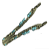
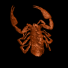
In this contribution a third new species of the rare genus Burmesescorpiops Lourenço, 2016 is described. The discovery of this new element belonging to the family Palaeoeuscorpiidae Lourenço, 2003 and to the subfamily Archaeoscorpiopinae Lourenço, 2015 brings further elements to support the validity of the genus Burmesescorpiops. This generic group remains however, poorly speciose. This is the latest discovery of Burmesescorpiops wunpawng, the name is derived from the Kachin Hilltribe peoples who are indigenous to the area. The data provided here is a 3D surface.
Burmesescorpiops wunpawng ps-gyi-01-25 View specimen
|
M3#18463d Surface Volume Type: "3D_surfaces"doi: 10.18563/m3.sf.1846 state:published |
Download 3D surface file |
The present 3D Dataset contains the 3D models analyzed in Mennecart, B., Duranthon, F., & Costeur, L. 2024. Systematic contribution of the auditory region to the knowledge of the oldest European Bovidae (Mammalia, Ruminantia). Journal of Anatomy XXX. https://doi.org/10.1111/joa.14132
Pusillutragus montrealensis MHNT.PAL.2015.0.2261.4 View specimen
|
M3#1522Right petrosal, bony labyrinth, stapes Type: "3D_surfaces"doi: 10.18563/m3.sf.1522 state:published |
Download 3D surface file |
Pusillutragus montrealensis MHNT.PAL.2015.0.2261.9 View specimen
|
M3#1523Left petrosal and left bony labyrinth Type: "3D_surfaces"doi: 10.18563/m3.sf.1523 state:published |
Download 3D surface file |
Eotragus artenensis SMNS-P-41625 View specimen
|
M3#1524Petrosal (right), bony labyrinth (left) Type: "3D_surfaces"doi: 10.18563/m3.sf.1524 state:published |
Download 3D surface file |
Eotragus clavatus NMB San.15056 View specimen
|
M3#1528Right petrosal and right bony labyrinth Type: "3D_surfaces"doi: 10.18563/m3.sf.1528 state:published |
Download 3D surface file |
Eotragus clavatus NMB San.15055 View specimen
|
M3#1526Left Petrosal Type: "3D_surfaces"doi: 10.18563/m3.sf.1526 state:published |
Download 3D surface file |
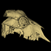
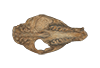
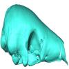
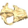
The present 3D Dataset contains the 3D model of the skull of the raoellid Indohyus indirae described in Patel et al. 2024.
Indohyus indirae RR 207 View specimen
|
M3#1259dorsoventrally crushed skull Type: "3D_surfaces"doi: 10.18563/m3.sf.1259 state:published |
Download 3D surface file |
Indohyus indirae RR 601 View specimen
|
M3#1268dorsoventrally crushed skull Type: "3D_surfaces"doi: 10.18563/m3.sf.1268 state:published |
Download 3D surface file |
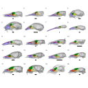
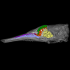
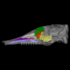
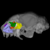
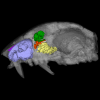
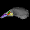
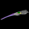
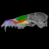
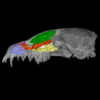
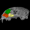
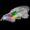
This contribution contains the 3D models described and figured in: Phylogenetic signal in anteater snout morphology: implications for interpreting rare vermilinguan fossils. Palaeobiodiversity and Palaeoenvironments.
Indet indet VPPLT 977 View specimen

|
M3#17933D surface models of the cranium, nasal bone and cranial canals Type: "3D_surfaces"doi: 10.18563/m3.sf.1793 state:in_press |
Download 3D surface file |
Cyclopes didactylus M 1525 View specimen
|
M3#17943D models of the cranium and internal cranial canals Type: "3D_surfaces"doi: 10.18563/m3.sf.1794 state:in_press |
Download 3D surface file |
Cyclopes didactylus M 1571 View specimen
|
M3#17953D surface models of the cranium, nasal bone and cranial canals Type: "3D_surfaces"doi: 10.18563/m3.sf.1795 state:in_press |
Download 3D surface file |
Myrmecophaga tridactyla M 3023 View specimen
|
M3#17963D surface models of the cranium, nasal bone and cranial canals Type: "3D_surfaces"doi: 10.18563/m3.sf.1796 state:in_press |
Download 3D surface file |
Tamandua tetradactyla NHMUK 3.7.7.135 View specimen
|
M3#17973D models of the cranium and internal cranial canals Type: "3D_surfaces"doi: 10.18563/m3.sf.1797 state:in_press |
Download 3D surface file |
Tamandua tetradactyla NHMUK 4.7.4.90 View specimen
|
M3#17983D surface models of the cranium, nasal bone and cranial canals Type: "3D_surfaces"doi: 10.18563/m3.sf.1798 state:in_press |
Download 3D surface file |
Tamandua tetradactyla UM 788N View specimen
|
M3#17993D models of the cranium and internal cranial canals Type: "3D_surfaces"doi: 10.18563/m3.sf.1799 state:in_press |
Download 3D surface file |
Cyclopes didactylus MVZ 121210 View specimen
|
M3#18003D models of the cranium and internal cranial canals Type: "3D_surfaces"doi: 10.18563/m3.sf.1800 state:in_press |
Download 3D surface file |
Myrmecophaga tridactyla MVZ 112943 View specimen
|
M3#18013D models of the cranium and internal cranial canals Type: "3D_surfaces"doi: 10.18563/m3.sf.1801 state:in_press |
Download 3D surface file |
Myrmecophaga tridactyla MVZ 185238 View specimen
|
M3#18023D models of the cranium and internal cranial canals Type: "3D_surfaces"doi: 10.18563/m3.sf.1802 state:in_press |
Download 3D surface file |
