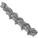
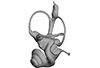
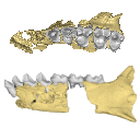
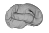
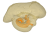
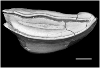
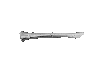
3D models of Cainotheriids Ossicular chain
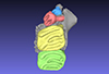
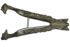
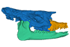
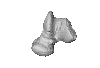
Explodable 3D Dog Skull for Veterinary Education
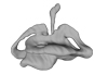
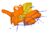
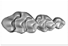
3D models of Kalakocetus, the earliest Cetacea
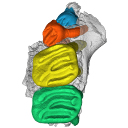
3D GM dataset of bird skeletal variation
Skeletal embryonic development in the catshark
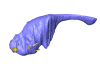
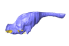
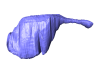
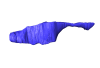
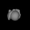
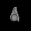
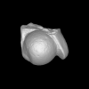
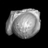
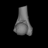
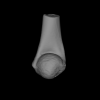
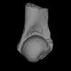
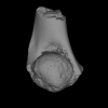
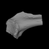
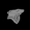
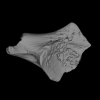
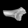
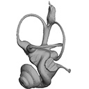
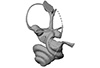
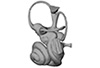
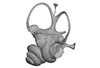
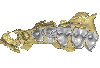
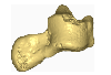
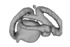
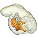
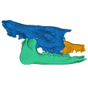
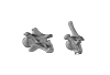
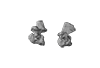
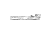
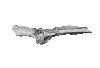
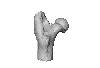
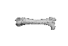
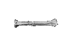
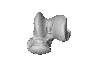
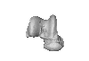
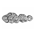
Bony connexions of the petrosal bone of extant hippos
bony labyrinth (14) , inner ear (11) , Eocene (11) , geometric morphometrics (10) , CT-scan (10) , Oligocene (9) , Micro-CT (9)
Lionel Hautier (25) , Maëva Judith Orliac (24) , Laurent Marivaux (18) , Renaud Lebrun (15) , Rodolphe Tabuce (15) , Pierre-Olivier Antoine (13) , Bastien Mennecart (13)
|
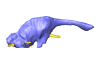
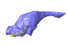
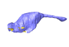
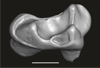
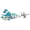
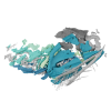
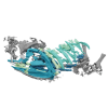
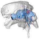
3D models related to the publication: Virtual endocasts of Clevosaurus brasiliensis and the tuatara: rhynchocephalian neuroanatomy and the oldest endocranial record for Lepidosauria
|
|
M3#10993D surface model of the cranial endocast of specimen CM 30660 (Sphenodon punctatus). Type: "3D_surfaces"doi: 10.18563/m3.sf.1099 state:published |
Download 3D surface file |
Sphenodon punctatus KCLZJ 001 View specimen
|
M3#11003D surface models of the cranial endocast and the initial trunks of the cranial nerves of specimen KCLZJ 001 (Sphenodon punctatus). Type: "3D_surfaces"doi: 10.18563/m3.sf.1100 state:published |
Download 3D surface file |
Sphenodon punctatus LDUCZ x0036 View specimen
|
M3#11013D surface models of the cranial endocast and the initial trunks of the cranial nerves of specimen LDUCZ x0036 (Sphenodon punctatus). Type: "3D_surfaces"doi: 10.18563/m3.sf.1101 state:published |
Download 3D surface file |
Sphenodon punctatus LDUCZ x1126 View specimen
|
M3#11023D surface model of the cranial endocast of specimen LDUCZ x1126 (Sphenodon punctatus). Type: "3D_surfaces"doi: 10.18563/m3.sf.1102 state:published |
Download 3D surface file |
Clevosaurus brasiliensis MCN PV 2852 View specimen
|
M3#11033D surface model of the cranial endocast of specimen MCN PV 2852 (Clevosaurus brasiliensis). Type: "3D_surfaces"doi: 10.18563/m3.sf.1103 state:published |
Download 3D surface file |
Sphenodon punctatus SAMA 70524 View specimen
|
M3#11043D surface models of the cranial endocast, brain, endosseous labyrinth and initial trunks of the cranial nerves of specimen SAMA 70524 (Sphenodon punctatus). Type: "3D_surfaces"doi: 10.18563/m3.sf.1104 state:published |
Download 3D surface file |
Sphenodon punctatus SU1 View specimen
|
M3#11053D surface models of the cranial endocast and the initial trunks of the cranial nerves of specimen SU1 (Sphenodon punctatus). Type: "3D_surfaces"doi: 10.18563/m3.sf.1105 state:published |
Download 3D surface file |
Sphenodon punctatus YPM HERR 009194 View specimen
|
M3#11063D surface models of the cranial endocast and the initial trunks of the cranial nerves of specimen YPM HERR 009194 (Sphenodon punctatus). Type: "3D_surfaces"doi: 10.18563/m3.sf.1106 state:published |
Download 3D surface file |

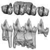
This contribution contains the three-dimensional digital model of one isolated fossil tooth of an anthropoid primate (Ashaninkacebus simpsoni), discovered in sedimentary deposits located on the upper Rio Juruá in State of Acre, Brazil (Western Amazonia). This fossil was described, figured and discussed in the following publication: Marivaux et al. (2023), An eosimiid primate of South Asian affinities in the Paleogene of Western Amazonia and the origin of New World monkeys. Proceedings of the National Academy of Sciences USA. https://doi.org/10.1073/pnas.2301338120
Ashaninkacebus simpsoni UFAC-CS 066 View specimen
|
M3#1114Right first upper molar (rM1), pristine. Type: "3D_surfaces"doi: 10.18563/m3.sf.1114 state:published |
Download 3D surface file |
This contribution contains the three-dimensional digital models of a part of the dental fossil material (the large specimens) of caviomorph rodents, discovered in late middle Miocene detrital deposits of the TAR-31 locality in Peruvian Amazonia (San Martín, Peru). These fossils were described, figured and discussed in the following publication: Boivin, Marivaux et al. (2021), Late middle Miocene caviomorph rodents from Tarapoto, Peruvian Amazonia. PLoS ONE 16(11): e0258455. https://doi.org/10.1371/journal.pone.0258455
Microscleromys paradoxalis MUSM 4643 View specimen
|
M3#1115Fragment of left mandibule preserving dp4, m1 and a portion of incisor Type: "3D_surfaces"doi: 10.18563/m3.sf.1115 state:published |
Download 3D surface file |
Ricardomys longidens MUSM 4375 View specimen
|
M3#1116Fragment of left maxillary preserving DP4 and M1 (or M1 and M2) Type: "3D_surfaces"doi: 10.18563/m3.sf.1116 state:published |
Download 3D surface file |
"Scleromys" sp. MUSM 4272 View specimen
|
M3#1117Isolated left upper molar Type: "3D_surfaces"doi: 10.18563/m3.sf.1117 state:published |
Download 3D surface file |
"Scleromys" sp. MUSM 4275 View specimen
|
M3#1118Isolated right upper molar Type: "3D_surfaces"doi: 10.18563/m3.sf.1118 state:published |
Download 3D surface file |
"Scleromys" sp. MUSM 4273 View specimen
|
M3#1119Isolated left upper molar Type: "3D_surfaces"doi: 10.18563/m3.sf.1119 state:published |
Download 3D surface file |
"Scleromys" sp. MUSM 4276 View specimen
|
M3#1120Isolated right upper molar Type: "3D_surfaces"doi: 10.18563/m3.sf.1120 state:published |
Download 3D surface file |
"Scleromys" sp. MUSM 4282 View specimen
|
M3#1121Isolated right lower molar Type: "3D_surfaces"doi: 10.18563/m3.sf.1121 state:published |
Download 3D surface file |
"Scleromys" sp. MUSM 4281 View specimen
|
M3#1122Isolated right lower molar Type: "3D_surfaces"doi: 10.18563/m3.sf.1122 state:published |
Download 3D surface file |
"Scleromys" sp. MUSM 4280 View specimen
|
M3#1123Isolated left p4 Type: "3D_surfaces"doi: 10.18563/m3.sf.1123 state:published |
Download 3D surface file |
"Scleromys" sp. MUSM 4277 View specimen
|
M3#1124Isolated left lower dp4 Type: "3D_surfaces"doi: 10.18563/m3.sf.1124 state:published |
Download 3D surface file |
"Scleromys" sp. MUSM 4279 View specimen
|
M3#1125Isolated right lower dp4 (mesial fragment) Type: "3D_surfaces"doi: 10.18563/m3.sf.1125 state:published |
Download 3D surface file |
gen.indet sp. indet MUSM 4283 View specimen
|
M3#1126Isolated right lower p4 Type: "3D_surfaces"doi: 10.18563/m3.sf.1126 state:published |
Download 3D surface file |
Microscleromys sp. MUSM 4658 View specimen
|
M3#1127Isolated left tarsal bone (astragalus) Type: "3D_surfaces"doi: 10.18563/m3.sf.1127 state:published |
Download 3D surface file |
The present contribution contains the 3D models of fossil humeri and ilia of anurans from various Eocene-Miocene deposits of Peruvian Amazonia. These fossils were described and figured in the following publication: Jansen et al. (2023), First Eocene–Miocene anuran fossils from Peruvian Amazonia: insights into Neotropical frog evolution and diversity. Papers in Palaeontology, The Palaeontological Association.
Indet. indet. MUSM 4746 View specimen
|
M3#1231Humeral fragment (distal end) Type: "3D_surfaces"doi: 10.18563/m3.sf.1231 state:published |
Download 3D surface file |
Indet. indet. MUSM 4747 View specimen
|
M3#1232Humeral fragment (distal end) Type: "3D_surfaces"doi: 10.18563/m3.sf.1232 state:published |
Download 3D surface file |
Indet. indet. MUSM 4748 View specimen
|
M3#1233Humeral fragment (distal end) Type: "3D_surfaces"doi: 10.18563/m3.sf.1233 state:published |
Download 3D surface file |
Indet. indet. MUSM 4755 View specimen
|
M3#1234Humeral fragment (distal end) Type: "3D_surfaces"doi: 10.18563/m3.sf.1234 state:published |
Download 3D surface file |
Indet. indet. MUSM 4756 View specimen
|
M3#1235Humeral fragment (distal end) Type: "3D_surfaces"doi: 10.18563/m3.sf.1235 state:published |
Download 3D surface file |
Indet. indet. MUSM 4757 View specimen
|
M3#1236Humeral fragment (distal end) Type: "3D_surfaces"doi: 10.18563/m3.sf.1236 state:published |
Download 3D surface file |
Indet. indet. MUSM 4761 View specimen
|
M3#1237Humeral fragment (distal end) Type: "3D_surfaces"doi: 10.18563/m3.sf.1237 state:published |
Download 3D surface file |
Indet. indet. MUSM 4763 View specimen
|
M3#1238Humeral fragment (distal end) Type: "3D_surfaces"doi: 10.18563/m3.sf.1238 state:published |
Download 3D surface file |
Indet. indet. MUSM 4765 View specimen
|
M3#1239Humeral fragment (distal end) Type: "3D_surfaces"doi: 10.18563/m3.sf.1239 state:published |
Download 3D surface file |
Indet. indet. MUSM 4766 View specimen
|
M3#1240Humeral fragment (distal end) Type: "3D_surfaces"doi: 10.18563/m3.sf.1240 state:published |
Download 3D surface file |
Indet. indet. MUSM 4775 View specimen
|
M3#1241Humeral fragment (distal end) Type: "3D_surfaces"doi: 10.18563/m3.sf.1241 state:published |
Download 3D surface file |
cf. Pipa sp. MUSM 4776 View specimen
|
M3#1242Humeral fragment (distal end) Type: "3D_surfaces"doi: 10.18563/m3.sf.1242 state:published |
Download 3D surface file |
Indet. indet. MUSM 4788 View specimen
|
M3#1243Ilial fragment Type: "3D_surfaces"doi: 10.18563/m3.sf.1243 state:published |
Download 3D surface file |
Indet. indet. MUSM 4789 View specimen
|
M3#1244Ilial fragment Type: "3D_surfaces"doi: 10.18563/m3.sf.1244 state:published |
Download 3D surface file |
Indet. indet. MUSM 4790 View specimen
|
M3#1245Ilial fragment Type: "3D_surfaces"doi: 10.18563/m3.sf.1245 state:published |
Download 3D surface file |
Indet. indet. MUSM 4792 View specimen
|
M3#1246Ilial fragment Type: "3D_surfaces"doi: 10.18563/m3.sf.1246 state:published |
Download 3D surface file |
Indet. indet. MUSM 4793 View specimen
|
M3#1247Ilial fragment Type: "3D_surfaces"doi: 10.18563/m3.sf.1247 state:published |
Download 3D surface file |
Indet. indet. MUSM 4794 View specimen
|
M3#1249Ilial fragment Type: "3D_surfaces"doi: 10.18563/m3.sf.1249 state:published |
Download 3D surface file |
Indet. indet. MUSM 4795 View specimen
|
M3#1250Ilial fragment Type: "3D_surfaces"doi: 10.18563/m3.sf.1250 state:published |
Download 3D surface file |
cf. Pipa sp. MUSM 4796 View specimen
|
M3#1251Ilial fragment Type: "3D_surfaces"doi: 10.18563/m3.sf.1251 state:published |
Download 3D surface file |
cf. Pipa sp. MUSM 4797 View specimen
|
M3#1252Ilial fragment Type: "3D_surfaces"doi: 10.18563/m3.sf.1252 state:published |
Download 3D surface file |

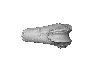
This contribution contains 3D models of the cranial endoskeleton of three specimens of the Permian ‘acanthodian’ stem-group chondrichthyan (cartilaginous fish) Acanthodes confusus, obtained using computed tomography. These datasets were described and analyzed in Dearden et al. (2024) “3D models related to the publication: The pharynx of the iconic stem-group chondrichthyan Acanthodes Agassiz, 1833 revisited with micro computed tomography.” Zoological Journal of the Linnean Society
Acanthodes confusus MNHN-F-SAA20 View specimen
|
M3#14703D surfaces representing the three-dimensionally fossilised head of Acanthodes confusus Type: "3D_surfaces"doi: 10.18563/m3.sf.1470 state:published |
Download 3D surface file |
Acanthodes confusus MNHN-F-SAA21 View specimen
|
M3#14713D surfaces representing the three-dimensionally fossilised head of Acanthodes confusus Type: "3D_surfaces"doi: 10.18563/m3.sf.1471 state:published |
Download 3D surface file |
Acanthodes confusus MNHN-F-SAA24 View specimen
|
M3#14723D surfaces representing the three-dimensionally fossilised head of Acanthodes confusus Type: "3D_surfaces"doi: 10.18563/m3.sf.1472 state:published |
Download 3D surface file |
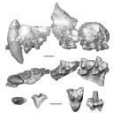
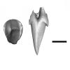
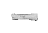
This contribution contains the three-dimensional models of the most complete and/or informative fossil materials attributed to Peradectes crocheti Gernelle, 2024, the earliest peradectid metatherian species of Europe, from its type locality (Palette, Provence, ~55 Ma). These specimens were analyzed and discussed in: Gernelle et al. (2024), Taxonomy and evolutionary history of peradectids (Metatheria): new data from the early Eocene of France. https://doi.org/10.1007/s10914-024-09724-5
Peradectes crocheti MHN.AIX.PV.2018.26.14 View specimen
|
M3#14993D surface model of MHN.AIX.PV.2018.26.14, fragmentary left maxilla with C-P1, anterior root of P2, and M1-M3 Type: "3D_surfaces"doi: 10.18563/m3.sf.1499 state:published |
Download 3D surface file |
Peradectes crocheti MHN.AIX.PV.2017.6.6 View specimen
|
M3#15003D surface model of MHN.AIX.PV.2017.6.6, left P2 Type: "3D_surfaces"doi: 10.18563/m3.sf.1500 state:published |
Download 3D surface file |
Peradectes crocheti MHN.AIX.PV.2017.6.7 View specimen
|
M3#15013D surface model of MHN.AIX.PV.2017.6.7, left M3 Type: "3D_surfaces"doi: 10.18563/m3.sf.1501 state:published |
Download 3D surface file |
Peradectes crocheti MHN.AIX.PV.2017.6.8 View specimen
|
M3#15023D surface model of MHN.AIX.PV.2017.6.8, right hemi-mandible fragment with canine alveolus, posterior root of p1, partial p2, p3, partial m1, and m2-m3 Type: "3D_surfaces"doi: 10.18563/m3.sf.1502 state:published |
Download 3D surface file |
Peradectes crocheti MHN.AIX.PV.2017.6.9 View specimen
|
M3#15033D surface model of MHN.AIX.PV.2017.6.9, leftm1-m4 row with fragments of dentary Type: "3D_surfaces"doi: 10.18563/m3.sf.1503 state:published |
Download 3D surface file |
Peradectes crocheti MHN.AIX.PV.2017.6.14 View specimen
|
M3#15043D surface model of MHN.AIX.PV.2017.6.14, right astragalus Type: "3D_surfaces"doi: 10.18563/m3.sf.1504 state:published |
Download 3D surface file |
This project presents a µCT dataset and an associated 3D surface model of the holotype of Donrussellia magna (UM PAT 17; Primates, Adapiformes). UM PAT17 is the only known specimen for the species and consists of a well-preserved left lower jaw with p4-m3. It documents one of the oldest European primates, eventually dated near the Paleocene Eocene Thermal Maximum.
Donrussellia magna UM PAT 17 View specimen

|
M3#173D surface file model of UM PAT 17 (type specimen of Donrussellia magna), which is a well preserved left lower jaw with p4-m3. The teeth (and roots) were manually segmented. Type: "3D_surfaces"doi: 10.18563/m3.sf17 state:published |
Download 3D surface file |
|
M3#18CT Scan Data of Donrussellia magna UM PAT 17. Voxel size (in µm): 36µm (isotropic voxels). Dimensions in x,y,z : 594 pixels, 294 pixels, 1038 pixels. Image type : 8-bit voxels. Image format : raw data format (no header). Type: "3D_CT"doi: 10.18563/m3.sf18 state:published |
Download CT data |
This contribution contains the 3D model described and figured in the following publication: Albino, A., Carrillo-Briceño, J. D. & Neenan, J. M. 2016. An enigmatic aquatic snake from the Cenomanian of northern South America. PeerJ 4:e2027 http://dx.doi.org/10.7717/peerj.2027
Lunaophis aquaticus MCNC-1827-F View specimen

|
M3#116Articulated precloacal vertebrae of Lunaophis aquaticus Type: "3D_surfaces"doi: 10.18563/m3.sf.116 state:published |
Download 3D surface file |
The present 3D Dataset contains the 3D models analyzed in the article Mennecart, B., and L. Costeur. 2016. A Dorcatherium (Mammalia, Ruminantia, Middle Miocene) petrosal bone and the tragulid ear region. Journal of Vertebrate Paleontology 36(6), 1211665(1)-1211665(7). DOI: 10.1080/02724634.2016.1211665.
Tragulus javanicus 10028 View specimen
|
M3#1193D surface of the left bony labyrinth of Tragulus javanicus NMB 10028 Type: "3D_surfaces"doi: 10.18563/m3.sf.119 state:published |
Download 3D surface file |
Moschiola meminna C.2453 View specimen
|
M3#1203D surface of the left bony labyrinth of Moschiola meminna NMB C.2453 Type: "3D_surfaces"doi: 10.18563/m3.sf.120 state:published |
Download 3D surface file |
Hyemoschus aquaticus C.1930 View specimen
|
M3#1223D surface of the right bony labyrinth of Hyemoschus aquaticus NMB C.1930 Type: "3D_surfaces"doi: 10.18563/m3.sf.122 state:published |
Download 3D surface file |
Dorcatherium crassum San.15053 View specimen
|
M3#1233D surface of the right bony labyrinth of Dorcatherium crassum NMB San.15053 Type: "3D_surfaces"doi: 10.18563/m3.sf.123 state:published |
Download 3D surface file |
The present 3D Dataset contains the 3D models analyzed in: "a giant dapediid from the Late Triassic of Switzerland and insights into neopterygian phylogeny", Royal Society Open Science, https://doi.org/10.1098/rsos.180497
Scopulipiscis saxciput PIMUZ A/I 3026 View specimen
|
M3#1773D surfaces of the skull and endocranial spaces inside neurocranium, including the aortic canal, braincase, fossa bridgei, lateral cranial canal, nerves and other passageways, notochord, posterior myodome, and right semicircular canals. Type: "3D_surfaces"doi: 10.18563/m3.sf.177 state:published |
Download 3D surface file |

|
M3#178Scan of the neurocranium of PIMUZ A/I 3026 Type: "3D_CT"doi: 10.18563/m3.sf.178 state:published |
Download CT data |
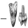
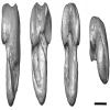
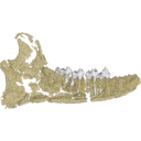
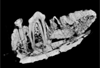
This contribution contains the 3D models of the fossil remains (maxilla, dentary, and talus) attributed to Djebelemur martinezi, a ca. 50 Ma primate from Tunisia (Djebel Chambi), described and figured in the following publication: Marivaux et al. (2013), Djebelemur, a tiny pre-tooth-combed primate from the Eocene of Tunisia: a glimpse into the origin of crown strepsirhines. PLoS ONE. https://doi.org/10.1371/journal.pone.0080778
Djebelemur martinezi CBI-1-544 View specimen
|
M3#365CBI-1-544, left maxilla preserving P3-M3 and alveoli for P2 and C1 Type: "3D_surfaces"doi: 10.18563/m3.sf.365 state:published |
Download 3D surface file |
Djebelemur martinezi CBI-1-567 View specimen
|
M3#363Isolated left upper P4 Type: "3D_surfaces"doi: 10.18563/m3.sf.363 state:published |
Download 3D surface file |
Djebelemur martinezi CBI-1-565-577-587-580 View specimen
|
M3#366- CBI-1-565, a damaged right mandible, which consists of three isolated pieces found together and reassembled here: the anterior part of the dentary bears the p3 and m1, and alveoli for p4, p2 and c, while the posterior part preserves m3 and a portion of the ascending ramus; the m2 was found isolated but in the same small calcareous block treated by acid processing. - CBI-1-577, isolated right lower p4. - CBI-1-587, isolated left lower p2 (reversed). - CBI-1-580, isolated left lower canine (reversed). Type: "3D_surfaces"doi: 10.18563/m3.sf.366 state:published |
Download 3D surface file |
Djebelemur martinezi CBI-1-545 View specimen
|
M3#364Right Talus Type: "3D_surfaces"doi: 10.18563/m3.sf.364 state:published |
Download 3D surface file |
The present 3D Dataset contains the 3D models analyzed in the publication ‘Ontogenetic development of the otic region in the new model organism, Leucoraja erinacea (Chondrichthyes; Rajidae)’, https://doi.org/10.1017/S1755691018000993
Leucoraja erinacea 2018.9.26.1 View specimen
|
M3#3673D model of the right skeletal labyrinth of the adult specimen of Leucoraja erincea. T Type: "3D_surfaces"doi: 10.18563/m3.sf.367 state:published |
Download 3D surface file |
Leucoraja erinacea 2018.9.25.2 View specimen
|
M3#3683D model of the right skeletal labyrinth of the stage 34 specimen of Leucoraja erincea. Type: "3D_surfaces"doi: 10.18563/m3.sf.368 state:published |
Download 3D surface file |
Leucoraja erinacea 2018.9.25.3 View specimen
|
M3#3693D model of the right skeletal labyrinth of the stage 32 specimen of Leucoraja erinacea. Type: "3D_surfaces"doi: 10.18563/m3.sf.369 state:published |
Download 3D surface file |
|
M3#3723D model of the right membranous system of stage 32 of Leucoraja erincea. Type: "3D_surfaces"doi: 10.18563/m3.sf.372 state:published |
Download 3D surface file |
Leucoraja erinacea 2018.9.25.4 View specimen
|
M3#3703D model of the right skeletal labyrinth of the stage 31 specimen of Leucoraja erinacea. Type: "3D_surfaces"doi: 10.18563/m3.sf.370 state:published |
Download 3D surface file |
Leucoraja erinacea 2018.9.26.5 View specimen

|
M3#3763D model of the right skeletal labyrinth of the stage 29 specimen of Leucoraja erinacea. Type: "3D_surfaces"doi: 10.18563/m3.sf.376 state:published |
Download 3D surface file |
This contribution contains the 3D model of the holotype of Simplomys hugi, the new dormouse species from the locality of Glovelier described and figured in the following publication: New data on the Miocene dormouse Simplomys García-Paredes, 2009 from the peri-alpin basins of Switzerland and Germany: palaeodiversity of a rare genus in Central Europe. https://doi.org/10.1007/s12549-018-0339-y
Simplomys hugi MJSN-GLM017-0001 View specimen
|
M3#385the left maxilla with four teeth ( DP4, P4, M1 and M2) Type: "3D_surfaces"doi: 10.18563/m3.sf.385 state:published |
Download 3D surface file |
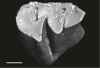
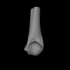
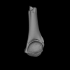
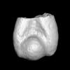
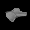
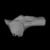
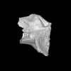
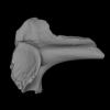
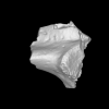
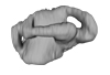
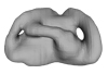
In this contribution, we describe the external and internal morphology of a delphinid petrosal bone collected from Ahu Tahai, a burial site located on the Southwestern coast of Easter Island, at Hangaroa. We discuss the taxonomic attribution of this archaeological item and describe its internal structures based on µCT data, including the bony labyrinth and the nerve and vein patterns. Identification of the nerves exists lead us to relocate the identification of the foramen singulare in delphinid petrosals.
indet. indet. AT1 View specimen
|
M3#420Stapes Type: "3D_surfaces"doi: 10.18563/m3.sf.420 state:published |
Download 3D surface file |
|
M3#421petrosal bone Type: "3D_surfaces"doi: 10.18563/m3.sf.421 state:published |
Download 3D surface file |
|
M3#422in situ bony labyrinth Type: "3D_surfaces"doi: 10.18563/m3.sf.422 state:published |
Download 3D surface file |
|
M3#423bony labyrinth and associated nerves and blood vessels Type: "3D_surfaces"doi: 10.18563/m3.sf.423 state:published |
Download 3D surface file |
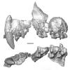
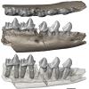
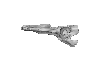
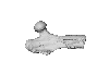
The present 3D Dataset contains two 3D models described in Tissier et al. (https://doi.org/10.1098/rsos.200633): the only known complete mandible of the early-branching rhinocerotoid Epiaceratherium magnum Uhlig, 1999, and a hypothetical reconstruction of the complete archetypic skull of Epiaceratherium Heissig, 1969, created by merging three cranial parts from three distinct Epiaceratherium species.
Epiaceratherium magnum NMB.O.B.928 View specimen
|
M3#5343D surface model of the mandible NMB.O.B.928 of Epiaceratherium magnum, with texture file. Type: "3D_surfaces"doi: 10.18563/m3.sf.534 state:published |
Download 3D surface file |
Epiaceratherium magnum NMB.O.B.928 + MJSN POI007–245 + NMB.I.O.43 View specimen
|
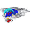
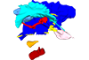
M3#535Archetypal reconstruction of the skull of Epiaceratherium, generated by 3D virtual association of the cranium of E. delemontense (MJSN POI007–245, in blue), mandible of E. magnum (NMB.O.B.928, green) and snout of E. bolcense (NMB.I.O.43, in orange). Type: "3D_surfaces"doi: 10.18563/m3.sf.535 state:published |
Download 3D surface file |
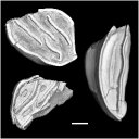
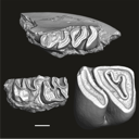
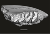
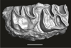
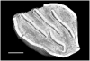
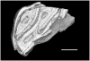
This contribution contains the 3D models of the fossil teeth of two chinchilloid caviomorph rodents (Borikenomys praecursor and Chinchilloidea gen. et sp. indet.) discovered from lower Oligocene deposits of Puerto Rico, San Sebastian Formation (locality LACM Loc. 8060). These fossils were described and figured in the following publication: Marivaux et al. (2020), Early Oligocene chinchilloid caviomorphs from Puerto Rico and the initial rodent colonization of the West Indies. Proceedings of the Royal Society B. http://dx.doi.org/10.1098/rspb.2019.2806
Borikenomys praecursor LACM 162447 View specimen
|
M3#638Right lower m3. This isolated tooth was scanned with a resolution of 6 µm using a μ-CT-scanning station EasyTom 150 / Rx Solutions (Montpellier RIO Imaging, ISE-M, Montpellier, France). AVIZO 7.1 (Visualization Sciences Group) software was used for visualization, segmentation, and 3D rendering. The specimen was prepared within a “labelfield” module of AVIZO, using the segmentation threshold selection tool. Type: "3D_surfaces"doi: 10.18563/m3.sf.638 state:published |
Download 3D surface file |
Borikenomys praecursor LACM 162446 View specimen
|
M3#639Fragment of lower molar (most of the mesial part). This isolated broken tooth was scanned with a resolution of 6 µm using a μ-CT-scanning station EasyTom 150 / Rx Solutions (Montpellier RIO Imaging, ISE-M, Montpellier, France). AVIZO 7.1 (Visualization Sciences Group) software was used for visualization, segmentation, and 3D rendering. The specimen was prepared within a “labelfield” module of AVIZO, using the segmentation threshold selection tool. Type: "3D_surfaces"doi: 10.18563/m3.sf.639 state:published |
Download 3D surface file |
indet indet LACM 162448 View specimen
|
M3#640Fragment of either an upper tooth (mesial laminae) or a lower tooth (distal laminae). The specimen was scanned with a resolution of 6 µm using a μ-CT-scanning station EasyTom 150 / Rx Solutions (Montpellier RIO Imaging, ISE-M, Montpellier, France). AVIZO 7.1 (Visualization Sciences Group) software was used for visualization, segmentation, and 3D rendering. This fragment of tooth was prepared within a “labelfield” module of AVIZO, using the segmentation threshold selection tool. Type: "3D_surfaces"doi: 10.18563/m3.sf.640 state:published |
Download 3D surface file |

This contribution contains the 3D models analyzed in Müller et al. (2021) “Pushing the boundary? Testing the ‘functional elongation hypothesis’ of the giraffe’s neck”.
Aepyceros melampus ZFMK 2001.278 View specimen

|
M3#643Vertebrae C7, T1 Type: "3D_surfaces"doi: 10.18563/m3.sf.643 state:published |
Download 3D surface file |
Giraffa camelopardalis ZMB 66393 View specimen

|
M3#644Vertebrae Type: "3D_surfaces"doi: 10.18563/m3.sf.644 state:published |
Download 3D surface file |
Giraffa camelopardalis ZSM 1967/17 View specimen
|
M3#645Vertebrae Type: "3D_surfaces"doi: 10.18563/m3.sf.645 state:published |
Download 3D surface file |
Giraffa camelopardalis ZSM 1981/19 View specimen

|
M3#646C3, C4, C5, C6, C7, T1, T2 Type: "3D_surfaces"doi: 10.18563/m3.sf.646 state:published |
Download 3D surface file |
Giraffa camelopardalis KMDA M-10861 View specimen

|
M3#647C3, C4, C5, C6, C7, T1, T2. Acquired via laser scanner. Type: "3D_surfaces"doi: 10.18563/m3.sf.647 state:published |
Download 3D surface file |
Giraffa camelopardalis SMF 84214 View specimen

|
M3#648C7, T1. Warning : photogrammetric models (unit scale is CM, not MM). Type: "3D_surfaces"doi: 10.18563/m3.sf.648 state:published |
Download 3D surface file |
Giraffa camelopardalis SMF 78299 View specimen

|
M3#649C7, T1. Warning : unscaled photogrammetric 3D models (unknown size). Type: "3D_surfaces"doi: 10.18563/m3.sf.649 state:published |
Download 3D surface file |
Giraffa camelopardalis SMF o. N View specimen

|
M3#650C7, T1. Warning : unscaled photogrammetric 3D models (unknown size). Type: "3D_surfaces"doi: 10.18563/m3.sf.650 state:published |
Download 3D surface file |
Giraffa camelopardalis SMNS 19138 View specimen

|
M3#671C7, T1. Warning : unscaled photogrammetric 3D models (unknown size). Type: "3D_surfaces"doi: 10.18563/m3.sf.671 state:published |
Download 3D surface file |
Okapia johnstoni ZMB 62086 View specimen

|
M3#651C3, C4, C5, C6, C7, T1, T2 Type: "3D_surfaces"doi: 10.18563/m3.sf.651 state:published |
Download 3D surface file |
Okapia johnstoni ZMB 70325 View specimen

|
M3#652C3, C4, C5, C6, C7, T1, T2 Type: "3D_surfaces"doi: 10.18563/m3.sf.652 state:published |
Download 3D surface file |
Sivatherium giganteum NHMUK 15707 View specimen

|
M3#653C7. Warning : unscaled photogrammetric 3D model (unknown size). Type: "3D_surfaces"doi: 10.18563/m3.sf.653 state:published |
Download 3D surface file |
Sivatherium giganteum NHMUK 15297 View specimen
|
M3#654T1. Warning : unscaled photogrammetric 3D model (unknown size). Type: "3D_surfaces"doi: 10.18563/m3.sf.654 state:published |
Download 3D surface file |
Cervus elaphus ZMB 47502 View specimen

|
M3#655C3, C4, C5, C6, C7, T1, T2 Type: "3D_surfaces"doi: 10.18563/m3.sf.655 state:published |
Download 3D surface file |
Axis axis SMF 1450 View specimen

|
M3#656C7, T1 Type: "3D_surfaces"doi: 10.18563/m3.sf.656 state:published |
Download 3D surface file |
Cervus nippon SMF 4368 View specimen

|
M3#657C7, T1 Type: "3D_surfaces"doi: 10.18563/m3.sf.657 state:published |
Download 3D surface file |
Capreolus capreolus SMF 79852 View specimen

|
M3#658C7, T1 Type: "3D_surfaces"doi: 10.18563/m3.sf.658 state:published |
Download 3D surface file |
Capreolus capreolus ZFMK 67.237 View specimen

|
M3#659C7, T1 Type: "3D_surfaces"doi: 10.18563/m3.sf.659 state:published |
Download 3D surface file |
Muntiacus reevesi SMF 92954 View specimen

|
M3#660C7, T1 Type: "3D_surfaces"doi: 10.18563/m3.sf.660 state:published |
Download 3D surface file |
Muntiacus reevesi SMF 92332 View specimen

|
M3#661C7, T1 Type: "3D_surfaces"doi: 10.18563/m3.sf.661 state:published |
Download 3D surface file |
Alces alces SMF 35549 View specimen

|
M3#662C7, T1 Type: "3D_surfaces"doi: 10.18563/m3.sf.662 state:published |
Download 3D surface file |
Dama dama ZFMK 86.125 View specimen

|
M3#663C7, T1 Type: "3D_surfaces"doi: 10.18563/m3.sf.663 state:published |
Download 3D surface file |
Antilope cervicapra ZMB 78829 View specimen

|
M3#664C3, C4, C5, C6, C7, T1, T2 Type: "3D_surfaces"doi: 10.18563/m3.sf.664 state:published |
Download 3D surface file |
Bison bonasus SMNS 2998 View specimen

|
M3#665C7, T1. Warning : unscaled photogrammetric 3D models (unknown size). Type: "3D_surfaces"doi: 10.18563/m3.sf.665 state:published |
Download 3D surface file |
Nanger dama SMF 74435 View specimen

|
M3#666C7, T1 Type: "3D_surfaces"doi: 10.18563/m3.sf.666 state:published |
Download 3D surface file |
Litocranius walleri SMF 23747 View specimen

|
M3#667C7, T1 Type: "3D_surfaces"doi: 10.18563/m3.sf.667 state:published |
Download 3D surface file |
Litocranius walleri SMF 23749 View specimen

|
M3#669C7, T1 Type: "3D_surfaces"doi: 10.18563/m3.sf.669 state:published |
Download 3D surface file |
Tragelaphus eurycerus SMF 95875 View specimen

|
M3#670C7, T1 Type: "3D_surfaces"doi: 10.18563/m3.sf.670 state:published |
Download 3D surface file |
Bos javanicus SMF 64934 View specimen

|
M3#672C7, T1 Type: "3D_surfaces"doi: 10.18563/m3.sf.672 state:published |
Download 3D surface file |
Ovis aries ZFMK 1982.338 View specimen

|
M3#673C7, T1 Type: "3D_surfaces"doi: 10.18563/m3.sf.673 state:published |
Download 3D surface file |
Rupicapra rupicapra ZFMK 72.367 View specimen

|
M3#674C7, T1 Type: "3D_surfaces"doi: 10.18563/m3.sf.674 state:published |
Download 3D surface file |
Kobus ellipsiprymnus SMNS 4443 View specimen

|
M3#675C7, T1 Type: "3D_surfaces"doi: 10.18563/m3.sf.675 state:published |
Download 3D surface file |
Sylvicapra grimmia SMNS 15292 View specimen

|
M3#676C7, T1 Type: "3D_surfaces"doi: 10.18563/m3.sf.676 state:published |
Download 3D surface file |
Syncerus caffer SMNS 7347 View specimen

|
M3#677C7, T1. Warning : unscaled photogrammetric 3D models (unknown size). Type: "3D_surfaces"doi: 10.18563/m3.sf.677 state:published |
Download 3D surface file |
Procapra gutturosa SMNS 5796 View specimen

|
M3#678C7, T1 Type: "3D_surfaces"doi: 10.18563/m3.sf.678 state:published |
Download 3D surface file |
Damaliscus pygargus SMNS 21617 View specimen

|
M3#679C7, T1 Type: "3D_surfaces"doi: 10.18563/m3.sf.679 state:published |
Download 3D surface file |
Madoqua kirkii SMNS 4432 View specimen

|
M3#680C7, T1 Type: "3D_surfaces"doi: 10.18563/m3.sf.680 state:published |
Download 3D surface file |
Bubalus mindorensis SMNS 2054 View specimen

|
M3#681C7, T1. Warning : unscaled photogrammetric 3D models (unknown size). Type: "3D_surfaces"doi: 10.18563/m3.sf.681 state:published |
Download 3D surface file |
Capra hircus SMNS 51328 View specimen

|
M3#682C7, T1 Type: "3D_surfaces"doi: 10.18563/m3.sf.682 state:published |
Download 3D surface file |
Connochaetes taurinus SMNS 4442 View specimen

|
M3#683C7, T1. Warning : unscaled photogrammetric 3D models (unknown size). Type: "3D_surfaces"doi: 10.18563/m3.sf.683 state:published |
Download 3D surface file |
Antilocapra americana ZSM 1964/218 View specimen

|
M3#684C3, C4, C5, C6, C7, T1, T2 Type: "3D_surfaces"doi: 10.18563/m3.sf.684 state:published |
Download 3D surface file |
Antilocapra americana ZMB 77281 View specimen

|
M3#685C7, T1 Type: "3D_surfaces"doi: 10.18563/m3.sf.685 state:published |
Download 3D surface file |
Moschus moschiferus ZMB 62080 View specimen

|
M3#686C3, C4, C5, C6, C7, T1, T2 Type: "3D_surfaces"doi: 10.18563/m3.sf.686 state:published |
Download 3D surface file |
Moschus moschiferus ZMB 60367 View specimen

|
M3#687C7, T1 Type: "3D_surfaces"doi: 10.18563/m3.sf.687 state:published |
Download 3D surface file |
Moschus moschiferus ZMB 51830 View specimen

|
M3#688C7, T1 Type: "3D_surfaces"doi: 10.18563/m3.sf.688 state:published |
Download 3D surface file |
Tragulus javanicus SMF 82179 View specimen

|
M3#689C7, T1 Type: "3D_surfaces"doi: 10.18563/m3.sf.689 state:published |
Download 3D surface file |
Tragulus javanicus ZMB 86222 View specimen

|
M3#690C7, T1 Type: "3D_surfaces"doi: 10.18563/m3.sf.690 state:published |
Download 3D surface file |
Tragulus sp. ZMB o. N. View specimen

|
M3#691C7, T1 Type: "3D_surfaces"doi: 10.18563/m3.sf.691 state:published |
Download 3D surface file |
Hyemoschus aquaticus ZMB 71071 View specimen

|
M3#692C7, T1 Type: "3D_surfaces"doi: 10.18563/m3.sf.692 state:published |
Download 3D surface file |
Hyemoschus aquaticus ZMB 103235 View specimen

|
M3#693C7, T1 Type: "3D_surfaces"doi: 10.18563/m3.sf.693 state:published |
Download 3D surface file |
Vicugna vicugna SMF 94752 View specimen

|
M3#694C7, T1 Type: "3D_surfaces"doi: 10.18563/m3.sf.694 state:published |
Download 3D surface file |
Camelus dromedarius SMF 70473 View specimen

|
M3#695C7, T1. Warning : unscaled photogrammetric 3D models (unknown size). Type: "3D_surfaces"doi: 10.18563/m3.sf.695 state:published |
Download 3D surface file |
Camelus bactrianus SMF 25542 View specimen

|
M3#696C7, T1. Warning : unscaled photogrammetric 3D models (unknown size). Type: "3D_surfaces"doi: 10.18563/m3.sf.696 state:published |
Download 3D surface file |
Lama glama SMNS 31175 View specimen

|
M3#697C7, T1 Type: "3D_surfaces"doi: 10.18563/m3.sf.697 state:published |
Download 3D surface file |
Vicugna pacos SMNS 46255 View specimen

|
M3#698C7, T1 Type: "3D_surfaces"doi: 10.18563/m3.sf.698 state:published |
Download 3D surface file |
Vicugna pacos SMNS 7349 View specimen

|
M3#699C7, T1 Type: "3D_surfaces"doi: 10.18563/m3.sf.699 state:published |
Download 3D surface file |
This contribution contains the 3D models described and figured in the following publication: Paulina-Carabajal A and Calvo JO 2021. Re-description of the braincase of the rebbachisaurid sauropod Limaysaurus tessonei and novel endocranial information based on CT scans. Anais da Academia Brasileira de Ciências 93(Suppl. 2): e20200762 https://doi.org/10.1590/0001-3765202120200762
Limaysaurus tessonei MUCPv-205 View specimen
|
M3#700Renderings of the virtually isolate braincase, brain, and right inner ear. Type: "3D_surfaces"doi: 10.18563/m3.sf.700 state:published |
Download 3D surface file |
This contribution contains the 3D models of postcranial bones (humerus, ulna, innominate, femur, tibia, astragalus, navicular, and metatarsal III) described and figured in the following publication: “Postcranial morphology of the extinct rodent Neoepiblema (Rodentia: Chinchilloidea): insights into the paleobiology of neoepiblemids”.
Neoepiblema acreensis UFAC 3549 View specimen
|
M3#719UFAC 3549, left humerus missing the proximal region. Type: "3D_surfaces"doi: 10.18563/m3.sf.719 state:published |
Download 3D surface file |
Neoepiblema acreensis UFAC 5076 View specimen
|
M3#720UFAC 5076, right humerus missing the proximal region. Type: "3D_surfaces"doi: 10.18563/m3.sf.720 state:published |
Download 3D surface file |
Neoepiblema acreensis UFAC 1939 View specimen
|
M3#721UFAC 1939, right ulna missing the olecranon epiphysis and the distal region. Type: "3D_surfaces"doi: 10.18563/m3.sf.721 state:published |
Download 3D surface file |
Neoepiblema acreensis UFAC 3697 View specimen
|
M3#722UFAC 3697, right innominate bone. Type: "3D_surfaces"doi: 10.18563/m3.sf.722 state:published |
Download 3D surface file |
Neoepiblema acreensis UFAC 2574 View specimen
|
M3#723UFAC 2574, proximal region of a left femur. Type: "3D_surfaces"doi: 10.18563/m3.sf.723 state:published |
Download 3D surface file |
Neoepiblema acreensis UFAC 2937 View specimen
|
M3#724UFAC 2937, right femur with damaged proximal region. Type: "3D_surfaces"doi: 10.18563/m3.sf.724 state:published |
Download 3D surface file |
Neoepiblema acreensis UFAC 2210 View specimen

|
M3#725UFAC 2210, distal region of a right femur. Type: "3D_surfaces"doi: 10.18563/m3.sf.725 state:published |
Download 3D surface file |
Neoepiblema acreensis UFAC 1887 View specimen
|
M3#726UFAC 1887, right tibia Type: "3D_surfaces"doi: 10.18563/m3.sf.726 state:published |
Download 3D surface file |
Neoepiblema acreensis UFAC 1840 View specimen
|
M3#727UFAC 1840, left astragalus. Type: "3D_surfaces"doi: 10.18563/m3.sf.727 state:published |
Download 3D surface file |
Neoepiblema acreensis UFAC 2549 View specimen
|
M3#728UFAC 2549, right astragalus. Type: "3D_surfaces"doi: 10.18563/m3.sf.728 state:published |
Download 3D surface file |
Neoepiblema acreensis UFAC 3672 View specimen
|
M3#729UFAC 3672, right navicular. Type: "3D_surfaces"doi: 10.18563/m3.sf.729 state:published |
Download 3D surface file |
Neoepiblema acreensis UFAC 2116 View specimen
|
M3#730UFAC 2116, left metatarsal III. Type: "3D_surfaces"doi: 10.18563/m3.sf.730 state:published |
Download 3D surface file |
Neoepiblema horridula UFAC 3260 View specimen
|
M3#731UFAC 3260, fragmented left innominate. Type: "3D_surfaces"doi: 10.18563/m3.sf.731 state:published |
Download 3D surface file |
Neoepiblema horridula UFAC 2620 View specimen
|
M3#732UFAC 2620, distal region of a right femur. Type: "3D_surfaces"doi: 10.18563/m3.sf.732 state:published |
Download 3D surface file |
Neoepiblema horridula UFAC 2737 View specimen
|
M3#733UFAC 2737, proximal region of right femur. Type: "3D_surfaces"doi: 10.18563/m3.sf.733 state:published |
Download 3D surface file |
Neoepiblema horridula UFAC 3202 View specimen
|
M3#734UFAC 3202, right tibia, missing the proximalmost and distal portions. Type: "3D_surfaces"doi: 10.18563/m3.sf.734 state:published |
Download 3D surface file |
Neoepiblema horridula UFAC 3212 View specimen
|
M3#735UFAC 3212, left astragalus. Type: "3D_surfaces"doi: 10.18563/m3.sf.735 state:published |
Download 3D surface file |
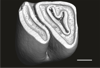
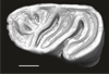
This contribution contains 3D models of upper molar rows of house mice (Mus musculus domesticus). The erupted part of the right row is presented for specimens belonging to four groups: wild-trapped mice, wild-derived lab offspring, a typical laboratory strain (Swiss) and hybrids between wild-derived and Swiss mice. These models are analyzed in the following publication: Savriama et al 2021: Wild versus lab house mice: Effects of age, diet, and genetics on molar geometry and topography. https://doi.org/10.1111/joa.13529
Mus musculus BW_03 View specimen

|
M3#736BW_03 Type: "3D_surfaces"doi: 10.18563/m3.sf.736 state:published |
Download 3D surface file |
Mus musculus BW_04 View specimen

|
M3#752BW_04 Type: "3D_surfaces"doi: 10.18563/m3.sf.752 state:published |
Download 3D surface file |
Mus musculus BW_06 View specimen

|
M3#753BW_06 Type: "3D_surfaces"doi: 10.18563/m3.sf.753 state:published |
Download 3D surface file |
Mus musculus BW_07 View specimen

|
M3#754BW_07 Type: "3D_surfaces"doi: 10.18563/m3.sf.754 state:published |
Download 3D surface file |
Mus musculus BW_08 View specimen

|
M3#755BW_08 Type: "3D_surfaces"doi: 10.18563/m3.sf.755 state:published |
Download 3D surface file |
Mus musculus BW_11 View specimen

|
M3#756BW_11 Type: "3D_surfaces"doi: 10.18563/m3.sf.756 state:published |
Download 3D surface file |
Mus musculus BW_12 View specimen

|
M3#757BW_12 Type: "3D_surfaces"doi: 10.18563/m3.sf.757 state:published |
Download 3D surface file |
Mus musculus Blab_035 View specimen

|
M3#758Blab_035 Type: "3D_surfaces"doi: 10.18563/m3.sf.758 state:published |
Download 3D surface file |
Mus musculus Blab_046 View specimen

|
M3#759Blab_046 Type: "3D_surfaces"doi: 10.18563/m3.sf.759 state:published |
Download 3D surface file |
Mus musculus Blab_054 View specimen

|
M3#760Blab_054 Type: "3D_surfaces"doi: 10.18563/m3.sf.760 state:published |
Download 3D surface file |
Mus musculus Blab_056 View specimen

|
M3#761Blab_056 Type: "3D_surfaces"doi: 10.18563/m3.sf.761 state:published |
Download 3D surface file |
Mus musculus Blab_082 View specimen

|
M3#762Blab_082 Type: "3D_surfaces"doi: 10.18563/m3.sf.762 state:published |
Download 3D surface file |
Mus musculus Blab_086 View specimen

|
M3#763Blab_086 Type: "3D_surfaces"doi: 10.18563/m3.sf.763 state:published |
Download 3D surface file |
Mus musculus Blab_092 View specimen

|
M3#764Blab_092 Type: "3D_surfaces"doi: 10.18563/m3.sf.764 state:published |
Download 3D surface file |
Mus musculus Blab_319 View specimen

|
M3#751Blab_319 Type: "3D_surfaces"doi: 10.18563/m3.sf.751 state:published |
Download 3D surface file |
Mus musculus Blab_325 View specimen

|
M3#750Blab_325 Type: "3D_surfaces"doi: 10.18563/m3.sf.750 state:published |
Download 3D surface file |
Mus musculus Blab_329 View specimen

|
M3#737Blab_329 Type: "3D_surfaces"doi: 10.18563/m3.sf.737 state:published |
Download 3D surface file |
Mus musculus Blab_330 View specimen
|
M3#738Blab_330 Type: "3D_surfaces"doi: 10.18563/m3.sf.738 state:published |
Download 3D surface file |
Mus musculus Blab_F2a View specimen

|
M3#739Blab_F2a Type: "3D_surfaces"doi: 10.18563/m3.sf.739 state:published |
Download 3D surface file |
Mus musculus Blab_F2b View specimen

|
M3#740Blab_F2b Type: "3D_surfaces"doi: 10.18563/m3.sf.740 state:published |
Download 3D surface file |
Mus musculus Blab_BB3w View specimen

|
M3#741Blab_BB3w Type: "3D_surfaces"doi: 10.18563/m3.sf.741 state:published |
Download 3D surface file |
Mus musculus hyb_BS01 View specimen

|
M3#742hyb_BS01 Type: "3D_surfaces"doi: 10.18563/m3.sf.742 state:published |
Download 3D surface file |
Mus musculus hyb_BS02 View specimen

|
M3#743hyb_BS02 Type: "3D_surfaces"doi: 10.18563/m3.sf.743 state:published |
Download 3D surface file |
Mus musculus hyb_SB01 View specimen

|
M3#744hyb_SB01 Type: "3D_surfaces"doi: 10.18563/m3.sf.744 state:published |
Download 3D surface file |
Mus musculus hyb_SB02 View specimen

|
M3#745hyb_SB02 Type: "3D_surfaces"doi: 10.18563/m3.sf.745 state:published |
Download 3D surface file |
Mus musculus SW_001 View specimen

|
M3#746SW_001 Type: "3D_surfaces"doi: 10.18563/m3.sf.746 state:published |
Download 3D surface file |
Mus musculus SW_002 View specimen

|
M3#747SW_002 Type: "3D_surfaces"doi: 10.18563/m3.sf.747 state:published |
Download 3D surface file |
Mus musculus SW_005 View specimen

|
M3#748SW_005 Type: "3D_surfaces"doi: 10.18563/m3.sf.748 state:published |
Download 3D surface file |
Mus musculus SW_0ter View specimen

|
M3#749SW_0ter Type: "3D_surfaces"doi: 10.18563/m3.sf.749 state:published |
Download 3D surface file |
Mus musculus SW_343 View specimen

|
M3#765SW_343 Type: "3D_surfaces"doi: 10.18563/m3.sf.765 state:published |
Download 3D surface file |