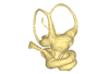
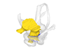
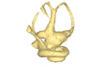
3D models of Cainotheriids Ossicular chain
Explodable 3D Dog Skull for Veterinary Education
3D models of Kalakocetus, the earliest Cetacea
3D GM dataset of bird skeletal variation
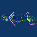
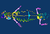
Skeletal embryonic development in the catshark
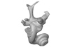
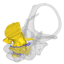
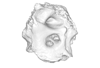
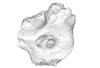
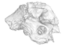
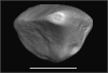
Bony connexions of the petrosal bone of extant hippos
bony labyrinth (14) , inner ear (11) , Eocene (11) , geometric morphometrics (10) , CT-scan (10) , Oligocene (9) , Micro-CT (9)
Lionel Hautier (25) , Maëva Judith Orliac (24) , Laurent Marivaux (18) , Renaud Lebrun (15) , Rodolphe Tabuce (15) , Pierre-Olivier Antoine (13) , Bastien Mennecart (13)
|
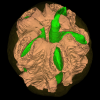
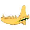
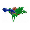
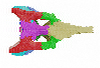
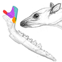
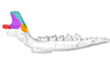
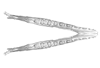
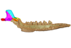
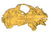
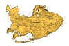
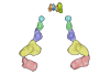
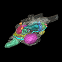
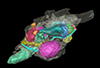
3D models related to the publication: The neuroanatomy and pneumaticity of Hamadasuchus from the Cretaceous of Morocco and its significance for the paleoecology of Peirosauridae and other altirostral crocodylomorphsYohan Pochat-Cottilloux
Published online: 14/06/2023 |
|
M3#10943D volume reconstruction of the braincase osteology Type: "3D_surfaces"doi: 10.18563/m3.sf.1094 state:published |
Download 3D surface file |
|
M3#10963D volume reconstruction of the endocast Type: "3D_surfaces"doi: 10.18563/m3.sf.1096 state:published |
Download 3D surface file |
|
M3#10973D volume reconstruction of the labyrinths Type: "3D_surfaces"doi: 10.18563/m3.sf.1097 state:published |
Download 3D surface file |
|
M3#10983D volume reconstruction of the pneumatic cavities Type: "3D_surfaces"doi: 10.18563/m3.sf.1098 state:published |
Download 3D surface file |
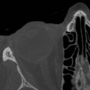
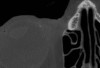
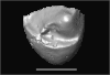
The present Dataset contains the micro-CT scan of the head of an anonymous 54 year old female donor, at a voxel resolution of 145µm. The skin of the face has been masked in order to avoid the donor to be recognized.
Homo sapiens UM_HS_2018_09_13 View specimen
|
M3#1152Micro-ct data set Type: "3D_CT"doi: 10.18563/m3.sf.1152 state:published |
Download CT data |
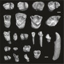
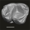
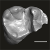
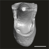
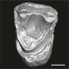
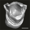
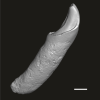
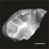
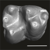
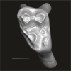
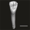
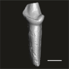
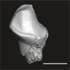
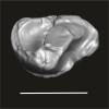
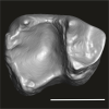
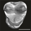
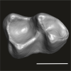
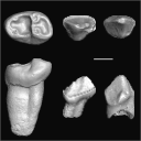
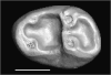
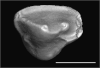
This contribution contains the three-dimensional digital models of the dental fossil material of anthropoid and strepsirrhine primates, discovered in Lower Oligocene detrital deposits outcropping in the Porto Rico and El Argoub areas, east of the Dakhla peninsula region (Atlantic Sahara; in the south of Morocco, near the northern border of Mauritania). These fossils were described, figured and discussed in the following publication: Marivaux et al. (2024), A new primate community from the earliest Oligocene of the Atlantic margin of Northwest Africa: Systematic, paleobiogeographic and paleoenvironmental implications. Journal of Human Evolution. https://doi.org/10.1016/j.jhevol.2024.103548
Catopithecus aff. browni DAK-Arg-087 View specimen
|
M3#1211Isolated right lower m3 (worn) Type: "3D_surfaces"doi: 10.18563/m3.sf.1211 state:published |
Download 3D surface file |
Catopithecus aff. browni DAK-Arg-088 View specimen
|
M3#1212Isolated right lower m2 (abraded/corroded) Type: "3D_surfaces"doi: 10.18563/m3.sf.1212 state:published |
Download 3D surface file |
Catopithecus aff. browni DAK-Arg-089 View specimen
|
M3#1213Isolated left lower m1 (worn) Type: "3D_surfaces"doi: 10.18563/m3.sf.1213 state:published |
Download 3D surface file |
Catopithecus aff. browni DAK-Pto-052 View specimen
|
M3#1214Isolated right lower m1 (pristine but lacking the mesiobuccal region) Type: "3D_surfaces"doi: 10.18563/m3.sf.1214 state:published |
Download 3D surface file |
Catopithecus aff. browni DAK-Arg-090 View specimen
|
M3#1215Isolated left upper P4 Type: "3D_surfaces"doi: 10.18563/m3.sf.1215 state:published |
Download 3D surface file |
Catopithecus aff. browni DAK-Arg-091 View specimen
|
M3#1216Isolated left upper M2 (worn and corroded) Type: "3D_surfaces"doi: 10.18563/m3.sf.1216 state:published |
Download 3D surface file |
Catopithecus aff. browni DAK-Pto-053 View specimen
|
M3#1217Isolated right upper M1 (lacking the buccal region) Type: "3D_surfaces"doi: 10.18563/m3.sf.1217 state:published |
Download 3D surface file |
Abuqatrania cf. basiodontos DAK-Arg-092 View specimen
|
M3#1218Isolated left lower c1 Type: "3D_surfaces"doi: 10.18563/m3.sf.1218 state:published |
Download 3D surface file |
?Propliopithecus sp. DAK-Pto-056 View specimen
|
M3#1219Isolated right lower m3 (fragment of talonid of a germ) Type: "3D_surfaces"doi: 10.18563/m3.sf.1219 state:published |
Download 3D surface file |
Abuqatrania cf. basiodontos DAK-Arg-093 View specimen
|
M3#1469Isolated right lower m1 Type: "3D_surfaces"doi: 10.18563/m3.sf.1469 state:published |
Download 3D surface file |
Abuqatrania cf. basiodontos DAK-Arg-094 View specimen
|
M3#1221Isolated left upper M1 or M2 (corroded, lacking the enamel cap [exposed dentine]) Type: "3D_surfaces"doi: 10.18563/m3.sf.1221 state:published |
Download 3D surface file |
Abuqatrania cf. basiodontos DAK-Arg-095 View specimen
|
M3#1222Isolated right lower i1 or i2 Type: "3D_surfaces"doi: 10.18563/m3.sf.1222 state:published |
Download 3D surface file |
Abuqatrania cf. basiodontos DAK-Arg-096 View specimen
|
M3#1223Isolated right lower p2 (worn apex) Type: "3D_surfaces"doi: 10.18563/m3.sf.1223 state:published |
Download 3D surface file |
Abuqatrania cf. basiodontos DAK-Arg-097 View specimen
|
M3#1224Isolated right lower p2 (worn apex and broken root) Type: "3D_surfaces"doi: 10.18563/m3.sf.1224 state:published |
Download 3D surface file |
Afrotarsius sp. DAK-Arg-098 View specimen
|
M3#1225Isolated left lower p3 Type: "3D_surfaces"doi: 10.18563/m3.sf.1225 state:published |
Download 3D surface file |
Afrotarsius sp. DAK-Pto-054 View specimen
|
M3#1226Isolated right lower m1 (abraded/corroded) Type: "3D_surfaces"doi: 10.18563/m3.sf.1226 state:published |
Download 3D surface file |
Orolemur mermozi DAK-Pto-055 View specimen
|
M3#1227Isolated right upper M1 or M2 (pristine, Holotype) Type: "3D_surfaces"doi: 10.18563/m3.sf.1227 state:published |
Download 3D surface file |
Wadilemur cf. elegans DAK-Arg-099 View specimen
|
M3#1228Isolated right lower m2 Type: "3D_surfaces"doi: 10.18563/m3.sf.1228 state:published |
Download 3D surface file |
cf. 'Anchomomys' milleri DAK-Arg-100 View specimen
|
M3#1229Isolated right lower c1 Type: "3D_surfaces"doi: 10.18563/m3.sf.1229 state:published |
Download 3D surface file |
Abuqatrania cf. basiodontos DAK-Arg-101 View specimen
|
M3#1396Isolated left upper M3 (abraded) Type: "3D_surfaces"doi: 10.18563/m3.sf.1396 state:published |
Download 3D surface file |
Orogalago saintexuperyi DAK-Arg-102 View specimen
|
M3#1397Isolated left lower m2 Type: "3D_surfaces"doi: 10.18563/m3.sf.1397 state:published |
Download 3D surface file |
Wadilemur cf. elegans DAK-Arg-103 View specimen
|
M3#1473Isolated right upper M1 or M2 (lacking the mesial and buccal regions) Type: "3D_surfaces"doi: 10.18563/m3.sf.1473 state:published |
Download 3D surface file |
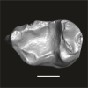
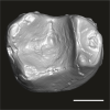
The present 3D Dataset contains the 3D models produced in the frame of the article Perthuis, A. de, Mennecart, B., Barrier, P., Chenot, É., Falconnet, J., Gagnaison, J.-C., Georgalis, G. L., Gilbert, C., Guevel, B., Langevin, D., Lapparent de Broin, F. de, Lemierre, A., Maubert, F., Ossó, À., Potel, S., Thivaiou, D., Tissier, J., Toullec, R., Xerri, S., Gagnaison, C. 2025. Révision des données sédimentologiques et biostratigraphiques des gisements à vertébrés des sables de l’Orléanais, à Beaugency, Tavers et Le Bardon (Miocène Moyen ; Loiret, France). Geodiversitas 47 (12): 2-76. https://doi.org/10.5252/geodiversitas2025v47a12
Bunolistriodon lockharti ULB-TAV-21 View specimen
|
M3#1837Left upper M3 Type: "3D_surfaces"doi: 10.18563/m3.sf.1837 state:published |
Download 3D surface file |
Megamphicyon giganteus ULB-TAV-13 View specimen
|
M3#1531Left first lower molar Type: "3D_surfaces"doi: 10.18563/m3.sf.1531 state:published |
Download 3D surface file |
Hispanotherium matritense ULB-TAV-17 View specimen
|
M3#1532Left first lower molar Type: "3D_surfaces"doi: 10.18563/m3.sf.1532 state:published |
Download 3D surface file |
Plesiaceratherium lumiarense ULB-TAV-18 View specimen
|
M3#1533Left third upper molar Type: "3D_surfaces"doi: 10.18563/m3.sf.1533 state:published |
Download 3D surface file |
Chelydropsis aff. sansaniensis ULB-TAV-23 View specimen
|
M3#1535Cast of a skull Type: "3D_surfaces"doi: 10.18563/m3.sf.1535 state:published |
Download 3D surface file |
Ronzotherium romani ULB-TAV-4 View specimen
|
M3#1556Right fourth upper premolar Type: "3D_surfaces"doi: 10.18563/m3.sf.1556 state:published |
Download 3D surface file |
Prodeinotherium bavaricum ULB-TAV-24 View specimen
|
M3#1557left hemimandibule Type: "3D_surfaces"doi: 10.18563/m3.sf.1557 state:published |
Download 3D surface file |
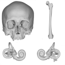
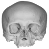
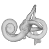
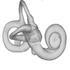
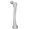
The present 3D Dataset contains the models analyzed in the publication: Menéndez L, Rios C, Acosta Morano C, Novellino P, Schmelzle T, Aguirre-Fernández G, Breidenstein A, Barquera R, Schuenemann VJ, Stafford TW, Sánchez-Villagra M, Barbieri C. (2025). A human skeleton from Última Esperanza, South-West Patagonia, Chile: Osteobiography, morphometric, and genetic analysis. The models include the skull, femur, and the segmented left and right inner ears of a late Holocene human skeleton from southern Patagonia. In the associated paper, we present the radiocarbon dating, an osteobiography profile evaluating some aspects of the life history of this individual, as well as genetic and morphometric analysis assessing biological relatedness to other individuals and populations.
Homo sapiens PIMUZ A/V 4612 View specimen
|
M3#1650Homo sapiens skull Type: "3D_surfaces"doi: 10.18563/m3.sf.1650 state:published |
Download 3D surface file |
|
M3#1652Homo sapiens left inner ear Type: "3D_surfaces"doi: 10.18563/m3.sf.1652 state:published |
Download 3D surface file |
|
M3#1653Homo sapiens right inner ear Type: "3D_surfaces"doi: 10.18563/m3.sf.1653 state:published |
Download 3D surface file |
Homo sapiens PIMUZ A/V 4613 View specimen
|
M3#1651Homo sapiens femur Type: "3D_surfaces"doi: 10.18563/m3.sf.1651 state:published |
Download 3D surface file |
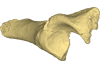
This contribution contains the 3D model described and figured in the following publication: Hautier L, Sarr R, Lihoreau F, Tabuce R, Marwan Hameh P. 2014. First record of the family Protocetidae in the Lutetian of Senegal (West Africa). Palaeovertebrata 38(2)-e2
indet. indet. SN103 View specimen
|
M3#5SN103, partial left innominate. Age and occurrence – Taïba Formation, Lutetian of the near Taïba Ndiaye, quarry of the Industries Chimiques du Sénégal (ICS) Type: "3D_surfaces"doi: 10.18563/m3.sf5 state:published |
Download 3D surface file |
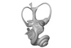
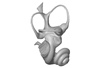
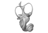
The present 3D Dataset contains the 3D models analyzed in Costeur L., Mennecart B., Müller B., Schulz G., 2016. Prenatal growth stages show the development of the ruminant bony labyrinth and petrosal bone. Journal of Anatomy. https://doi.org/10.1111/joa.12549
Bos taurus NMB3038 View specimen
|
M3#124Right bony labyrinth of a Bos taurus foetus (gestational age 115 days) Type: "3D_surfaces"doi: 10.18563/m3.sf.124 state:published |
Download 3D surface file |
Bos taurus NMB3367 View specimen
|
M3#125Right bony labyrinth of a Bos taurus foetus (gestational age 165 days) Type: "3D_surfaces"doi: 10.18563/m3.sf.125 state:published |
Download 3D surface file |
Bos taurus NMB3365 View specimen
|
M3#126Right bony labyrinth of a Bos taurus foetus (gestational age 210 days) Type: "3D_surfaces"doi: 10.18563/m3.sf.126 state:published |
Download 3D surface file |
Bos taurus NMB2855 View specimen
|
M3#127Right bony labyrinth of a Bos taurus foetus (gestational age 260 days) Type: "3D_surfaces"doi: 10.18563/m3.sf.127 state:published |
Download 3D surface file |
Bos taurus NMB1037 View specimen
|
M3#128Left bony labyrinth of an adult Bos taurus Type: "3D_surfaces"doi: 10.18563/m3.sf.128 state:published |
Download 3D surface file |
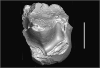
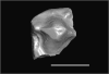
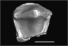
This contribution contains the 3D models described and figured in the publication entitled "The petrosal and bony labyrinth of Diplobune minor, an enigmatic Artiodactyla from the Oligocene of Western Europe" by Orliac, Araújo, and Lihoreau published in Journal of Morphology (Orliac et al. 2017) https://doi.org/10.1002/jmor.20702.
Diplobune minor UM ITD 1079 View specimen
|
M3#138right bony labyrinth of Diplobune minor from Itardies, France Type: "3D_surfaces"doi: 10.18563/m3.sf.138 state:published |
Download 3D surface file |
|
M3#139right isolated petrosal of Diplobune minor from Itardies, France Type: "3D_surfaces"doi: 10.18563/m3.sf.139 state:published |
Download 3D surface file |
Diplobune minor UM ITD 1080 View specimen
|
M3#140left bony labyrinth of Diplobune minor from Itardies, France Type: "3D_surfaces"doi: 10.18563/m3.sf.140 state:published |
Download 3D surface file |
|
M3#141left isolated petrosal of Diplobune minor from Itardies, France Type: "3D_surfaces"doi: 10.18563/m3.sf.141 state:published |
Download 3D surface file |
Diplobune minor UM ITD 1081 View specimen
|
M3#142right bony labyrinth and associated nerves and veins of Diplobune minor from Itardies, France Type: "3D_surfaces"doi: 10.18563/m3.sf.142 state:published |
Download 3D surface file |
|
M3#143right isolated petrosal of Diplobune minor from Itardies, France Type: "3D_surfaces"doi: 10.18563/m3.sf.143 state:published |
Download 3D surface file |
Diplobune minor UM ITD 1083 View specimen
|
M3#144left bony labyrinth of Diplobune minor from Itardies, France Type: "3D_surfaces"doi: 10.18563/m3.sf.144 state:published |
Download 3D surface file |
|
M3#145left petrosal of Diplobune minor from Itardies, France Type: "3D_surfaces"doi: 10.18563/m3.sf.145 state:published |
Download 3D surface file |
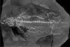
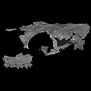
The present contribution contains the 3D model and dataset analyzed in the following publication: Scheyer, T. M., J. M. Neenan, T. Bodogan, H. Furrer, C. Obrist, and M. Plamondon. 2017. A new, exceptionally preserved juvenile specimen of Eusaurosphargis dalsassoi (Diapsida) and implications for Mesozoic marine diapsid phylogeny. Scientific Reports, https://doi.org/10.1038/s41598-017-04514-x .
Eusaurosphargis dalsassoi PIMUZ A/III 4380 View specimen
|
M3#17994 extracted surfaces of skeletal elements of PIMUZ A/III 4380 Type: "3D_surfaces"doi: 10.18563/m3.sf.179 state:published |
Download 3D surface file |
|
M3#180Accompanying CT scan dataset Type: "3D_CT"doi: 10.18563/m3.sf.180 state:published |
Download CT data |
This contribution contains the 3D models described and figured in the following publication: Mennecart B., de Perthuis Ad., Rössner G.E., Guzmán J.A., de Perthuis Au., Costeur L. The first French tragulid skull (Mammalia, Ruminantia, Tragulidae) and associated tragulid remains from the Middle Miocene of Contres (Loir-et-Cher, France). Comptes Rendus Palévol. https://doi.org/10.1016/j.crpv.2017.08.004
Dorcatherium crassum NMB Fa.213.abg View specimen
|
M3#181The 3D surface files of the specimen NMB Fa.213 are the reconstructions of the main skull fragments, the right petrosal bone, and the left bony labyrinth. Type: "3D_surfaces"doi: 10.18563/m3.sf.181 state:published |
Download 3D surface file |
The present 3D Dataset contains the 3D models analyzed in Velazco P. M., Grohé C. 2017. Comparative anatomy of the bony labyrinth of the bats Platalina genovensium (Phyllostomidae, Lonchophyllinae) and Tomopeas ravus (Molossidae, Tomopeatinae). Biotempo 14(2).
Platalina genovensium 278520 View specimen

|
M3#276Right bony labyrinth surface positioned (.PLY) Labels associated (.FLG) Type: "3D_surfaces"doi: 10.18563/m3.sf.276 state:published |
Download 3D surface file |
Tomopeas ravus 278525 View specimen

|
M3#277Right bony labyrinth surface (.PLY) Labels associated (.FLG) Type: "3D_surfaces"doi: 10.18563/m3.sf.277 state:published |
Download 3D surface file |
This contribution contains the 3D models of the isolated teeth attributed to stem representatives of the Cebuella and Cebus lineages (Cebuella sp. and Cebus sp.), described and figured in the following publication: Marivaux et al. (2016), Dental remains of cebid platyrrhines from the earliest late Miocene of Western Amazonia, Peru: macroevolutionary implications on the extant capuchin and marmoset lineages. American Journal of Physical Anthropology. http://dx.doi.org/10.1002/ajpa.23052
Cebus sp. MUSM-3243 View specimen
|
M3#2823D model of left lower m1 (lingual part) Type: "3D_surfaces"doi: 10.18563/m3.sf.282 state:published |
Download 3D surface file |
Cebuella sp. MUSM-3239 View specimen
|
M3#2833D model of left lower p4 Type: "3D_surfaces"doi: 10.18563/m3.sf.283 state:published |
Download 3D surface file |
Cebuella sp. MUSM-3240 View specimen
|
M3#2943D model of right upper P3 or P4 (buccal part) Type: "3D_surfaces"doi: 10.18563/m3.sf.294 state:published |
Download 3D surface file |
Cebuella sp. MUSM-3241 View specimen
|
M3#2953D model of right upper P2 Type: "3D_surfaces"doi: 10.18563/m3.sf.295 state:published |
Download 3D surface file |
Cebuella sp. MUSM-3242 View specimen
|
M3#2963D model of upper I2 Type: "3D_surfaces"doi: 10.18563/m3.sf.296 state:published |
Download 3D surface file |
The present 3D Dataset contains the 3D models of the holotype (NMB Sth. 833) of the new species Micromeryx? eiselei analysed in the article Aiglstorfer, M., Costeur, L., Mennecart, B., Heizmann, E.P.J.. 2017. Micromeryx? eiselei - a new moschid species from Steinheim am Albuch, Germany, and the first comprehensive description of moschid cranial material from the Miocene of Central Europe. PlosOne https://doi.org/10.1371/journal.pone.0185679
Micromeryx? eiselei NMB Sth. 833 View specimen
|
M3#284The 3 D surfaces comprises the skull, petrosal, and bony labyrinth of NMB Sth.833, the holotype of Micromeryx? eiselei Type: "3D_surfaces"doi: 10.18563/m3.sf.284 state:published |
Download 3D surface file |
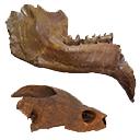
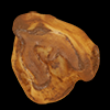
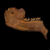
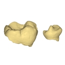
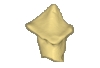
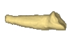
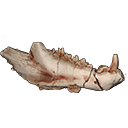
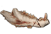
This note presents the 3D model of the hemi-mandible UM-PAT 159 of the MP7 Diacodexis species D. cf. gigasei and 3D models corresponding to the restoration of the ascending ramus, broken on the original specimen, and to a restoration of a complete mandible based on the preserved left hemi-mandible.
Diacodexis cf. gigasei UMPAT159 View specimen
|
M3#3153D models of UM PAT 159 after the restoration of the ascending ramus Type: "3D_surfaces"doi: 10.18563/m3.sf.315 state:published |
Download 3D surface file |
|
M3#316restoration of a complete mandible based on the preserved left hemi-mandible UM PAT 159 Type: "3D_surfaces"doi: 10.18563/m3.sf.316 state:published |
Download 3D surface file |
|
M3#3173D model of the hemi-mandible UM PAT 159 Type: "3D_surfaces"doi: 10.18563/m3.sf.317 state:published |
Download 3D surface file |
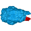
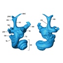
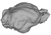
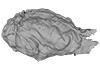
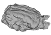
The present 3D Dataset contains the 3D models analyzed in: Amson et al., Under review. Evolutionary Adaptation to Aquatic Lifestyle in Extinct Sloths Can Lead to Systemic Alteration of Bone Structure doi:10.1098/rspb.2018.0270.
Bradypus tridactylus MNHN ZM-MO-1999-1065 View specimen

|
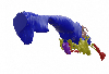
M3#337Brain endocast Type: "3D_surfaces"doi: 10.18563/m3.sf.337 state:published |
Download 3D surface file |
Choloepus didactylus MNHN-ZM-MO-1996-594 View specimen
|
M3#338Brain endocast Type: "3D_surfaces"doi: 10.18563/m3.sf.338 state:published |
Download 3D surface file |
Thalassocnus natans MNHN-F-SAS-734 View specimen
|
M3#339Brain endocast Type: "3D_surfaces"doi: 10.18563/m3.sf.339 state:published |
Download 3D surface file |
Thalassocnus littoralis MNHN-F-SAS-1610 View specimen
|
M3#340Brain endocast Type: "3D_surfaces"doi: 10.18563/m3.sf.340 state:published |
Download 3D surface file |
Thalassocnus littoralis MNHN-F-SAS-1615 View specimen
|
M3#341Brain endocast Type: "3D_surfaces"doi: 10.18563/m3.sf.341 state:published |
Download 3D surface file |
Thalassocnus carolomartini SMNK-3814 View specimen
|
M3#342Brain endocast lacking right olfactory bulb Type: "3D_surfaces"doi: 10.18563/m3.sf.342 state:published |
Download 3D surface file |
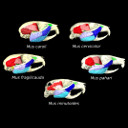
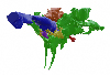
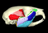
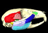
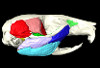
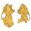
The present 3D Dataset contains the 3D models analyzed in the article entitled "One skull to rule them all? Descriptive and comparative anatomy of the masticatory apparatus in five mice species based on traditional and digital dissections" (Ginot et al. 2018, Journal of Morphology, https://doi.org/10.1002/jmor.20845).
Mus cervicolor R7314 View specimen
|
M3#343.ply surfaces of the skull and masticatory muscles of Mus cervicolor. Created with MorphoDig, .pos and .ntw files also included. Scans were obtained thanks to the Institut des Sciences de l'Evolution de Montpellier MRI platform. Type: "3D_surfaces"doi: 10.18563/m3.sf.343 state:published |
Download 3D surface file |
Mus caroli R7264 View specimen
|
M3#344.ply surfaces of the skull and masticatory muscles of Mus caroli. Created with MorphoDig, .pos and .ntw files also included. Scans were obtained thanks to the Institut des Sciences de l'Evolution de Montpellier MRI platform. Type: "3D_surfaces"doi: 10.18563/m3.sf.344 state:published |
Download 3D surface file |
Mus fragilicauda R7260 View specimen
|
M3#345.ply surfaces of the skull and masticatory muscles of Mus fragilicauda. Created with MorphoDig, .pos and .ntw files also included. Scans were obtained thanks to the Institut des Sciences de l'Evolution de Montpellier MRI platform. Type: "3D_surfaces"doi: 10.18563/m3.sf.345 state:published |
Download 3D surface file |
Mus pahari R7226 View specimen
|
M3#346.ply surfaces of the skull and masticatory muscles of Mus pahari. Created with MorphoDig, .pos and .ntw files also included. Scans were obtained thanks to the Institut des Sciences de l'Evolution de Montpellier MRI platform. Type: "3D_surfaces"doi: 10.18563/m3.sf.346 state:published |
Download 3D surface file |
Mus minutoides minutoides-1 View specimen

|
M3#347.ply surfaces of the skull and masticatory muscles of Mus minutoides. Created with MorphoDig, .pos and .ntw files also included. Scans were obtained thanks to the Institut des Sciences de l'Evolution de Montpellier MRI platform. Type: "3D_surfaces"doi: 10.18563/m3.sf.347 state:published |
Download 3D surface file |
The present 3D Dataset contains the 3D models described and figured in the following publication: Grohé C., Bonis L. de, Chaimanee Y., Chavasseau O., Rugbumrung M., Yamee C., Suraprasit K., Gibert C., Surault J., Blondel C., Jaeger J.-J. 2020. The late middle Miocene Mae Moh Basin of northern Thailand: the richest Neogene assemblage of Carnivora from Southeast Asia and a paleobiogeographic analysis of Miocene Asian carnivorans. American Museum Novitates. http://digitallibrary.amnh.org/handle/2246/7223
Siamogale bounosa MM-54 View specimen
|
M3#5053D model of the skull of Siamogale bounosa The zip file contains: - the 3D surface in PLY - the orientation files in .pos and .ori - the project in .ntw Type: "3D_surfaces"doi: 10.18563/m3.sf.505 state:published |
Download 3D surface file |
Vishnuonyx maemohensis MM-78 View specimen
|
M3#5063D model of the skull of Vishnuonyx maemohensis The zip file contains: - the 3D surface in PLY - the orientation files in .pos and .ori - the project in .ntw Type: "3D_surfaces"doi: 10.18563/m3.sf.506 state:published |
Download 3D surface file |
|
M3#5073D model of the reconstructed upper teeth of Vishnuonyx maemohensis The zip file contains: - the 3D surface in PLY - the orientation files in .pos and .ori - the project in .ntw Type: "3D_surfaces"doi: 10.18563/m3.sf.507 state:published |
Download 3D surface file |
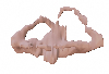
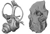
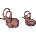
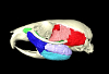
The present Dataset contains the 3D model of the male genital organs of greater horseshoe bat, Rhinolophus ferrumequinum. This is the first detailed 3D structure of the soft-tissue genital organs of bats. The 3D model was generated using microCT and techniques of virtual reconstruction.
Rhinolophus ferrumequinum JP18-006 View specimen
|
M3#521The genital organs of male greater horseshoe bat. Type: "3D_surfaces"doi: 10.18563/m3.sf.521 state:published |
Download 3D surface file |
This contribution contains the 3D models of the fossil teeth of a small-bodied platyrrhine primate, Neosaimiri cf. fieldsi (Cebinae, Cebidae, Platyrrhini) discovered from Laventan deposits (late Middle Miocene) of Peruvian Amazonia, San Martín Department (TAR-31: Tarapoto/Juan Guerra vertebrate fossil-bearing locus n°31). These fossils were described and figured in the following publication: Marivaux et al. (2020), New record of Neosaimiri (Cebidae, Platyrrhini) from the late Middle Miocene of Peruvian Amazonia. Journal of Human Evolution. https://doi.org/10.1016/j.jhevol.2020.102835
Neosaimiri cf. fieldsi MUSM-3888 View specimen
|
M3#538MUSM-3888, right m3 of Neosaimiri cf. fieldsi. Type: "3D_surfaces"doi: 10.18563/m3.sf.538 state:published |
Download 3D surface file |
Neosaimiri cf. fieldsi MUSM-3890 View specimen
|
M3#540MUSM-3890, left dp2 of Neosaimiri cf. fieldsi. Type: "3D_surfaces"doi: 10.18563/m3.sf.540 state:published |
Download 3D surface file |
Neosaimiri cf. fieldsi MUSM-3895 View specimen
|
M3#541MUSM-3895, right DC1 of Neosaimiri cf. fieldsi. Type: "3D_surfaces"doi: 10.18563/m3.sf.541 state:published |
Download 3D surface file |
Neosaimiri cf. fieldsi MUSM-3891 View specimen
|
M3#542MUSM-3891, lingual part of a fragmentary right M1 or M2 of Neosaimiri cf. fieldsi. Type: "3D_surfaces"doi: 10.18563/m3.sf.542 state:published |
Download 3D surface file |
Neosaimiri cf. fieldsi MUSM-3892 View specimen
|
M3#543MUSM-3892, distobuccal part of a fragmentary right upper molar (metacone region) of Neosaimiri cf. fieldsi. Type: "3D_surfaces"doi: 10.18563/m3.sf.543 state:published |
Download 3D surface file |
Neosaimiri cf. fieldsi MUSM-3893 View specimen
|
M3#544MUSM-3893, buccal part of a fragmentary right P3 or P4 of Neosaimiri cf. fieldsi. Type: "3D_surfaces"doi: 10.18563/m3.sf.544 state:published |
Download 3D surface file |
Neosaimiri cf. fieldsi MUSM-3894 View specimen
|
M3#545MUSM-3894, lingual part of a fragmentary left P3 or P4 of Neosaimiri cf. fieldsi. Type: "3D_surfaces"doi: 10.18563/m3.sf.545 state:published |
Download 3D surface file |
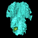
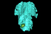
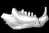
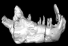
This contribution contains the 3D model(s) described and figured in the following publication: The present 3D Dataset contains the 3D models and CT-Scan slices of the lower jaws and teeth analyzed in “A new prozostrodontian cynodont (Eucynodontia, Probainognathia) from the Upper Triassic of southern Brazil”. https://doi.org/10.1080/02724634.2020.1782415
Agudotherium gassenae CAPPA/UFSM 0262 View specimen
|
M3#546Left lower jaw and cheek teeth Type: "3D_surfaces"doi: 10.18563/m3.sf.546 state:published |
Download 3D surface file |
|
M3#5471578 slices Type: "3D_CT"doi: 10.18563/m3.sf.547 state:published |
Download CT data |
Agudotherium gassenae CAPPA/UFSM 0208 View specimen
|
M3#548right lower jaw Type: "3D_surfaces"doi: 10.18563/m3.sf.548 state:published |
Download 3D surface file |
|
M3#549CT data of CAPPA_UFSM_0208 Type: "3D_CT"doi: 10.18563/m3.sf.549 state:published |
Download CT data |