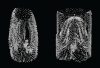
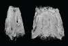
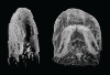
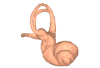
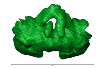
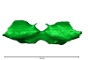
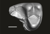
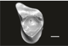
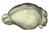
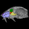
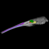
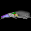
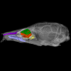
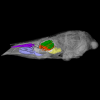
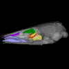
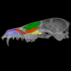
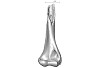
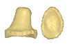
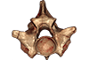
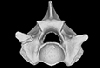
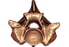
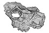
3D models of Cainotheriids Ossicular chain
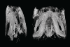
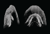
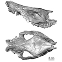
Explodable 3D Dog Skull for Veterinary Education
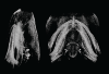
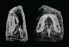
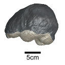
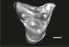
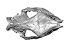
3D models of Kalakocetus, the earliest Cetacea
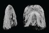
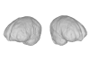
3D GM dataset of bird skeletal variation
Skeletal embryonic development in the catshark
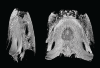
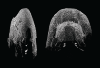
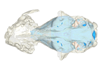
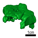
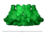
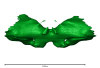
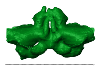
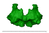
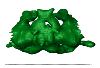
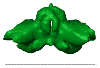
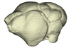
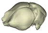
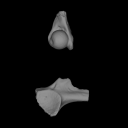
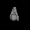
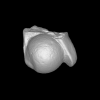
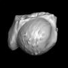
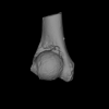
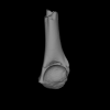
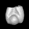
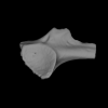
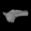
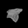
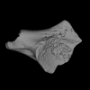
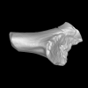
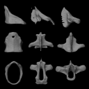
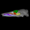
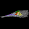
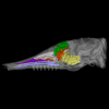
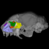
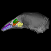
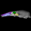
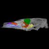
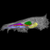
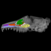
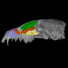
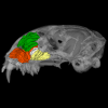
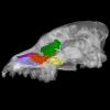
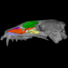
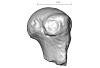
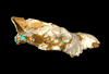
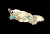
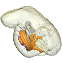
Bony connexions of the petrosal bone of extant hippos
bony labyrinth (14) , inner ear (11) , Eocene (11) , geometric morphometrics (10) , CT-scan (10) , Oligocene (9) , Micro-CT (9)
Lionel Hautier (25) , Maëva Judith Orliac (24) , Laurent Marivaux (18) , Renaud Lebrun (15) , Rodolphe Tabuce (15) , Pierre-Olivier Antoine (13) , Bastien Mennecart (13)
|
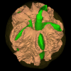
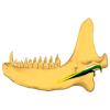
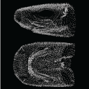
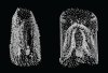
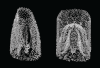
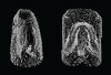
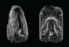
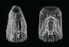
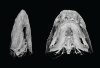
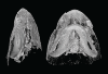
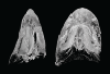
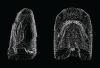
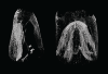
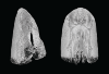
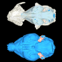
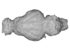
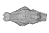
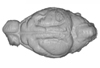
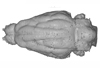
3D models related to the publication: Hide and seek shark teeth in Random Forests: Machine learning applied to Scyliorhinus caniculaFidji Berio
Published online: 24/05/2022 |
|
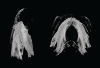
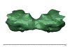
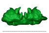
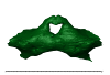
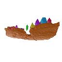
M3#941Head of a 10.6 cm long Scyliorhinus canicula female from a North Atlantic population. Type: "3D_surfaces"doi: 10.18563/m3.sf.941 state:published |
Download 3D surface file |
Scyliorhinus canicula 081118B View specimen
|
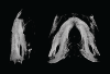
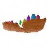
M3#942Head of a 11.0 cm long Scyliorhinus canicula female from a North Atlantic population. Type: "3D_surfaces"doi: 10.18563/m3.sf.942 state:published |
Download 3D surface file |
Scyliorhinus canicula 200118I View specimen
|
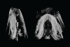
M3#959Head of a 45.0 cm long Scyliorhinus canicula female from a Mediterranean population. Type: "3D_surfaces"doi: 10.18563/m3.sf.959 state:published |
Download 3D surface file |
Scyliorhinus canicula 200118H View specimen
|
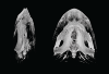
M3#958Head of a 47.0 cm long Scyliorhinus canicula female from a Mediterranean population. Type: "3D_surfaces"doi: 10.18563/m3.sf.958 state:published |
Download 3D surface file |
Scyliorhinus canicula 200118G View specimen
|
M3#957Head of a 40.0 cm long Scyliorhinus canicula female from a Mediterranean population. Type: "3D_surfaces"doi: 10.18563/m3.sf.957 state:published |
Download 3D surface file |
Scyliorhinus canicula 081118C View specimen
|
M3#940Head of a 11.2 cm long Scyliorhinus canicula female from a North Atlantic population. Type: "3D_surfaces"doi: 10.18563/m3.sf.940 state:published |
Download 3D surface file |
Scyliorhinus canicula 081118D View specimen
|
M3#939Head of a 10.2 cm long Scyliorhinus canicula female from a North Atlantic population. Type: "3D_surfaces"doi: 10.18563/m3.sf.939 state:published |
Download 3D surface file |
Scyliorhinus canicula 081118E View specimen
|
M3#938Head of a 12.0 cm long Scyliorhinus canicula male from a North Atlantic population. Type: "3D_surfaces"doi: 10.18563/m3.sf.938 state:published |
Download 3D surface file |
Scyliorhinus canicula 081118F View specimen
|
M3#937Head of a 10.7 cm long Scyliorhinus canicula male from a North Atlantic population. Type: "3D_surfaces"doi: 10.18563/m3.sf.937 state:published |
Download 3D surface file |
Scyliorhinus canicula 081118G View specimen
|
M3#936Head of a 10.8 cm long Scyliorhinus canicula male from a North Atlantic population. Type: "3D_surfaces"doi: 10.18563/m3.sf.936 state:published |
Download 3D surface file |
Scyliorhinus canicula 200118F View specimen
|
M3#935Head of a 41.5 cm long Scyliorhinus canicula female from a Mediterranean population. Type: "3D_surfaces"doi: 10.18563/m3.sf.935 state:published |
Download 3D surface file |
Scyliorhinus canicula 200118E View specimen
|
M3#934Head of a 40.0 cm long Scyliorhinus canicula female from a Mediterranean population. Type: "3D_surfaces"doi: 10.18563/m3.sf.934 state:published |
Download 3D surface file |
Scyliorhinus canicula 200118D View specimen
|
M3#933Head of a 42.0 cm long Scyliorhinus canicula male from a Mediterranean population. Type: "3D_surfaces"doi: 10.18563/m3.sf.933 state:published |
Download 3D surface file |
Scyliorhinus canicula 200118C View specimen
|
M3#943Head of a 41.0 cm long Scyliorhinus canicula male from a Mediterranean population. Type: "3D_surfaces"doi: 10.18563/m3.sf.943 state:published |
Download 3D surface file |
Scyliorhinus canicula 200118B View specimen
|
M3#945Head of a 44.0 cm long Scyliorhinus canicula male from a Mediterranean population. Type: "3D_surfaces"doi: 10.18563/m3.sf.945 state:published |
Download 3D surface file |
Scyliorhinus canicula 200118A View specimen
|
M3#944Head of a 46.0 cm long Scyliorhinus canicula male from a Mediterranean population. Type: "3D_surfaces"doi: 10.18563/m3.sf.944 state:published |
Download 3D surface file |
Scyliorhinus canicula 030418A View specimen
|
M3#956Head of a 13.9 cm long Scyliorhinus canicula female from a North Atlantic population. Type: "3D_surfaces"doi: 10.18563/m3.sf.956 state:published |
Download 3D surface file |
Scyliorhinus canicula 030418B View specimen
|
M3#955Head of a 13.6 cm long Scyliorhinus canicula female from a North Atlantic population. Type: "3D_surfaces"doi: 10.18563/m3.sf.955 state:published |
Download 3D surface file |
Scyliorhinus canicula 030418C View specimen
|
M3#954Head of a 13.4 cm long Scyliorhinus canicula male from a North Atlantic population. Type: "3D_surfaces"doi: 10.18563/m3.sf.954 state:published |
Download 3D surface file |
Scyliorhinus canicula 030418D View specimen
|
M3#953Head of a 13.2 cm long Scyliorhinus canicula male from a North Atlantic population. Type: "3D_surfaces"doi: 10.18563/m3.sf.953 state:published |
Download 3D surface file |
Scyliorhinus canicula 071118A View specimen
|
M3#952Head of a 36.0 cm long Scyliorhinus canicula female from a North Atlantic population. Type: "3D_surfaces"doi: 10.18563/m3.sf.952 state:published |
Download 3D surface file |
Scyliorhinus canicula 071118B View specimen
|
M3#951Head of a 33.0 cm long Scyliorhinus canicula female from a North Atlantic population. Type: "3D_surfaces"doi: 10.18563/m3.sf.951 state:published |
Download 3D surface file |
Scyliorhinus canicula 071118C View specimen
|
M3#950Head of a 32.0 cm long Scyliorhinus canicula female from a North Atlantic population. Type: "3D_surfaces"doi: 10.18563/m3.sf.950 state:published |
Download 3D surface file |
Scyliorhinus canicula 071118D View specimen
|
M3#949Head of a 35.0 cm long Scyliorhinus canicula male from a North Atlantic population. Type: "3D_surfaces"doi: 10.18563/m3.sf.949 state:published |
Download 3D surface file |
Scyliorhinus canicula 071118E View specimen
|
M3#948Head of a 35.0 cm long Scyliorhinus canicula male from a North Atlantic population. Type: "3D_surfaces"doi: 10.18563/m3.sf.948 state:published |
Download 3D surface file |
Scyliorhinus canicula 071118F View specimen
|
M3#947Head of a 33.0 cm long Scyliorhinus canicula male from a North Atlantic population. Type: "3D_surfaces"doi: 10.18563/m3.sf.947 state:published |
Download 3D surface file |
Scyliorhinus canicula 121118G View specimen
|
M3#946Head of a 36.0 cm long Scyliorhinus canicula female from a North Atlantic population. Type: "3D_surfaces"doi: 10.18563/m3.sf.946 state:published |
Download 3D surface file |
Scyliorhinus canicula 121118H View specimen
|
M3#932Head of a 35.0 cm long Scyliorhinus canicula female from a North Atlantic population. Type: "3D_surfaces"doi: 10.18563/m3.sf.932 state:published |
Download 3D surface file |
Scyliorhinus canicula 121118I View specimen
|
M3#931Head of a 33.0 cm long Scyliorhinus canicula male from a North Atlantic population. Type: "3D_surfaces"doi: 10.18563/m3.sf.931 state:published |
Download 3D surface file |
Scyliorhinus canicula 121118J View specimen
|
M3#917Head of a 36.0 cm long Scyliorhinus canicula male from a North Atlantic population. Type: "3D_surfaces"doi: 10.18563/m3.sf.917 state:published |
Download 3D surface file |
Scyliorhinus canicula 180118A View specimen
|
M3#916Head of a 57.0 cm long Scyliorhinus canicula female from a North Atlantic population. Type: "3D_surfaces"doi: 10.18563/m3.sf.916 state:published |
Download 3D surface file |
Scyliorhinus canicula 180118B View specimen
|
M3#915Head of a 58.0 cm long Scyliorhinus canicula female from a North Atlantic population. Type: "3D_surfaces"doi: 10.18563/m3.sf.915 state:published |
Download 3D surface file |
Scyliorhinus canicula 180118C View specimen
|
M3#911Head of a 58.5 cm long Scyliorhinus canicula female from a North Atlantic population. Type: "3D_surfaces"doi: 10.18563/m3.sf.911 state:published |
Download 3D surface file |
Scyliorhinus canicula 180118D View specimen
|
M3#914Head of a 56.0 cm long Scyliorhinus canicula male from a North Atlantic population. Type: "3D_surfaces"doi: 10.18563/m3.sf.914 state:published |
Download 3D surface file |
Scyliorhinus canicula 180118E View specimen
|
M3#913Head of a 58.0 cm long Scyliorhinus canicula male from a North Atlantic population. Type: "3D_surfaces"doi: 10.18563/m3.sf.913 state:published |
Download 3D surface file |
Scyliorhinus canicula 180118F View specimen
|
M3#912Head of a 59.0 cm long Scyliorhinus canicula male from a North Atlantic population. Type: "3D_surfaces"doi: 10.18563/m3.sf.912 state:published |
Download 3D surface file |
Scyliorhinus canicula 270918A View specimen
|
M3#910Head of a 56.0 cm long Scyliorhinus canicula male from a North Atlantic population. Type: "3D_surfaces"doi: 10.18563/m3.sf.910 state:published |
Download 3D surface file |
Scyliorhinus canicula 270918B View specimen
|
M3#908Head of a 59.5 cm long Scyliorhinus canicula male from a North Atlantic population. Type: "3D_surfaces"doi: 10.18563/m3.sf.908 state:published |
Download 3D surface file |
Scyliorhinus canicula 270918C View specimen
|
M3#909Head of a 63.0 cm long Scyliorhinus canicula female from a North Atlantic population. Type: "3D_surfaces"doi: 10.18563/m3.sf.909 state:published |
Download 3D surface file |
Scyliorhinus canicula 270918D View specimen
|
M3#907Head of a 64.0 cm long Scyliorhinus canicula female from a North Atlantic population. Type: "3D_surfaces"doi: 10.18563/m3.sf.907 state:published |
Download 3D surface file |
Scyliorhinus canicula 12111931 View specimen
|
M3#905Head of a 9.5 cm long Scyliorhinus canicula male from a Mediterranean population. Type: "3D_surfaces"doi: 10.18563/m3.sf.905 state:published |
Download 3D surface file |
Scyliorhinus canicula 12111933 View specimen
|
M3#906Head of a 9.5 cm long Scyliorhinus canicula female from a Mediterranean population. Type: "3D_surfaces"doi: 10.18563/m3.sf.906 state:published |
Download 3D surface file |
Scyliorhinus canicula 190118A View specimen
|
M3#918Head of a 8.8 cm long Scyliorhinus canicula female from a Mediterranean population. Type: "3D_surfaces"doi: 10.18563/m3.sf.918 state:published |
Download 3D surface file |
Scyliorhinus canicula 190118C View specimen
|
M3#930Head of a 9.0 cm long Scyliorhinus canicula female from a Mediterranean population. Type: "3D_surfaces"doi: 10.18563/m3.sf.930 state:published |
Download 3D surface file |
Scyliorhinus canicula 190118D View specimen
|
M3#929Head of a 8.9 cm long Scyliorhinus canicula male from a Mediterranean population. Type: "3D_surfaces"doi: 10.18563/m3.sf.929 state:published |
Download 3D surface file |
Scyliorhinus canicula 190118F View specimen
|
M3#928Head of a 9.1 cm long Scyliorhinus canicula male from a Mediterranean population. Type: "3D_surfaces"doi: 10.18563/m3.sf.928 state:published |
Download 3D surface file |
Scyliorhinus canicula 060718A View specimen
|
M3#927Head of a 25.5 cm long Scyliorhinus canicula male from a Mediterranean population. Type: "3D_surfaces"doi: 10.18563/m3.sf.927 state:published |
Download 3D surface file |
Scyliorhinus canicula 060718B View specimen
|
M3#926Head of a 23.0 cm long Scyliorhinus canicula female from a Mediterranean population. Type: "3D_surfaces"doi: 10.18563/m3.sf.926 state:published |
Download 3D surface file |
Scyliorhinus canicula 060718C View specimen
|
M3#925Head of a 28.0 cm long Scyliorhinus canicula male from a Mediterranean population. Type: "3D_surfaces"doi: 10.18563/m3.sf.925 state:published |
Download 3D surface file |
Scyliorhinus canicula 060718D View specimen
|
M3#924Head of a 21.0 cm long Scyliorhinus canicula male from a Mediterranean population. Type: "3D_surfaces"doi: 10.18563/m3.sf.924 state:published |
Download 3D surface file |
Scyliorhinus canicula 060718E View specimen

|
M3#923Head of a 23.5 cm long Scyliorhinus canicula male from a Mediterranean population. Type: "3D_surfaces"doi: 10.18563/m3.sf.923 state:published |
Download 3D surface file |
Scyliorhinus canicula 060718F View specimen
|
M3#922Head of a 22.5 cm long Scyliorhinus canicula female from a Mediterranean population. Type: "3D_surfaces"doi: 10.18563/m3.sf.922 state:published |
Download 3D surface file |
Scyliorhinus canicula 121218A View specimen
|
M3#921Head of a 31.0 cm long Scyliorhinus canicula female from a Mediterranean population. Type: "3D_surfaces"doi: 10.18563/m3.sf.921 state:published |
Download 3D surface file |
Scyliorhinus canicula 121218B View specimen
|
M3#920Head of a 31.0 cm long Scyliorhinus canicula female from a Mediterranean population. Type: "3D_surfaces"doi: 10.18563/m3.sf.920 state:published |
Download 3D surface file |
Scyliorhinus canicula 121218C View specimen
|
M3#919Head of a 31.0 cm long Scyliorhinus canicula female from a Mediterranean population. Type: "3D_surfaces"doi: 10.18563/m3.sf.919 state:published |
Download 3D surface file |
Scyliorhinus canicula 121218D View specimen
|
M3#904Head of a 31.0 cm long Scyliorhinus canicula male from a Mediterranean population. Type: "3D_surfaces"doi: 10.18563/m3.sf.904 state:published |
Download 3D surface file |
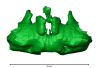
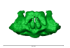
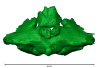
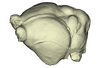
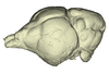
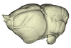
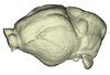
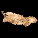
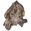
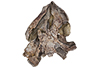
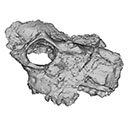
This contribution contains the 3D models described and figured in the following publication: Bonis, L. de, Grohé, C., Surault, J., Gardin, A. 2022. Description of the first cranium and endocranial structures of Stenoplesictis minor (Mammalia, Carnivora), an early aeluroid from the Oligocene of the Quercy Phosphorites (southwestern France). Historical Biology. https://doi.org/10.1080/08912963.2022.2045980
Stenoplesictis minor UM-ACQ 6705 View specimen
|
M3#961Endocranium Type: "3D_surfaces"doi: 10.18563/m3.sf.961 state:published |
Download 3D surface file |
|
M3#962Right bony labyrinth Type: "3D_surfaces"doi: 10.18563/m3.sf.962 state:published |
Download 3D surface file |

|
M3#963Left bony labyrinth Type: "3D_surfaces"doi: 10.18563/m3.sf.963 state:published |
Download 3D surface file |
|
M3#964Cranium in transparency with endocranial structures Type: "3D_surfaces"doi: 10.18563/m3.sf.964 state:published |
Download 3D surface file |
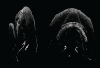
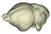
This contribution contains the 3D model of an endocranial cast analyzed in “A 10 ka intentionally deformed human skull from Northeast Asia”. There are many studies on the morphological characteristics of intentional cranial deformation (ICD), but few related 3D models were published. Here, we present the surface model of an intentionally deformed 10 ka human cranium for further research on ICD practice. The 3D model of the endocranial cast of this ICD cranium was discovered near Harbin City, Province Heilongjiang, Northeast China. The fossil preserved only the frontal, parietal, and occipital bones. To complete the endocast model of the specimen, we printed a 3D model and used modeling clay to reconstruct the missing part based on the general form of the modern human endocast morphology.
Homo sapiens IVPP-PA1616 View specimen
|
M3#972The frontal region of the endocast is flattened, probably formed by the constant pressure on the frontal bone during growth. There is a well-developed frontal crest on the endocranial surface. The endocast widens posteriorly from the frontal lobe. The widest point of the endocast is at the lateral border of the parietal lobe. The lower parietal areas display a marked lateral expansion. The overall shape of the endocast is asymmetrical, with the left side of the parietal lobe being more laterally expanded than the right side. Like the frontal lobe, the occipital lobe is also anteroposteriorly flattened. Type: "3D_surfaces"doi: 10.18563/m3.sf.972 state:published |
Download 3D surface file |
|
M3#976The original endocranial cast model (with texture) of IVPP-PA1616. It shows the original structures of the specimen, and was not altered in any way. Type: "3D_surfaces"doi: 10.18563/m3.sf.976 state:published |
Download 3D surface file |
The present 3D Dataset contains the 3D models analyzed in: Perrichon et al. 2023. Ontogenetic variability of the intertympanic sinus distinguishes lineages within Crocodylia.
Mecistops sp. ag SVSTUA 022001 View specimen
|
M3#980Intertympanic sinus system (expressed in meters) Type: "3D_surfaces"doi: 10.18563/m3.sf.980 state:published |
Download 3D surface file |
Crocodylus niloticus ag SVSTUA 022002 View specimen
|
M3#981Intertympanic sinus system Type: "3D_surfaces"doi: 10.18563/m3.sf.981 state:published |
Download 3D surface file |
Mecistops sp. AMU Zoo 04721 View specimen
|
M3#982Intertympanic sinus system Type: "3D_surfaces"doi: 10.18563/m3.sf.982 state:published |
Download 3D surface file |
Crocodylus sp. MHNL QV14 View specimen
|
M3#983Intertympanic sinus system Type: "3D_surfaces"doi: 10.18563/m3.sf.983 state:published |
Download 3D surface file |
Crocodylus rhombifer MHNL 42006506 View specimen

|
M3#1008Intertympanic sinus system (incomplete) Type: "3D_surfaces"doi: 10.18563/m3.sf.1008 state:published |
Download 3D surface file |
Crocodylus rhombifer MHNL 42006507 View specimen
|
M3#1007Intertympanic sinus system (incomplete) Type: "3D_surfaces"doi: 10.18563/m3.sf.1007 state:published |
Download 3D surface file |
Crocodylus niloticus MHNL 50001387 View specimen
|
M3#1006Intertympanic sinus system Type: "3D_surfaces"doi: 10.18563/m3.sf.1006 state:published |
Download 3D surface file |
Crocodylus palustris MHNL 50001388 View specimen
|
M3#1004Intertympanic sinus system Type: "3D_surfaces"doi: 10.18563/m3.sf.1004 state:published |
Download 3D surface file |
Crocodylus porosus MHNL 50001389 View specimen

|
M3#1009Intertympanic sinus system Type: "3D_surfaces"doi: 10.18563/m3.sf.1009 state:published |
Download 3D surface file |
Mecistops sp. MHNL 50001393 View specimen
|
M3#1010Intertympanic sinus system Type: "3D_surfaces"doi: 10.18563/m3.sf.1010 state:published |
Download 3D surface file |
Crocodylus niloticus MHNL 50001397 View specimen
|
M3#1018Intertympanic sinus system Type: "3D_surfaces"doi: 10.18563/m3.sf.1018 state:published |
Download 3D surface file |
Crocodylus porosus MHNL 50001398 View specimen
|
M3#1017Intertympanic sinus system Type: "3D_surfaces"doi: 10.18563/m3.sf.1017 state:published |
Download 3D surface file |
Crocodylus niloticus MHNL 50001405 View specimen
|
M3#1016Intertympanic sinus system Type: "3D_surfaces"doi: 10.18563/m3.sf.1016 state:published |
Download 3D surface file |
Gavialis gangeticus MHNL 50001407 View specimen
|
M3#1015Intertympanic sinus system Type: "3D_surfaces"doi: 10.18563/m3.sf.1015 state:published |
Download 3D surface file |
Crocodylus niloticus MHNL 90001850 View specimen
|
M3#1014Intertympanic sinus system Type: "3D_surfaces"doi: 10.18563/m3.sf.1014 state:published |
Download 3D surface file |
Crocodylus niloticus MHNL 90001851 View specimen
|
M3#1013Intertympanic sinus system Type: "3D_surfaces"doi: 10.18563/m3.sf.1013 state:published |
Download 3D surface file |
Crocodylus niloticus MHNL 90001855 View specimen
|
M3#1012Intertympanic sinus system Type: "3D_surfaces"doi: 10.18563/m3.sf.1012 state:published |
Download 3D surface file |
Osteolaemus tetraspis MHNM.9095.0 View specimen
|
M3#1011Intertympanic sinus system Type: "3D_surfaces"doi: 10.18563/m3.sf.1011 state:published |
Download 3D surface file |
Voay robustus MNHN F.1908-5 View specimen
|
M3#1003Intertympanic sinus system Type: "3D_surfaces"doi: 10.18563/m3.sf.1003 state:published |
Download 3D surface file |
Crocodylus sp. MNHN-F.1908-5-2 View specimen
|
M3#1005Intertympanic sinus system Type: "3D_surfaces"doi: 10.18563/m3.sf.1005 state:published |
Download 3D surface file |
Osteolaemus tetraspis MZS Cro 040 View specimen
|
M3#1002Intertympanic sinus system Type: "3D_surfaces"doi: 10.18563/m3.sf.1002 state:published |
Download 3D surface file |
Crocodylus acutus MZS Cro 055 View specimen
|
M3#991Intertympanic sinus system Type: "3D_surfaces"doi: 10.18563/m3.sf.991 state:published |
Download 3D surface file |
Melanosuchus niger MZS Cro 073 View specimen
|
M3#989Intertympanic sinus system Type: "3D_surfaces"doi: 10.18563/m3.sf.989 state:published |
Download 3D surface file |
Mecistops sp. MZS Cro 083 View specimen
|
M3#990Intertympanic sinus system Type: "3D_surfaces"doi: 10.18563/m3.sf.990 state:published |
Download 3D surface file |
Tomistoma schlegelii MZS Cro 094 View specimen
|
M3#988Intertympanic sinus system Type: "3D_surfaces"doi: 10.18563/m3.sf.988 state:published |
Download 3D surface file |
Gavialis gangeticus NHMUK 1846.1.7.3 View specimen
|
M3#987Intertympanic sinus system Type: "3D_surfaces"doi: 10.18563/m3.sf.987 state:published |
Download 3D surface file |
Osteolaemus tetraspis NHMUK 1862.6.30.5 View specimen
|
M3#986Intertympanic sinus system Type: "3D_surfaces"doi: 10.18563/m3.sf.986 state:published |
Download 3D surface file |
Gavialis gangeticus NHMUK 1873 View specimen
|
M3#985intertympanic sinus system Type: "3D_surfaces"doi: 10.18563/m3.sf.985 state:published |
Download 3D surface file |
Tomistoma schlegelii NHMUK 1893.3.6.14 View specimen
|
M3#984Intertympanic sinus system Type: "3D_surfaces"doi: 10.18563/m3.sf.984 state:published |
Download 3D surface file |
Mecistops sp. NHMUK 1924.5.10.1 View specimen
|
M3#992Intertympanic sinus system Type: "3D_surfaces"doi: 10.18563/m3.sf.992 state:published |
Download 3D surface file |
Voay robustus NHMUK PV R 36684 View specimen
|
M3#993Intertympanic sinus system Type: "3D_surfaces"doi: 10.18563/m3.sf.993 state:published |
Download 3D surface file |
Voay robustus NHMUK PV R 36685 View specimen
|
M3#1001Intertympanic sinus system Type: "3D_surfaces"doi: 10.18563/m3.sf.1001 state:published |
Download 3D surface file |
Crocodylus niloticus UCBL FSL 532077 View specimen

|
M3#1000Intertympanic sinus system Type: "3D_surfaces"doi: 10.18563/m3.sf.1000 state:published |
Download 3D surface file |
Crocodylus porosus/siamensis UCBLZ 2019-1-237 View specimen
|
M3#999Intertympanic sinus system Type: "3D_surfaces"doi: 10.18563/m3.sf.999 state:published |
Download 3D surface file |
Osteolaemus tetraspis UCBLZ 2019-1-236 View specimen
|
M3#998Intertympanic sinus system Type: "3D_surfaces"doi: 10.18563/m3.sf.998 state:published |
Download 3D surface file |
Alligator mississipiensis UCBLZ WB35 View specimen
|
M3#997Intertympanic sinus system Type: "3D_surfaces"doi: 10.18563/m3.sf.997 state:published |
Download 3D surface file |
Crocodylus siamensis UCBLZ WB41 View specimen
|
M3#996Intertympanic sinus system Type: "3D_surfaces"doi: 10.18563/m3.sf.996 state:published |
Download 3D surface file |
Tomistoma schlegelii UM 1097 View specimen
|
M3#995Intertympanic sinus system Type: "3D_surfaces"doi: 10.18563/m3.sf.995 state:published |
Download 3D surface file |
Crocodylus niloticus UM 2001-1756-1-434 NR View specimen
|
M3#994Intertympanic sinus system Type: "3D_surfaces"doi: 10.18563/m3.sf.994 state:published |
Download 3D surface file |
Mecistops sp. UM N89 View specimen
|
M3#1019Intertympanic sinus system Type: "3D_surfaces"doi: 10.18563/m3.sf.1019 state:published |
Download 3D surface file |
The present 3D Dataset contains the 3D models analyzed in the publication Fossils from the Montceau-les-Mines Lagerstätte (305 Ma) shed light on the anatomy, ecology and phylogeny of Carboniferous millipedes. Authors: Lheritier Mickael, Perroux Maëva, Vannier Jean, Escarguel Gilles, Wesener Thomas, Moritz Leif, Chabard Dominique, Adrien Jerome and Perrier Vincent. Journal of Systematics Palaeontology. https://doi.org/10.1080/14772019.2023.2169891
Amynilyspes fatimae MNHN.F.SOT.2134 View specimen

|
M3#1073Nearly complete specimen. Type: "3D_surfaces"doi: 10.18563/m3.sf.1073 state:published |
Download 3D surface file |
Amynilyspes fatimae MNHN.F.SOT.14983 View specimen
|
M3#1074Nearly complete specimen. Type: "3D_surfaces"doi: 10.18563/m3.sf.1074 state:published |
Download 3D surface file |
Amynilyspes fatimae MNHN.F.SOT.2129 View specimen

|
M3#1075Nearly complete specimen. Type: "3D_surfaces"doi: 10.18563/m3.sf.1075 state:published |
Download 3D surface file |
Blanzilius parriati MNHN.F.SOT.2114A View specimen
|
M3#1076Front part. Type: "3D_surfaces"doi: 10.18563/m3.sf.1076 state:published |
Download 3D surface file |
Blanzilius parriati MNHN.F.SOT.5148 View specimen
|
M3#1077Front part. Type: "3D_surfaces"doi: 10.18563/m3.sf.1077 state:published |
Download 3D surface file |
Blanzilius parriati MNHN.F.SOT.2113 View specimen
|
M3#1078Fragment with legs, sternites and possible tracheal openings. Type: "3D_surfaces"doi: 10.18563/m3.sf.1078 state:published |
Download 3D surface file |
Blanzilius parriati MNHN.F.SOT.81522 View specimen
|
M3#1079Nealry complete specimen. Type: "3D_surfaces"doi: 10.18563/m3.sf.1079 state:published |
Download 3D surface file |
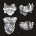
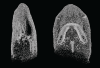
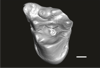
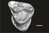
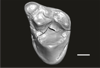
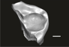
This contribution contains the three-dimensional digital models of eleven isolated fossil teeth of a merialine paroxyclaenid (Welcommoides gurki), discovered from lower Oligocene deposits of the Bugti Hills (Balochistan, Pakistan). These fossils were described, figured and discussed in the following publication: Solé et al. (2024), An unexpected late paroxyclaenid (Mammalia, Cimolesta) out of Europe: dental evidence from the Oligocene of the Bugti Hills, Pakistan. Papers in Palaeontology. https://doi.org/10.1002/spp2.1599
Welcommoides gurki UM-DBC 2225 View specimen
|
M3#1083Left m3 Type: "3D_surfaces"doi: 10.18563/m3.sf.1083 state:published |
Download 3D surface file |
Welcommoides gurki UM-DBC 2226 View specimen
|
M3#1084Right m3 Type: "3D_surfaces"doi: 10.18563/m3.sf.1084 state:published |
Download 3D surface file |
Welcommoides gurki UM-DBC 2227 View specimen
|
M3#1085Trigonid of a right lower molar Type: "3D_surfaces"doi: 10.18563/m3.sf.1085 state:published |
Download 3D surface file |
Welcommoides gurki UM-DBC 2230 View specimen
|
M3#1086Right DP4 Type: "3D_surfaces"doi: 10.18563/m3.sf.1086 state:published |
Download 3D surface file |
Welcommoides gurki UM-DBC 2228 View specimen
|
M3#1093Right M1 Type: "3D_surfaces"doi: 10.18563/m3.sf.1093 state:published |
Download 3D surface file |
Welcommoides gurki UM-DBC 2229 View specimen
|
M3#1087Right M2 Type: "3D_surfaces"doi: 10.18563/m3.sf.1087 state:published |
Download 3D surface file |
Welcommoides gurki UM-DBC 2236 View specimen
|
M3#1088Left M2 Type: "3D_surfaces"doi: 10.18563/m3.sf.1088 state:published |
Download 3D surface file |
Welcommoides gurki UM-DBC 2231 View specimen
|
M3#1089Left M3 Type: "3D_surfaces"doi: 10.18563/m3.sf.1089 state:published |
Download 3D surface file |
Welcommoides gurki UM-DBC 2232 View specimen
|
M3#1090Left M3 Type: "3D_surfaces"doi: 10.18563/m3.sf.1090 state:published |
Download 3D surface file |
Welcommoides gurki UM-DBC 2234 View specimen
|
M3#1091Left M3 Type: "3D_surfaces"doi: 10.18563/m3.sf.1091 state:published |
Download 3D surface file |
Welcommoides gurki UM-DBC 2233 View specimen
|
M3#1092Left M3 Type: "3D_surfaces"doi: 10.18563/m3.sf.1092 state:published |
Download 3D surface file |
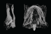
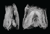
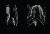
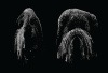
The present 3D Dataset contains the 3D models of extant Chiropteran endocranial casts, documenting 16 of the 19 extant bat families. They are used by Maugoust & Orliac (2023) to assess the correspondences between the brain and brain-surrounding tissues (i.e., neural tissues, blood vessels, meninges) and their imprint on the braincase, allowing for eventually proposing a Chiroptera-scale nomenclature of the endocast.
Balantiopteryx plicata UMMZ 102659 View specimen
|
M3#1132Endocranial cast of the corresponding cranium of Balantiopteryx plicata Type: "3D_surfaces"doi: 10.18563/m3.sf.1132 state:published |
Download 3D surface file |
Idiurus macrotis AMNH M-187705 View specimen
|
M3#1133Endocranial cast of the corresponding cranium of Nycteris macrotis Type: "3D_surfaces"doi: 10.18563/m3.sf.1133 state:published |
Download 3D surface file |
Thyroptera tricolor UMMZ 53240 View specimen
|
M3#1134Endocranial cast of the corresponding cranium of Thyroptera tricolor Type: "3D_surfaces"doi: 10.18563/m3.sf.1134 state:published |
Download 3D surface file |
Noctilio albiventris UMMZ 105827 View specimen
|
M3#1135Endocranial cast of the corresponding cranium of Noctilio albiventris Type: "3D_surfaces"doi: 10.18563/m3.sf.1135 state:published |
Download 3D surface file |
Mormoops blainvillii AMNH M-271513 View specimen
|
M3#1136Endocranial cast of the corresponding cranium of Mormoops blainvillii Type: "3D_surfaces"doi: 10.18563/m3.sf.1136 state:published |
Download 3D surface file |
Macrotus waterhousii UMMZ 95718 View specimen
|
M3#1137Endocranial cast of the corresponding cranium of Macrotus waterhousii Type: "3D_surfaces"doi: 10.18563/m3.sf.1137 state:published |
Download 3D surface file |
Nyctiellus lepidus UMMZ 105767 View specimen
|
M3#1138Endocranial cast of the corresponding cranium of Nyctiellus lepidus Type: "3D_surfaces"doi: 10.18563/m3.sf.1138 state:published |
Download 3D surface file |
Cheiromeles torquatus AMNH M-247585 View specimen
|
M3#1139Endocranial cast of the corresponding cranium of Cheiromeles torquatus Type: "3D_surfaces"doi: 10.18563/m3.sf.1139 state:published |
Download 3D surface file |
Miniopterus schreibersii UMMZ 156998 View specimen
|
M3#1140Endocranial cast of the corresponding cranium of Miniopterus schreibersii Type: "3D_surfaces"doi: 10.18563/m3.sf.1140 state:published |
Download 3D surface file |
Kerivoula pellucida UMMZ 161396 View specimen
|
M3#1141Endocranial cast of the corresponding cranium of Kerivoula pellucida Type: "3D_surfaces"doi: 10.18563/m3.sf.1141 state:published |
Download 3D surface file |
Scotophilus kuhlii UMMZ 157013 View specimen
|
M3#1142Endocranial cast of the corresponding cranium of Scotophilus kuhlii Type: "3D_surfaces"doi: 10.18563/m3.sf.1142 state:published |
Download 3D surface file |
Rhinolophus luctus MNHN CG-2006-87 View specimen
|
M3#1143Endocranial cast of the corresponding cranium of Rhinolophus luctus Type: "3D_surfaces"doi: 10.18563/m3.sf.1143 state:published |
Download 3D surface file |
Triaenops persicus AM RG-38552 View specimen
|
M3#1144Endocranial cast of the corresponding cranium of Triaenops persicus Type: "3D_surfaces"doi: 10.18563/m3.sf.1144 state:published |
Download 3D surface file |
Hipposideros armiger UM ZOOL-762-V View specimen
|
M3#1145Endocranial cast of the corresponding cranium of Hipposideros armiger Type: "3D_surfaces"doi: 10.18563/m3.sf.1145 state:published |
Download 3D surface file |
Lavia frons AM RG-12268 View specimen
|
M3#1146Endocranial cast of the corresponding cranium of Lavia frons Type: "3D_surfaces"doi: 10.18563/m3.sf.1146 state:published |
Download 3D surface file |
Rhinopoma hardwickii AM RG-M31166 View specimen
|
M3#1147Endocranial cast of the corresponding cranium of Rhinopoma hardwickii Type: "3D_surfaces"doi: 10.18563/m3.sf.1147 state:published |
Download 3D surface file |
Sphaerias blanfordi AMNH M-274330 View specimen
|
M3#1148Endocranial cast of the corresponding cranium of Sphaerias blanfordi Type: "3D_surfaces"doi: 10.18563/m3.sf.1148 state:published |
Download 3D surface file |
Rousettus aegyptiacus UMMZ 161026 View specimen
|
M3#1149Endocranial cast of the corresponding cranium of Rousettus aegyptiacus Type: "3D_surfaces"doi: 10.18563/m3.sf.1149 state:published |
Download 3D surface file |
Pteropus pumilus UMMZ 162253 View specimen
|
M3#1150Endocranial cast of the corresponding cranium of Pteropus pumilus Type: "3D_surfaces"doi: 10.18563/m3.sf.1150 state:published |
Download 3D surface file |
The present contribution contains the 3D models of fossil humeri and ilia of anurans from various Eocene-Miocene deposits of Peruvian Amazonia. These fossils were described and figured in the following publication: Jansen et al. (2023), First Eocene–Miocene anuran fossils from Peruvian Amazonia: insights into Neotropical frog evolution and diversity. Papers in Palaeontology, The Palaeontological Association.
Indet. indet. MUSM 4746 View specimen
|
M3#1231Humeral fragment (distal end) Type: "3D_surfaces"doi: 10.18563/m3.sf.1231 state:published |
Download 3D surface file |
Indet. indet. MUSM 4747 View specimen
|
M3#1232Humeral fragment (distal end) Type: "3D_surfaces"doi: 10.18563/m3.sf.1232 state:published |
Download 3D surface file |
Indet. indet. MUSM 4748 View specimen
|
M3#1233Humeral fragment (distal end) Type: "3D_surfaces"doi: 10.18563/m3.sf.1233 state:published |
Download 3D surface file |
Indet. indet. MUSM 4755 View specimen
|
M3#1234Humeral fragment (distal end) Type: "3D_surfaces"doi: 10.18563/m3.sf.1234 state:published |
Download 3D surface file |
Indet. indet. MUSM 4756 View specimen
|
M3#1235Humeral fragment (distal end) Type: "3D_surfaces"doi: 10.18563/m3.sf.1235 state:published |
Download 3D surface file |
Indet. indet. MUSM 4757 View specimen
|
M3#1236Humeral fragment (distal end) Type: "3D_surfaces"doi: 10.18563/m3.sf.1236 state:published |
Download 3D surface file |
Indet. indet. MUSM 4761 View specimen
|
M3#1237Humeral fragment (distal end) Type: "3D_surfaces"doi: 10.18563/m3.sf.1237 state:published |
Download 3D surface file |
Indet. indet. MUSM 4763 View specimen
|
M3#1238Humeral fragment (distal end) Type: "3D_surfaces"doi: 10.18563/m3.sf.1238 state:published |
Download 3D surface file |
Indet. indet. MUSM 4765 View specimen
|
M3#1239Humeral fragment (distal end) Type: "3D_surfaces"doi: 10.18563/m3.sf.1239 state:published |
Download 3D surface file |
Indet. indet. MUSM 4766 View specimen
|
M3#1240Humeral fragment (distal end) Type: "3D_surfaces"doi: 10.18563/m3.sf.1240 state:published |
Download 3D surface file |
Indet. indet. MUSM 4775 View specimen
|
M3#1241Humeral fragment (distal end) Type: "3D_surfaces"doi: 10.18563/m3.sf.1241 state:published |
Download 3D surface file |
cf. Pipa sp. MUSM 4776 View specimen
|
M3#1242Humeral fragment (distal end) Type: "3D_surfaces"doi: 10.18563/m3.sf.1242 state:published |
Download 3D surface file |
Indet. indet. MUSM 4788 View specimen
|
M3#1243Ilial fragment Type: "3D_surfaces"doi: 10.18563/m3.sf.1243 state:published |
Download 3D surface file |
Indet. indet. MUSM 4789 View specimen
|
M3#1244Ilial fragment Type: "3D_surfaces"doi: 10.18563/m3.sf.1244 state:published |
Download 3D surface file |
Indet. indet. MUSM 4790 View specimen
|
M3#1245Ilial fragment Type: "3D_surfaces"doi: 10.18563/m3.sf.1245 state:published |
Download 3D surface file |
Indet. indet. MUSM 4792 View specimen
|
M3#1246Ilial fragment Type: "3D_surfaces"doi: 10.18563/m3.sf.1246 state:published |
Download 3D surface file |
Indet. indet. MUSM 4793 View specimen
|
M3#1247Ilial fragment Type: "3D_surfaces"doi: 10.18563/m3.sf.1247 state:published |
Download 3D surface file |
Indet. indet. MUSM 4794 View specimen
|
M3#1249Ilial fragment Type: "3D_surfaces"doi: 10.18563/m3.sf.1249 state:published |
Download 3D surface file |
Indet. indet. MUSM 4795 View specimen
|
M3#1250Ilial fragment Type: "3D_surfaces"doi: 10.18563/m3.sf.1250 state:published |
Download 3D surface file |
cf. Pipa sp. MUSM 4796 View specimen
|
M3#1251Ilial fragment Type: "3D_surfaces"doi: 10.18563/m3.sf.1251 state:published |
Download 3D surface file |
cf. Pipa sp. MUSM 4797 View specimen
|
M3#1252Ilial fragment Type: "3D_surfaces"doi: 10.18563/m3.sf.1252 state:published |
Download 3D surface file |
The present 3D Dataset contains the 3D models analyzed in Brualla et al., 2024: Comparative anatomy of the vocal apparatus in bats and implication for the diversity of laryngeal echolocation. Zoological Journal of the Linnean Society, vol. zlad180. (https://doi.org/10.1093/zoolinnean/zlad180). Bat larynges are understudied in the previous anatomical studies. The description and comparison of the different morphological traits might provide important proxies to investigate the evolutionary origin of laryngeal echolocation in bats.
Eonycteris spelaea VN18-026 View specimen

|
M3#1305Laryngeal cartilages and muscles of the cave nectar bat Type: "3D_surfaces"doi: 10.18563/m3.sf.1305 state:published |
Download 3D surface file |
Macroglossus sobrinus VN15-017 View specimen

|
M3#1306Laryngeal anatomy of Macroglossus sobrinus Type: "3D_surfaces"doi: 10.18563/m3.sf.1306 state:published |
Download 3D surface file |
Aselliscus dongbacana VTTu15-013 View specimen
|
M3#1307Laryngeal anatomy of Aselliscus dongbacana Type: "3D_surfaces"doi: 10.18563/m3.sf.1307 state:published |
Download 3D surface file |
Coelops frithii VN19-196 View specimen
|
M3#1308Laryngeal anatomy of Coelops frithii Type: "3D_surfaces"doi: 10.18563/m3.sf.1308 state:published |
Download 3D surface file |
Hipposideros larvatus VN18-209 View specimen
|
M3#1309Laryngeal anatomy of Hipposideros larvatus Type: "3D_surfaces"doi: 10.18563/m3.sf.1309 state:published |
Download 3D surface file |
Rhinolophus cornutus JP21-025 View specimen

|
M3#14763D surfaces of Rhinolophus cornutus Type: "3D_surfaces"doi: 10.18563/m3.sf.1476 state:published |
Download 3D surface file |
Rhinolophus macrotis VN11-089 View specimen
|
M3#1477Laryngeal cartilages and muscles of Rhinolophus macrotis Type: "3D_surfaces"doi: 10.18563/m3.sf.1477 state:published |
Download 3D surface file |
Lyroderma lyra VN17-535 View specimen
|
M3#1312Laryngeal anatomy of Lyroderma lyra Type: "3D_surfaces"doi: 10.18563/m3.sf.1312 state:published |
Download 3D surface file |
Saccolaimus mixtus A3257 View specimen
|
M3#1478Laryngeal components of Saccolaimus mixtus Type: "3D_surfaces"doi: 10.18563/m3.sf.1478 state:published |
Download 3D surface file |
Taphozous melanopogon VN17-0252 View specimen
|
M3#1479Laryngeal cartilages and muscles of Taphozous melanopogon Type: "3D_surfaces"doi: 10.18563/m3.sf.1479 state:published |
Download 3D surface file |
Artibeus jamaicensis AJ001 View specimen
|
M3#1316Laryngeal anatomy of Artibeus jamaicensis Type: "3D_surfaces"doi: 10.18563/m3.sf.1316 state:published |
Download 3D surface file |
Kerivoula hardwickii VN11-0043 View specimen
|
M3#1317Laryngeal anatomy of Kerivoula hardwickii Type: "3D_surfaces"doi: 10.18563/m3.sf.1317 state:published |
Download 3D surface file |
Myotis ater VN19-016 View specimen
|
M3#1318Laryngeal anatomy of Myotis ater Type: "3D_surfaces"doi: 10.18563/m3.sf.1318 state:published |
Download 3D surface file |
Myotis siligorensis VTTu14-018 View specimen
|
M3#1319Laryngeal anatomy of Myotis siligorensis Type: "3D_surfaces"doi: 10.18563/m3.sf.1319 state:published |
Download 3D surface file |
Suncus murinus KATS_835A View specimen
|
M3#1395Laryngeal anatomy of Suncus murinus Type: "3D_surfaces"doi: 10.18563/m3.sf.1395 state:published |
Download 3D surface file |
The holotype of Hamadasuchus rebouli Buffetaut 1994 from the Kem Kem beds of Morocco (Late Albian – Cenomanian) consists of a left dentary which is limited, fragmentary and reconstructed in some areas. To aid in assessing if the original diagnosis can be considered as valid, the specimen was CT scanned for the first time. This is especially important to resolve the taxonomic status of certain specimens that have been assigned to Hamadasuchus rebouli since then. The reconstructed structures in this contribution are in agreement with the original description, notably in terms of alveolar count; thus the original diagnosis of this taxon remains valid and some specimens are not referable to H. rebouli anymore.
Hamadasuchus rebouli MDE C001 View specimen
|
M3#1402Dentary and teeth Type: "3D_surfaces"doi: 10.18563/m3.sf.1402 state:published |
Download 3D surface file |
|
M3#1403Toothmarks Type: "3D_surfaces"doi: 10.18563/m3.sf.1403 state:published |
Download 3D surface file |
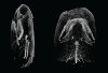
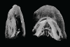
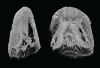
This contribution contains the three-dimensional models of the turbinal complex of 10 myrmecophagous and 10 non-myrmecophagous placental species. These specimens were analyzed and discussed in: Wright et. al (2024), Sniffing out morphological convergence in the turbinal complex of myrmecophagous placentals. https://doi.org/10.1002/ar.25603
Priodontes maximus NHMUK 732-a View specimen
|
M3#1536Turbinals of Priodontes maximus Type: "3D_surfaces"doi: 10.18563/m3.sf.1536 state:published |
Download 3D surface file |
Dasypus pilosus NHMUK 94-10-1-13 View specimen
|
M3#1537Turbinals of Dasypus pilosus Type: "3D_surfaces"doi: 10.18563/m3.sf.1537 state:published |
Download 3D surface file |
Dasypus novemcinctus AMNH 263287 View specimen
|
M3#1538Turbinals of Dasypus novemcinctus Type: "3D_surfaces"doi: 10.18563/m3.sf.1538 state:published |
Download 3D surface file |
Bradypus tridactylus UM 789N View specimen
|
M3#1539Turbinals of Bradypus tridactylus Type: "3D_surfaces"doi: 10.18563/m3.sf.1539 state:published |
Download 3D surface file |
Choloepus didactylus UM 767V View specimen
|
M3#1540Turbinals of Choloepus didactylus Type: "3D_surfaces"doi: 10.18563/m3.sf.1540 state:published |
Download 3D surface file |
Cyclopes didactylus NHMUK 88-8-8-14 View specimen
|
M3#1541Turbinals of Cyclopes didactylus Type: "3D_surfaces"doi: 10.18563/m3.sf.1541 state:published |
Download 3D surface file |
Myrmecophaga tridactyla UM 065V View specimen
|
M3#1542Turbinals of Myrmecophaga tridactyla Type: "3D_surfaces"doi: 10.18563/m3.sf.1542 state:published |
Download 3D surface file |
Tamandua tetradactyla NHMUK 3-7-7-135 View specimen
|
M3#1543Turbinals of Tamandua tetradactyla Type: "3D_surfaces"doi: 10.18563/m3.sf.1543 state:published |
Download 3D surface file |
Tamandua mexicana NHMUK 79-1-6-1 View specimen
|
M3#1544Turbinals of Tamandua mexicana Type: "3D_surfaces"doi: 10.18563/m3.sf.1544 state:published |
Download 3D surface file |
Orycteropus afer NHMUK 2-9-9-58 View specimen
|
M3#1545Turbinals of Orycteropus afer Type: "3D_surfaces"doi: 10.18563/m3.sf.1545 state:published |
Download 3D surface file |
Tenrec eucaudatus UM N439 View specimen
|
M3#1546Turbinals of Tenrec eucaudatus Type: "3D_surfaces"doi: 10.18563/m3.sf.1546 state:published |
Download 3D surface file |
Elephantulus rozeti UM N227 View specimen
|
M3#1547Turbinals of Elephantulus rozeti Type: "3D_surfaces"doi: 10.18563/m3.sf.1547 state:published |
Download 3D surface file |
Phataginus tetradactyla NHMUK 1-11-21-35 View specimen
|
M3#1548Turbinals of Phataginus tetradactyla Type: "3D_surfaces"doi: 10.18563/m3.sf.1548 state:published |
Download 3D surface file |
Smutsia gigantea KMMA 25479 View specimen
|
M3#1549Turbinals of Smutsia gigantea Type: "3D_surfaces"doi: 10.18563/m3.sf.1549 state:published |
Download 3D surface file |
Manis culionensis MNHN ZM-MO 1884-1822 View specimen
|
M3#1550Turbinals of Manis culionensis Type: "3D_surfaces"doi: 10.18563/m3.sf.1550 state:published |
Download 3D surface file |
Vulpes vulpes UM N140 View specimen
|
M3#1551Turbinals of Vulpes vulpes Type: "3D_surfaces"doi: 10.18563/m3.sf.1551 state:published |
Download 3D surface file |
Alopex lagopus UM N110 View specimen
|
M3#1552Turbinals of Alopex lagopus Type: "3D_surfaces"doi: 10.18563/m3.sf.1552 state:published |
Download 3D surface file |
Felis silvestris UM N149 View specimen
|
M3#1553Turbinals of Felis sylvestris Type: "3D_surfaces"doi: 10.18563/m3.sf.1553 state:published |
Download 3D surface file |
Hyaena hyaena UM N109 View specimen
|
M3#1554Turbinals of Hyaena hyaena Type: "3D_surfaces"doi: 10.18563/m3.sf.1554 state:published |
Download 3D surface file |
Proteles cristata NHMUK 4-3-1-58 View specimen
|
M3#1555Turbinals of Proteles cristata Type: "3D_surfaces"doi: 10.18563/m3.sf.1555 state:published |
Download 3D surface file |
The present 3D Dataset contains the 3D models of the skull of the holotype of Miocaperea pulchra.
Miocaperea pulchra SMNS-P-46978 View specimen
|
M3#1656Blender file containing two models (the skull being preserved in two parts) Type: "3D_surfaces"doi: 10.18563/m3.sf.1656 state:published |
Download 3D surface file |
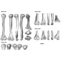
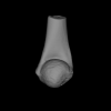
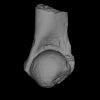
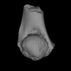
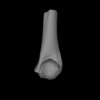
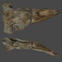
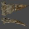
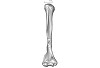
The present 3D Dataset contains the 3D models analyzed in the publication “Systematic and locomotor diversification of the Adapis group (Primates, Adapiformes) in the late Eocene of the Quercy (Southwest France), revealed by humeral remains”. In this paper, twenty humeral specimens from the old and new Quercy collections attributed to the fossil primates Adapis and Palaeolemur are described and analysed together. In this dataset only the scans of the fossils belonging to the collections of Université de Montpellier are provided.
In our paper (Marigó et al., 2019) we provide a qualitative and quantitative analysis of the different humeri, revealing that high variability is present within the “Adapis group” sample. Six different morphotypes are identified, confirming that what has often been called “Adapis parisiensis” is a mix of different species that present different locomotor adaptations.
Adapis sp. UM ROS 2-95 View specimen
|
M3#356Complete right humerus ROS 2-95 attributed to the Adapis group Type: "3D_surfaces"doi: 10.18563/m3.sf.356 state:published |
Download 3D surface file |
Adapis sp. UM ROS 2-536 View specimen
|
M3#357Proximal end of the right humerus ROS 2-536 attributed to the Adapis group Type: "3D_surfaces"doi: 10.18563/m3.sf.357 state:published |
Download 3D surface file |
Adapis sp. UM ROS 2-534 View specimen
|
M3#358Distal end of the left humerus ROS 2-534 attributed to the Adapis group Type: "3D_surfaces"doi: 10.18563/m3.sf.358 state:published |
Download 3D surface file |
Adapis sp. UM ROS 2-535 View specimen
|
M3#359Distal end of the left humerus ROS 2-535 attributed to the Adapis group Type: "3D_surfaces"doi: 10.18563/m3.sf.359 state:published |
Download 3D surface file |
Adapis sp. UM ROS 2-80 View specimen
|
M3#360Proximal end of the right humerus ROS 2-80 attributed to the Adapis group Type: "3D_surfaces"doi: 10.18563/m3.sf.360 state:published |
Download 3D surface file |
Adapis sp. UM ROS 2-79 View specimen
|
M3#361Distal end of the right humerus ROS 2-79 attributed to the Adapis group Type: "3D_surfaces"doi: 10.18563/m3.sf.361 state:published |
Download 3D surface file |
Adapis sp. UM ECA 1364 View specimen

|
M3#362Distal end of the left humerus ECA 1364 attributed to the Adapis group Type: "3D_surfaces"doi: 10.18563/m3.sf.362 state:published |
Download 3D surface file |
Adapis sp. UM ACQ-262 View specimen

|
M3#3733D model of ACQ 262. Humerus Type: "3D_surfaces"doi: 10.18563/m3.sf373 state:published |
Download 3D surface file |
This contribution provides for the first time the 3D model of the type specimen of Molassitherium delemontense (Mammalia, Rhinocerotidae) described in the following publication: Becker et al. (2013), Journal of Systematic Palaeontology, Vol. 11, Issue 8, 947–972, https://doi.org/10.1080/14772019.2012.699007. Conservation issues of the specimen and solutions using 3D model and 3D prints are detailed.
Molassitherium delemontense MJSN POI007–245 View specimen
|
M3#384Skull of Molassitherium delemontense Becker and Antoine, 2013 (in Becker et al. 2013): holotype Type: "3D_surfaces"doi: 10.18563/m3.sf.384 state:published |
Download 3D surface file |
The present 3D Dataset contains the 3D model analyzed in the article : Dubied et al. (2021), Endocranium and ecology of Eurotherium theriodis, a European hyaenodont mammal from the Lutetian. Acta Palaeontologica Polonica 2021, https://doi.org/10.4202/app.00771.2020
Eurotherium theriodis NMB.Em12 View specimen
|
M3#381NMB.Em12 unprepared specimen Type: "3D_surfaces"doi: 10.18563/m3.sf.381 state:published |
Download 3D surface file |
|
M3#382NMB.Em12 cranium Type: "3D_surfaces"doi: 10.18563/m3.sf.382 state:published |
Download 3D surface file |
|
M3#383NMB.Em12 endocast Type: "3D_surfaces"doi: 10.18563/m3.sf.383 state:published |
Download 3D surface file |
In this contribution, we describe the external and internal morphology of a delphinid petrosal bone collected from Ahu Tahai, a burial site located on the Southwestern coast of Easter Island, at Hangaroa. We discuss the taxonomic attribution of this archaeological item and describe its internal structures based on µCT data, including the bony labyrinth and the nerve and vein patterns. Identification of the nerves exists lead us to relocate the identification of the foramen singulare in delphinid petrosals.
indet. indet. AT1 View specimen
|
M3#420Stapes Type: "3D_surfaces"doi: 10.18563/m3.sf.420 state:published |
Download 3D surface file |
|
M3#421petrosal bone Type: "3D_surfaces"doi: 10.18563/m3.sf.421 state:published |
Download 3D surface file |
|
M3#422in situ bony labyrinth Type: "3D_surfaces"doi: 10.18563/m3.sf.422 state:published |
Download 3D surface file |
|
M3#423bony labyrinth and associated nerves and blood vessels Type: "3D_surfaces"doi: 10.18563/m3.sf.423 state:published |
Download 3D surface file |
The present 3D Dataset contains the 3D models analyzed in the following publication: Georgalis, G. L., and T. M. Scheyer. A new species of Palaeopython (Serpentes) and other extinct squamates from the Eocene of Dielsdorf (Zurich, Switzerland). Swiss Journal of Geosciences (in press). https://doi.org/10.1007/s00015-019-00341-6
Palaeopython helveticus PIMUZ A/III 631 View specimen
|
M3#399ZIP file containing .ply of vertebra PIMUZ A/III 631 from Palaeopython helveticus n. sp. Type: "3D_surfaces"doi: 10.18563/m3.sf.399 state:published |
Download 3D surface file |
|
M3#403dataset of snake vertebra PIMUZ A/III 631 Type: "3D_CT"doi: 10.18563/m3.sf.403 state:published |
Download CT data |
Palaeopython helveticus PIMUZ A/III 634 View specimen
|
M3#400ZIP file containing .ply of vertebra PIMUZ A/III 634 from Palaeopython helveticus n. sp. (holotype) Type: "3D_surfaces"doi: 10.18563/m3.sf.400 state:published |
Download 3D surface file |
|
M3#404dataset of snake vertbra PIMUZ A/III 634 (holotype) Type: "3D_CT"doi: 10.18563/m3.sf.404 state:published |
Download CT data |
Palaeopython helveticus PIMUZ A/III 636 View specimen
|
M3#401ZIP file containing .ply of vertebra PIMUZ A/III 636 from Palaeopython helveticus n. sp. Type: "3D_surfaces"doi: 10.18563/m3.sf.401 state:published |
Download 3D surface file |
|
M3#406dataset of snake vertebra PIMUZ A/III 636 Type: "3D_CT"doi: 10.18563/m3.sf.406 state:published |
Download CT data |
Palaeovaranus sp. PIMUZ A/III 234 View specimen
|
M3#402ZIP file containing .ply of dentary PIMUZ A/III 234 of Palaeovaranus sp. Type: "3D_surfaces"doi: 10.18563/m3.sf.402 state:published |
Download 3D surface file |

|
M3#405dataset of dentary of Palaeovaranus sp. (PIMUZ A/III 234) Type: "3D_CT"doi: 10.18563/m3.sf.405 state:published |
Download CT data |
This contribution contains the 3D surface model of the holotype cranium of the Late Jurassic thalassochelydian turtle Solnhofia brachyrhyncha described and figured in the publication of Anquetin and Püntener (2020).
Solnhofia brachyrhyncha MJSN BAN001-2.1 View specimen
|
M3#536Textured 3D surface model of the holotype cranium of the Late Jurassic turtle Solnhofia brachyrhyncha Type: "3D_surfaces"doi: 10.18563/m3.sf.536 state:published |
Download 3D surface file |
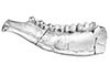
The present 3D Dataset contains the 3D models of the holotype mandible and referred fragmented skull of the new species Amphimoschus xishuiensis analyzed in the article Li, Y.-K., Mennecart, B., Aiglstorfer, M., Ni, X.-J., Li, Q., Deng, T. 2021. The early evolution of cranial appendages in Bovoidea revealed by new species of Amphimoschus (Mammalia: Ruminantia) from China. Zoological Journal of the Linnean Society https://doi.org/10.1093/zoolinnean/zlab053
Amphimoschus xishuiensis IVPP V 25521.1 View specimen
|
M3#803the holotype, a right hemimandible with tooth row p2 to m3 Type: "3D_surfaces"doi: 10.18563/m3.sf.803 state:published |
Download 3D surface file |
Amphimoschus xishuiensis IVPP V 25521.2 View specimen
|
M3#804referred material, anterior part of a skull with right P4-M3 and left P3-M2 Type: "3D_surfaces"doi: 10.18563/m3.sf.804 state:published |
Download 3D surface file |
The present 3D Dataset contains the 3D models illustrated and described in the chapter “Paleoneurology of Artiodactyla, an overview of the evolution of the artiodactyl brain” (Orliac et al. 2022) published in "Paleoneurology of amniotes: new directions in the study of fossil endocasts", edited by Dozo, Paulina-Carabajal, Macrini and Walsh.
Homacodon vagans AMNH 12695 View specimen
|
M3#1063Endocranial cast Type: "3D_surfaces"doi: 10.18563/m3.sf.1063 state:published |
Download 3D surface file |
Helohyus sp. AMNH 13079 View specimen
|
M3#1064Endocranial cast Type: "3D_surfaces"doi: 10.18563/m3.sf.1064 state:published |
Download 3D surface file |
Leptauchenia sp. AMNH 45508 View specimen
|
M3#1065endocranial cast Type: "3D_surfaces"doi: 10.18563/m3.sf.1065 state:published |
Download 3D surface file |
Agriochoerus sp. AMNH 95330 View specimen
|
M3#1067endocranial cast Type: "3D_surfaces"doi: 10.18563/m3.sf.1067 state:published |
Download 3D surface file |
Mouillacitherium elegans UM ACQ 6625 View specimen
|
M3#1068endocranial cast Type: "3D_surfaces"doi: 10.18563/m3.sf.1068 state:published |
Download 3D surface file |
Caenomeryx filholi UM PDS 2570 View specimen
|
M3#1069endocranial cast Type: "3D_surfaces"doi: 10.18563/m3.sf.1069 state:published |
Download 3D surface file |
Dichobune leporina MNHN.F.QU16586 View specimen
|
M3#1070endocranial cast Type: "3D_surfaces"doi: 10.18563/m3.sf.1070 state:published |
Download 3D surface file |
Anoplotherium sp. not numbered View specimen
|
M3#1071endocranial cast Type: "3D_surfaces"doi: 10.18563/m3.sf.1071 state:published |
Download 3D surface file |