Explodable 3D Dog Skull for Veterinary Education
3D models of a Sheep and Goat Skull and Inner ear
3D models of Miocene vertebrates from Tavers
3D GM dataset of bird skeletal variation
Skeletal embryonic development in the catshark
Bony connexions of the petrosal bone of extant hippos
bony labyrinth (11) , inner ear (10) , Eocene (8) , South America (8) , Paleobiogeography (7) , skull (7) , phylogeny (6)
Lionel Hautier (23) , Maëva Judith Orliac (21) , Laurent Marivaux (16) , Rodolphe Tabuce (14) , Bastien Mennecart (13) , Pierre-Olivier Antoine (12) , Renaud Lebrun (11)

|
3D models related to the publication: Morphogenesis of the inner ear at different stages of normal human developmentSaki Toyoda, Naoto Shiraki, Shigehito Yamada
Published online: 22/10/2015 |
|

|
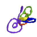
M3#42Computationally reconstructed membranous labyrinth of a human embryo (KC-CS23IER15919) at Carnegie Stage 23 (Crown Rump Length= 32.3mm). Type: "3D_surfaces"doi: 10.18563/m3.sf42 Data citation: Saki Toyoda, Naoto Shiraki, Shigehito Yamada, Chigako Uwabe, Hirohiko Imai, Tetsuya Matsuda, Akio Yoneyama, Tohoru Takeda and Tetsuya Takakuwa, 2015. M3#42. doi: 10.18563/m3.sf42 state:published |
Download 3D surface file |