|
Skeletogenesis during the late embryonic development of the catshark Scyliorhinus canicula (Chondrichthyes; Neoselachii)
Published online: 25/04/2016
Keywords:
Chondrichthyes; development; mineralization; Scyliorhinus canicula; skeleton
https://doi.org/10.18563/m3.1.4.e2
Cite this article:
Sébastien Enault, Sylvain Adnet and Mélanie Debiais-Thibaud, 2016. Skeletogenesis during the late embryonic development of the catshark Scyliorhinus canicula (Chondrichthyes; Neoselachii). MorphoMuseuM 1 (4)-e2. doi: 10.18563/m3.1.4.e2
Export citation
Abstract
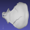
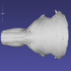
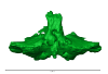
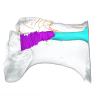
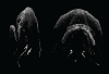
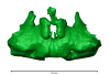
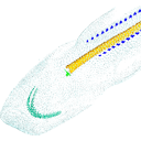
Current knowledge on the skeletogenesis of Chondrichthyes is scarce compared with their extant sister group, the bony fishes. Most of the previously described developmental tables in Chondrichthyes have focused on embryonic external morphology only. Due to its small body size and relative simplicity to raise eggs in laboratory conditions, the small-spotted catshark Scyliorhinus canicula has emerged as a reference species to describe developmental mechanisms in the Chondrichthyes lineage. Here we investigate the dynamic of mineralization in a set of six embryonic specimens using X-ray microtomography and describe the developing units of both the dermal skeleton (teeth and dermal scales) and endoskeleton (vertebral axis). This preliminary data on skeletogenesis in the catshark sets the first bases to a more complete investigation of the skeletal developmental in Chondrichthyes. It should provide comparison points with data known in osteichthyans and could thus be used in the broader context of gnathostome skeletal evolution.
M3 article infos
Published in Volume 01, Issue 04 (2016)
|
PDF
|