3D models of Cainotheriids Ossicular chain
3D models of Kalakocetus, the earliest Cetacea
The specimens of Speothos pacivorus
3D GM dataset of bird skeletal variation
Skeletal embryonic development in the catshark
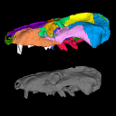
Bony connexions of the petrosal bone of extant hippos
bony labyrinth (14) , inner ear (11) , Eocene (11) , geometric morphometrics (10) , CT-scan (10) , Oligocene (9) , Micro-CT (9)
Lionel Hautier (25) , Maëva Judith Orliac (24) , Laurent Marivaux (18) , Renaud Lebrun (15) , Rodolphe Tabuce (15) , Pierre-Olivier Antoine (13) , Bastien Mennecart (13)
Page 1 of 1, showing 5 record(s) out of 5 total
|
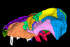
3D model related to the publication: Anatomy of the holotype of “Probelesodon” kitchingi revisited, a chiniquodontid cynodont (Synapsida, Probainognathia) from the early Late Triassic of southern BrazilCarolina Hoffmann
Published online: 23/05/2023 |
|
M3#11513D models of the skull with segmented bones and without the segmentation. colormap and orientation files also added. Type: "3D_surfaces"doi: 10.18563/m3.sf.1151 state:published |
Download 3D surface file |
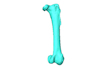
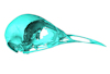
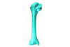
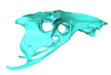
Macroevolution is integral to understanding the patterns of the diversification of life. As the life sciences increasingly use big data approaches, large multivariate datasets are required to test fundamental macroevolutionary hypotheses. In vertebrate evolution, large datasets have been created to quantify morphological variation, largely focusing on particular areas of the skeleton. We provide a landmarking protocol to quantify morphological variation in skeletal elements across the head, trunk, hindlimb and forelimb using 3-dimensional landmarks and semilandmarks, and present a large pan-skeletal database of bird morphology for 149 taxa across avian phylogeny using CT scan data. This large collection of 3D models and geometric morphometric data is open access and can be used in the future for new research, teaching and outreach. The 3D models and CT scans of the 149 specimens related to this project can be downloaded at MorphoSource (https://www.morphosource.org/projects/00000C420)
Menura novaehollandiae FMNH 336751 View specimen
|
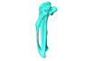
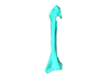
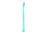
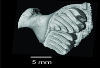
M3#5613D model of the left carpometacarpus of the superb lyrebird, Menura novaehollandia (displayed as a mirror image in the 3DHOP viewer). Type: "3D_surfaces"doi: 10.18563/m3.sf.561 state:published |
Download 3D surface file |

|
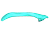
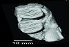
M3#5623D model of the mandible of the superb lyrebird, Menura novaehollandiae. Type: "3D_surfaces"doi: 10.18563/m3.sf.562 state:published |
Download 3D surface file |
|
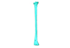
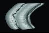
M3#5633D model of the right coracoid of the superb lyrebird, Menura novaehollandiae. Type: "3D_surfaces"doi: 10.18563/m3.sf.563 state:published |
Download 3D surface file |
|
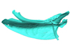
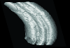
M3#5643D model of the right scapula of the superb lyrebird, Menura novaehollandiae. Type: "3D_surfaces"doi: 10.18563/m3.sf.564 state:published |
Download 3D surface file |
|
M3#5653D model of the right tarsometatarsus of the superb lyrebird, Menura novaehollandiae. Type: "3D_surfaces"doi: 10.18563/m3.sf.565 state:published |
Download 3D surface file |
|
M3#5663D model of the sternum of the superb lyrebird, Menura novaehollandiae. Type: "3D_surfaces"doi: 10.18563/m3.sf.566 state:published |
Download 3D surface file |
|
M3#5673D model of the left femur of the superb lyrebird, Menura novaehollandiae (displayed as a mirror image in the 3DHOP viewer). Type: "3D_surfaces"doi: 10.18563/m3.sf.567 state:published |
Download 3D surface file |
|
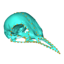
M3#5683D model of the skull of the superb lyrebird, Menura novaehollandiae. Type: "3D_surfaces"doi: 10.18563/m3.sf.568 state:published |
Download 3D surface file |
|
M3#5693D model of the left humerus of the superb lyrebird, Menura novaehollandiae (displayed as a mirror image in the 3DHOP viewer). Type: "3D_surfaces"doi: 10.18563/m3.sf.569 state:published |
Download 3D surface file |
|
M3#5703D model of the synsacrum of the superb lyrebird, Menura novaehollandiae. Type: "3D_surfaces"doi: 10.18563/m3.sf.570 state:published |
Download 3D surface file |
|
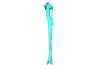
M3#5713D model of the left radius of the superb lyrebird, Menura novaehollandiae (displayed as a mirror image in the 3DHOP viewer). Type: "3D_surfaces"doi: 10.18563/m3.sf.571 state:published |
Download 3D surface file |
|
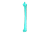
M3#5723D model of the left tibiotarsus of the superb lyrebird, Menura novaehollandiae (displayed as a mirror image in the 3DHOP viewer). Type: "3D_surfaces"doi: 10.18563/m3.sf.572 state:published |
Download 3D surface file |
|
M3#5733D model of the left ulna of the superb lyrebird, Menura novaehollandiae (displayed as a mirror image in the 3DHOP viewer). Type: "3D_surfaces"doi: 10.18563/m3.sf.573 state:published |
Download 3D surface file |
The present 3D Dataset contains the 3D model analyzed in the following publication: Paulina-Carabajal, A., Sterli, J., Werneburg, I., 2019. The endocranial anatomy of the stem turtle Naomichelys speciosa from the Early Cretaceous of North America. Acta Palaeontologica Polonica, https://doi.org/10.4202/app.00606.2019
Naomichelys speciosa FMNH PR273 View specimen
|
M3#428FMNH_PR273_1 - Naomichlys speciosa - skull Type: "3D_surfaces"doi: 10.18563/m3.sf.428 state:published |
Download 3D surface file |
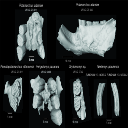
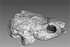
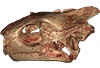
This contribution contains 3D models of extinct rodents Dinomyidae from Miocene and Quaternary of Brazil. The Miocene specimens that were digitalized include the holotypes of Potamarchus adamiae, Pseudopotamarchus villanuevai, and Ferigolomys pacarana collected in the Solimões Formation (Upper Miocene), northern Brazil. The Quaternary specimens are the holotype and paratype of Niedemys piauiensis, found in Upper Pleistocene deposits from northeast Brazil.
Potamarchus adamiae UFAC-CS 011 View specimen
|
M3#410UFAC-CS 011 – holotype, palatal region of the skull with cheek teeth Type: "3D_surfaces"doi: 10.18563/m3.sf.410 state:published |
Download 3D surface file |
Potamarchus adamiae UFAC-CS 043 View specimen
|
M3#411UFAC-CS 043, left dentary with cheek teeth Type: "3D_surfaces"doi: 10.18563/m3.sf.411 state:published |
Download 3D surface file |
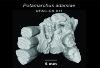
Pseudopotamarchus villanuevai UFAC 4762 View specimen
|
M3#412UFAC 4762 – holotype, incomplete right maxilla with cheek teeth Type: "3D_surfaces"doi: 10.18563/m3.sf.412 state:published |
Download 3D surface file |
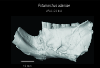
Ferigolomys pacarana UFAC 6460 View specimen
|
M3#413UFAC 6460 – holotype, palatal region of the skull with cheek teeth Type: "3D_surfaces"doi: 10.18563/m3.sf.413 state:published |
Download 3D surface file |
Drytomomys sp. UFAC 2742 View specimen

|
M3#414UFAC 2742, right dentary with cheek teeth Type: "3D_surfaces"doi: 10.18563/m3.sf.414 state:published |
Download 3D surface file |
Niedemys piauiensis FUMDHAM 113-146365-2 View specimen
|
M3#418FUMDHAM 113-146365-2 - holotype, upper right tooth Type: "3D_surfaces"doi: 10.18563/m3.sf.418 state:published |
Download 3D surface file |
Niedemys piauiensis FUMDHAM 113-145304-2 View specimen
|
M3#419FUMDHAM 113-145304-2 - paratype, left lower molar Type: "3D_surfaces"doi: 10.18563/m3.sf.419 state:published |
Download 3D surface file |
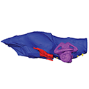
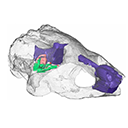
The present publication contains the µCT dataset and the 3D models analyzed in the following publication: Mautner, A.-K., A. E. Latimer, U. Fritz, and T. M. Scheyer. An updated description of the osteology of the pancake tortoise Malacochersus tornieri (Testudines: Testudinidae) with special focus on intraspecific variation. Journal of Morphology. https://doi.org/10.1002/jmor.20640
Malacochersus tornieri ZM 100.102 View specimen
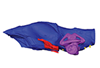
|
M3#129Virtual brain and inner ear endocast of Malacochersus tornieri (ZM 100.102; Zoological Museum of The University of Zurich). This virtual model is accompanied by the 3D dataset. Blue, endocranium; red, blood vessels; purple, semicircular canals; yellow, cranial nerves. Type: "3D_surfaces"doi: 10.18563/m3.sf.129 state:published |
Download 3D surface file |
|
M3#1303D dataset of skull of Malacochersus tornieri (ZM 100.102) Type: "3D_CT"doi: 10.18563/m3.sf.130 state:published |
Download CT data |
Page 1 of 1, showing 5 record(s) out of 5 total


