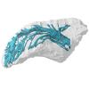
3D models of Cainotheriids Ossicular chain
Explodable 3D Dog Skull for Veterinary Education
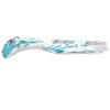
3D models of teeth and neurovascular system in the rostrum of Spinosauridae (theropod dinosaurs)
3D GM dataset of bird skeletal variation
Skeletal embryonic development in the catshark
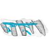
Bony connexions of the petrosal bone of extant hippos
bony labyrinth (14) , inner ear (11) , Eocene (11) , geometric morphometrics (10) , CT-scan (10) , Oligocene (9) , Micro-CT (9)
Lionel Hautier (25) , Maëva Judith Orliac (24) , Laurent Marivaux (19) , Bastien Mennecart (16) , Renaud Lebrun (15) , Rodolphe Tabuce (15) , Pierre-Olivier Antoine (13)
|
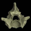
3D data related to the publication: A new species of Palaeopython (Serpentes) and other extinct squamates from the Eocene of Dielsdorf (Zurich, Switzerland)
|
|
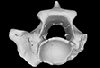
M3#399ZIP file containing .ply of vertebra PIMUZ A/III 631 from Palaeopython helveticus n. sp. Type: "3D_surfaces"doi: 10.18563/m3.sf.399 state:published |
Download 3D surface file |
|
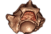
M3#403dataset of snake vertebra PIMUZ A/III 631 Type: "3D_CT"doi: 10.18563/m3.sf.403 state:published |
Download CT data |
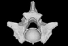
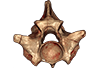
Palaeopython helveticus PIMUZ A/III 634 View specimen
|
M3#400ZIP file containing .ply of vertebra PIMUZ A/III 634 from Palaeopython helveticus n. sp. (holotype) Type: "3D_surfaces"doi: 10.18563/m3.sf.400 state:published |
Download 3D surface file |
|
M3#404dataset of snake vertbra PIMUZ A/III 634 (holotype) Type: "3D_CT"doi: 10.18563/m3.sf.404 state:published |
Download CT data |
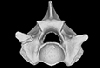
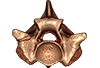
Palaeopython helveticus PIMUZ A/III 636 View specimen
|
M3#401ZIP file containing .ply of vertebra PIMUZ A/III 636 from Palaeopython helveticus n. sp. Type: "3D_surfaces"doi: 10.18563/m3.sf.401 state:published |
Download 3D surface file |
|
M3#406dataset of snake vertebra PIMUZ A/III 636 Type: "3D_CT"doi: 10.18563/m3.sf.406 state:published |
Download CT data |
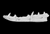
Palaeovaranus sp. PIMUZ A/III 234 View specimen
|
M3#402ZIP file containing .ply of dentary PIMUZ A/III 234 of Palaeovaranus sp. Type: "3D_surfaces"doi: 10.18563/m3.sf.402 state:published |
Download 3D surface file |

|
M3#405dataset of dentary of Palaeovaranus sp. (PIMUZ A/III 234) Type: "3D_CT"doi: 10.18563/m3.sf.405 state:published |
Download CT data |
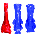
 , Leonardo Kerber and Sérgio Dias-da-Silva
 The present 3D Dataset contains the 3D models of brain endocast of traversodontid cynodonts studied in: Pavanatto et al. 2019. Virtual reconstruction of cranial endocasts of traversodontid cynodonts (Eucynodontia: Gomphodontia) from the upper Triassic of Southern Brazil. Journal of Morphology. https://doi.org/10.1002/jmor.21029
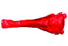
Siriusgnathus niemeyerorum CAPPA/UFSM 0032 View specimen
|
M3#4253D model of the brain endocast Type: "3D_surfaces"doi: 10.18563/m3.sf.425 state:published |
Download 3D surface file |
Exaeretodon riograndensis CAPPA/UFSM 0030 View specimen

|
M3#4263D model of the brain endocast Type: "3D_surfaces"doi: 10.18563/m3.sf.426 state:published |
Download 3D surface file |
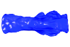
Exaeretodon riograndensis CAPPA/UFSM 0227 View specimen
|
M3#4273D model of the brain endocast Type: "3D_surfaces"doi: 10.18563/m3.sf.427 state:published |
Download 3D surface file |
 , Atila A. Stock Da-Rosa , Marcel, B. Lacerda and Flávio A. Pretto
 The present 3D Dataset contains the 3D models of an ilium, a vertebra, and a partial scapula of Prestosuchus sp. that were analyzed in “New Loricata remains from the Pinheiros-Chiniquá Sequence (Middle-Upper Triassic), southern Brazil”.
Prestosuchus sp. UFSM11603 View specimen
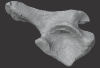
|
M3#1080Surface scan of a right ilium of Prestosuchus sp. with a 0.4 mm resolution. Type: "3D_surfaces"doi: 10.18563/m3.sf.1080 state:published |
Download 3D surface file |
Prestosuchus sp. UFSM11233 View specimen
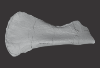
|
M3#1081Surface scan of a partial right scapula of Prestosuchus sp. with a 0.4mm resolution. Type: "3D_surfaces"doi: 10.18563/m3.sf.1081 state:published |
Download 3D surface file |
Prestosuchus sp. UFSM11602a View specimen
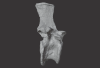
|
M3#1082Surface scan of a anterior dorsal vertebra of Prestosuchus sp. with a 0.2 mm resolution. Type: "3D_surfaces"doi: 10.18563/m3.sf.1082 state:published |
Download 3D surface file |
 , Fabiani Herrera , Steven Manchester , Maëva J. Orliac , Renaud Lebrun and Cédric Del Rio
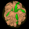
 The present 3D Dataset contains the 3D models of two endocarps from the Oligocene of Baraval Quercy locality. These endocarps document new fossil genera within the Anacardiaceae family and illustrate the morphological diversity of this family during the Palaeogene. The CT-scan data were processed with ImageJ and Mimics Innovation Suite version 1.13 to reconstruct the specimens. Here we provide .stl files easy to read with the software Meshlab.
Palaeochoerospondias sheppeyensis UM-BAV-675 View specimen
|
M3#1895Surfaces of the endocarp, seeds and lacunae Type: "3D_surfaces"doi: 10.18563/m3.sf.1895 state:published |
Download 3D surface file |
Baravalosphaera operculata UM-BAV-689 View specimen
|
M3#1896Surfaces of the endocarp Type: "3D_surfaces"doi: 10.18563/m3.sf.1896 state:published |
Download 3D surface file |
 and Christian Püntener
 This contribution contains the 3D surface model of the holotype cranium of the Late Jurassic thalassochelydian turtle Solnhofia brachyrhyncha described and figured in the publication of Anquetin and Püntener (2020).
Solnhofia brachyrhyncha MJSN BAN001-2.1 View specimen
|
M3#536Textured 3D surface model of the holotype cranium of the Late Jurassic turtle Solnhofia brachyrhyncha Type: "3D_surfaces"doi: 10.18563/m3.sf.536 state:published |
Download 3D surface file |
 , Olivier Maridet and Jérôme Priéto
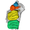
 This contribution contains the 3D model of the holotype of Simplomys hugi, the new dormouse species from the locality of Glovelier described and figured in the following publication: New data on the Miocene dormouse Simplomys García-Paredes, 2009 from the peri-alpin basins of Switzerland and Germany: palaeodiversity of a rare genus in Central Europe. https://doi.org/10.1007/s12549-018-0339-y
Simplomys hugi MJSN-GLM017-0001 View specimen
|
M3#385the left maxilla with four teeth ( DP4, P4, M1 and M2) Type: "3D_surfaces"doi: 10.18563/m3.sf.385 state:published |
Download 3D surface file |
 and Thomas Lehmann
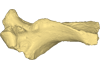
 This contribution contains the 3D model described and figured in the following publication: Crochet, J.-Y., Hautier, L., Lehmann, T., 2015. A pangolin (Manidae, Pholidota, Mammalia) from the French Quercy phosphorites (Pech du Fraysse, Saint-Projet, Tarn-et-Garonne, late Oligocene, MP 28). Palaeovertebrata 39(2)-e4. doi: 10.18563/pv.39.2.e4
Necromanis franconica UM PFY 4051 View specimen
|
M3#12A partial left humerus from Pech du Fraysse (Saint-Projet, Tarn-et-Garonne, France), MP 28 (late Oligocene) Type: "3D_surfaces"doi: 10.18563/m3.sf12 state:published |
Download 3D surface file |
 , Maëva J. Orliac , Renaud Lebrun and Cédric Del Rio
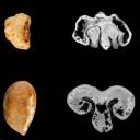
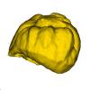
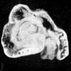
 The present 3D dataset contains the 3D models of two fossil seeds of Vitaceae from the Quercy fossiliferous area, southwestern France. One seed comes from the Eocene locality of Fontoffre 2, and the other from the Oligocene locality of Baraval. These seeds document new fossil species within the Vitaceae family and illustrate the morphological diversity of this family during the Palaeogene. The CT scan data were processed with ImageJ and Mimics Innovation Suite version 1.13 to reconstruct the specimens. Here we provide .stl files that can be easily opened with the software MeshLab.
Vitis quercyensis UM-FTF2-167 View specimen
|
M3#1930Surface of the seed Type: "3D_surfaces"doi: 10.18563/m3.sf.1930 state:published |
Download 3D surface file |
|
M3#1933Seed : 8.43 μm µCT scan Type: "3D_CT"doi: 10.18563/m3.sf.1933 state:published |
Download CT data |
Vitis praerotundifolia UM-BAV-660 View specimen
|
M3#1931Seed : 8.43 μm µCT scan Type: "3D_CT"doi: 10.18563/m3.sf.1931 state:published |
Download CT data |
|
M3#1932Surface of the seed Type: "3D_surfaces"doi: 10.18563/m3.sf.1932 state:published |
Download 3D surface file |
 and Juan D. Porfiri
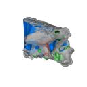
 The present 3D Dataset contains the 3D models analyzed in the following publication: Paulina-Carabajal, A., and Porfiri, J.D. 2026. Novel information on the braincase of Megaraptor namunhuaiquii (Dinosauria: Theropoda) using X-ray tomography: pneumaticity, paleoneurology and their paleobiological implications. Ameghiniana 63(1), 16-32
Megaraptor namunhuaiquii MUC-PV 595 View specimen
|
M3#18733D models of the braincase, lateral wall of the braincase, brain, right and left inner ears Type: "3D_surfaces"doi: 10.18563/m3.sf.1873 state:published |
Download 3D surface file |
 , Andreas Lang and Thomas Martin
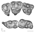
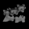
The present dataset contains the 3D models of the cheek teeth of eight raccoons analyzed in Koomen, S. E., Lang, A. J. & Martin, T. (2026). Tooth Function of the Northern Raccoon (Procyon lotor) and Adaptations to Omnivory in the Order Carnivora. Journal of Morphology.
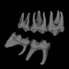
Procyon lotor ZFMK-MAM-2013.0341 View specimen
|
M3#1838Cheek teeth of P. lotor specimen ZFMK-MAM-2013.0341 Type: "3D_surfaces"doi: 10.18563/m3.sf.1838 state:in_press |
Download 3D surface file |
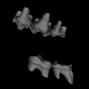
Procyon lotor ZFMK-MAM-1993.0289 View specimen
|
M3#1839Cheek teeth of P. lotor specimen ZFMK-MAM-1993.0289 Type: "3D_surfaces"doi: 10.18563/m3.sf.1839 state:in_press |
Download 3D surface file |
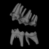
Procyon lotor ZFMK-MAM-2016.0912 View specimen
|
M3#1840Cheek teeth of P. lotor specimen ZFMK-MAM-2016.0912 Type: "3D_surfaces"doi: 10.18563/m3.sf.1840 state:in_press |
Download 3D surface file |
Procyon lotor ZFMK-MAM-2016.0914 View specimen
|
M3#1841Cheek teeth of P. lotor specimen ZFMK-MAM-2016.0914 Type: "3D_surfaces"doi: 10.18563/m3.sf.1841 state:in_press |
Download 3D surface file |
Procyon lotor ZFMK-MAM-2016.0923 View specimen
|
M3#1842Cheek teeth of P. lotor specimen ZFMK-MAM-2016.0923 Type: "3D_surfaces"doi: 10.18563/m3.sf.1842 state:in_press |
Download 3D surface file |
Procyon lotor ZFMK-MAM-2016.0937 View specimen
|
M3#1843Cheek teeth of P. lotor specimen ZFMK-MAM-2016.0937 Type: "3D_surfaces"doi: 10.18563/m3.sf.1843 state:in_press |
Download 3D surface file |
Procyon lotor ZFMK-MAM-2016.0938 View specimen
|
M3#1844Cheek teeth of P. lotor specimen ZFMK-MAM-2016.0938 Type: "3D_surfaces"doi: 10.18563/m3.sf.1844 state:in_press |
Download 3D surface file |
Procyon lotor ZFMK-MAM-2016.0967 View specimen
|
M3#1845Cheek teeth of P. lotor specimen ZFMK-MAM-2016.0967 Type: "3D_surfaces"doi: 10.18563/m3.sf.1845 state:in_press |
Download 3D surface file |
 , Marcos A. Alves , Léa Da Cunha and Benoit Moison
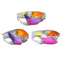
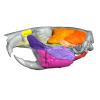
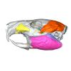
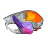
 This contribution contains the 3D models described and figured in the following publication: Hautier L, Da Cunha L, Alves Filho M, Moison B, Besson L, Fabre P-H. 20XX. A 3D atlas of the trigeminal nerve and its relevance for comparative studies of the masticatory apparatus in rodents. Journal of Anatomy
Rattus norvegicus UM-ZOOL-3105 View specimen
|
M3#19363D reconstruction of the masticatory musculature, trigeminal nerve and skull of the brown rat Rattus norvegicus Type: "3D_surfaces"doi: 10.18563/m3.sf.1936 state:in_press |
Download 3D surface file |
Cavia porcellus UM-ZOOL-499V View specimen
|
M3#19353D reconstruction of the masticatory musculature, trigeminal nerve and skull of the Guinea pig Cavia porcellus Type: "3D_surfaces"doi: 10.18563/m3.sf.1935 state:in_press |
Download 3D surface file |
Sciurus vulgaris BOUM-2022.2.12 View specimen
|
M3#19373D reconstruction of the masticatory musculature, trigeminal nerve and skull of the red squirrel Sciurus vulgaris Type: "3D_surfaces"doi: 10.18563/m3.sf.1937 state:in_press |
Download 3D surface file |
 , Sandrine Ladevèze and Rodolphe Tabuce
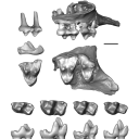
 This contribution contains three-dimensional models of the holotypes of Europeradectes marivauxi Gernelle, 2026, and E. williamsoni Gernelle, 2026, as well as other fossil material referred to these species from their type locality, Prémontré (Paris Basin; late early Eocene, 50.4-50.3 Ma). These specimens, among the youngest known for Europeradectes Gernelle, 2026, were analyzed and discussed in: Gernelle et al. (2026), A comprehensive evolutionary history of Peradectidae (Mammalia: Metatheria) revealed by a new dental morphology-based phylogenetic framework, with insights from late early Eocene French specimens.
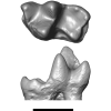
Europeradectes williamsoni MNHN.F.PRE26 View specimen
|
M3#1889right m3 (holotype) Type: "3D_surfaces"doi: 10.18563/m3.sf.1889 state:in_press |
Download 3D surface file |
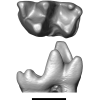
Europeradectes williamsoni MNHN.F.PRE643 View specimen
|
M3#1890left m2 Type: "3D_surfaces"doi: 10.18563/m3.sf.1890 state:in_press |
Download 3D surface file |
Europeradectes williamsoni MNHN.F.PRE272 View specimen
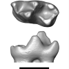
|
M3#1891right m1 Type: "3D_surfaces"doi: 10.18563/m3.sf.1891 state:in_press |
Download 3D surface file |
Europeradectes williamsoni MNHN.F.PMT167 View specimen
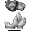
|
M3#1892left m4 Type: "3D_surfaces"doi: 10.18563/m3.sf.1892 state:in_press |
Download 3D surface file |
Europeradectes marivauxi MNHN.F.PMT169 View specimen
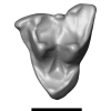
|
M3#1893left M2 Type: "3D_surfaces"doi: 10.18563/m3.sf.1893 state:in_press |
Download 3D surface file |
Europeradectes marivauxi MNHN.F.PMT166 View specimen
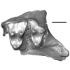
|
M3#1894partial right maxilla with M3-M4 (holotype) Type: "3D_surfaces"doi: 10.18563/m3.sf.1894 state:in_press |
Download 3D surface file |
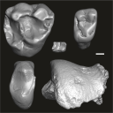 , Francisco R. Negri and Ana M. Ribeiro
 This contribution contains the three-dimensional digital models of fossil remains of platyrrhine primates from the late Middle to early Late Miocene (Upper Solimões Formation) of the State of Acre, Brazil (Western Amazonia). These fossils were described, figured and discussed in the following publication: Marivaux et al. (2026), New records of platyrrhine primates (Atelidae and Cebidae) from the Miocene of Western Amazonia, State of Acre, Brazil. Journal of Mammalian Evolution https://doi.org/10.1007/s10914-026-09829-z
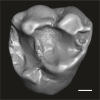
Parabrachyteles vesperus UFAC-CS 488 View specimen
|
M3#1865Isolated right M1 or M2 Type: "3D_surfaces"doi: 10.18563/m3.sf.1865 state:in_press |
Download 3D surface file |
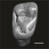
Migmacebus juruaensis UFAC-CS 487 View specimen
|
M3#1866Isolated right M1 or M2 Type: "3D_surfaces"doi: 10.18563/m3.sf.1866 state:in_press |
Download 3D surface file |
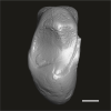
Gen indet sp. indet UFAC-CS 076 View specimen
|
M3#1867Isolated right P3 or P4 Type: "3D_surfaces"doi: 10.18563/m3.sf.1867 state:in_press |
Download 3D surface file |
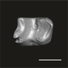
Gen indet sp. indet UFAC-CS 428 View specimen
|
M3#1868Isolated left m1 or m2 Type: "3D_surfaces"doi: 10.18563/m3.sf.1868 state:in_press |
Download 3D surface file |
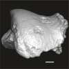
Gen indet sp. indet UFAC-CS 213 View specimen
|
M3#1869Fragment of left talus Type: "3D_surfaces"doi: 10.18563/m3.sf.1869 state:in_press |
Download 3D surface file |
 , Fanny Celestin, Sylvain Adnet and Guillaume Guinot
 The present 3D Dataset contains the 3D models produced for the paper "Szczygielski, T., Celestin, F., Adnet, S., Guinot, G., 2026. Triassic turtles of France: the first European Rhaetian stem turtle taxon (clades Testudinata, Proterochersidae). Swiss Journal of Palaeontology". The digitized material consists of shell and appendicular skeleton fragments of the first diagnostic stem turtle from the European Rhaetian and showcases a number of autapomorphic characters.
Bartassochersis lopezi UM-PDE2-01 View specimen
|
M3#1907Left axillary region and base of the anterior plastral lobe (left hyoplastron, small medial fragment of the right hyoplastron, part of the posterior process of the entoplastron) Type: "3D_surfaces"doi: 10.18563/m3.sf.1907 state:in_press |
Download 3D surface file |
Bartassochersis lopezi UM-PDE2-02 View specimen
|
M3#1908Posterior plastral lobe Type: "3D_surfaces"doi: 10.18563/m3.sf.1908 state:in_press |
Download 3D surface file |
Bartassochersis lopezi UM-PDE2-03 View specimen
|
M3#1909First vertebral scute region, right side Type: "3D_surfaces"doi: 10.18563/m3.sf.1909 state:in_press |
Download 3D surface file |
Bartassochersis lopezi UM-PDE2-04 View specimen
|
M3#1910?Carapace fragment Type: "3D_surfaces"doi: 10.18563/m3.sf.1910 state:in_press |
Download 3D surface file |
Bartassochersis lopezi UM-PDE2-05 View specimen
|
M3#1911Left scapula Type: "3D_surfaces"doi: 10.18563/m3.sf.1911 state:in_press |
Download 3D surface file |
Bartassochersis lopezi UM-PDE2-06 View specimen
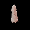
|
M3#1912Ungual Type: "3D_surfaces"doi: 10.18563/m3.sf.1912 state:in_press |
Download 3D surface file |
Bartassochersis lopezi UM-PDE2-08 View specimen
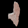
|
M3#1913Right hypoplastron Type: "3D_surfaces"doi: 10.18563/m3.sf.1913 state:in_press |
Download 3D surface file |
Bartassochersis lopezi UM-PDE2-09 View specimen
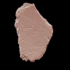
|
M3#1914Indeterminate Type: "3D_surfaces"doi: 10.18563/m3.sf.1914 state:in_press |
Download 3D surface file |
Bartassochersis lopezi UM-PDE2-10 View specimen
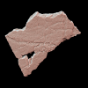
|
M3#1915?Mesoplastron/hypoplastron Type: "3D_surfaces"doi: 10.18563/m3.sf.1915 state:in_press |
Download 3D surface file |
Bartassochersis lopezi UM-PDE2-11 View specimen
|
M3#1916Left inguinal periphery (peripheral, hyoplastron) Type: "3D_surfaces"doi: 10.18563/m3.sf.1916 state:in_press |
Download 3D surface file |
Bartassochersis lopezi UM-PDE2-12 View specimen
|
M3#1917?Left side of a vertebral scute area with two partial pleural scute areas Type: "3D_surfaces"doi: 10.18563/m3.sf.1917 state:in_press |
Download 3D surface file |
Bartassochersis lopezi UM-PDE2-13 View specimen
|
M3#1918?Posterior pleural and supramarginal of marginal areas Type: "3D_surfaces"doi: 10.18563/m3.sf.1918 state:in_press |
Download 3D surface file |
Bartassochersis lopezi UM-PDE2-14 View specimen
|
M3#1919Posterior peripherals Type: "3D_surfaces"doi: 10.18563/m3.sf.1919 state:in_press |
Download 3D surface file |
Bartassochersis lopezi UM-PDE2-15A View specimen
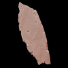
|
M3#1920?Left edge of the anterior plastral lobe Type: "3D_surfaces"doi: 10.18563/m3.sf.1920 state:in_press |
Download 3D surface file |
Bartassochersis lopezi UM-PDE2-15B View specimen
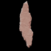
|
M3#1921Indeterminate Type: "3D_surfaces"doi: 10.18563/m3.sf.1921 state:in_press |
Download 3D surface file |
Bartassochersis lopezi UM-PDE2-16 View specimen
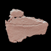
|
M3#1922Indeterminate Type: "3D_surfaces"doi: 10.18563/m3.sf.1922 state:in_press |
Download 3D surface file |
Bartassochersis lopezi UM-PDE2-17 View specimen
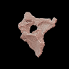
|
M3#1923?Bridge fragment Type: "3D_surfaces"doi: 10.18563/m3.sf.1923 state:in_press |
Download 3D surface file |
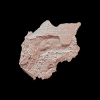
Bartassochersis lopezi UM-PDE2-19 View specimen
|
M3#1924Right bridge region Type: "3D_surfaces"doi: 10.18563/m3.sf.1924 state:in_press |
Download 3D surface file |
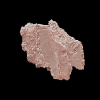
Bartassochersis lopezi UM-PDE2-20 View specimen
|
M3#1925Left femoral bulge Type: "3D_surfaces"doi: 10.18563/m3.sf.1925 state:in_press |
Download 3D surface file |
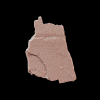
Bartassochersis lopezi UM-PDE2-21 View specimen
|
M3#1926Indeterminate Type: "3D_surfaces"doi: 10.18563/m3.sf.1926 state:in_press |
Download 3D surface file |
 , Cyril Gagnaison , Bastien Mennecart , Réjanne Le Bivic and Maxime Denis
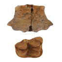
 The present 3D Dataset contains 3D models (1 turtle and 7 mammals) from the french Lower Miocene described in: Gagnaison C., Mennecart B., Bailleul J., Barrier P., Chenot É., Toullec R., Potel S., Martin H., Millet A. & Memeteau D. 2023. — Nouvelles données géologiques et biostratigraphiques du gisement paléontologique à vertébrés de Mauvières, à Marcilly-sur-Maulne (Miocène inférieur et moyen ; Indre-et-Loire, France). Geodiversitas 45 (16): 449-478. https://doi.org/10.5252/geodiversitas2023v45a16.
Testudo promarginata ULB-MAUV-5 View specimen
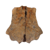
|
M3#1941Fragment of turtle shell of Testudo promarginata Type: "3D_surfaces"doi: 10.18563/m3.sf.1941 state:in_press |
Download 3D surface file |
Plesiaceratherium sp. ULB-MAUV-20 View specimen
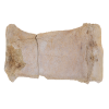
|
M3#1942right metacarpus III of Plesiaceratherium sp. Type: "3D_surfaces"doi: 10.18563/m3.sf.1942 state:in_press |
Download 3D surface file |
Brachyodus intermedius ULB-MAUV-21 View specimen
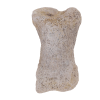
|
M3#1943phalanx II of the lateral finger (III or IV) of Brachyodus intermedius (Mayet, 1908) Type: "3D_surfaces"doi: 10.18563/m3.sf.1943 state:in_press |
Download 3D surface file |
Brachyodus intermedius ULB-MAUV-31 View specimen
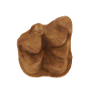
|
M3#1944cast of the right M3 of Brachyodus intermedius (Mayet, 1908) Type: "3D_surfaces"doi: 10.18563/m3.sf.1944 state:in_press |
Download 3D surface file |
Ligeromeryx praestans ULB-MAUV-24 View specimen
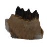
|
M3#1945right lower jaw bearing p3-m1 of Ligeromeryx praestans (Stehlin, 1937) Type: "3D_surfaces"doi: 10.18563/m3.sf.1945 state:in_press |
Download 3D surface file |
Ligeromeryx praestans ULB-MAUV-25 View specimen
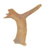
|
M3#1946cast of the right antler of Ligeromeryx praestans (Stehlin, 1937) Type: "3D_surfaces"doi: 10.18563/m3.sf.1946 state:in_press |
Download 3D surface file |
Megamphicyon carnutense ULB-MAUV-16 View specimen
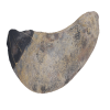
|
M3#1947right I1 of Megamphicyon carnutense (Antunes & Ginsburg, 1977) Type: "3D_surfaces"doi: 10.18563/m3.sf.1947 state:in_press |
Download 3D surface file |
Hemicyon gargan ULB-MAUV-18 View specimen
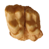
|
M3#1948cast of a left maxilla bearing M1-2 of Hemicyon gargan (Ginsburg & Morales, 1998) Type: "3D_surfaces"doi: 10.18563/m3.sf.1948 state:in_press |
Download 3D surface file |
 , Juan C. Sotomayor Chacón , María R. Arcaria , Nahuel Vega, Juan M. Leardi , Federico D. Seoane and Alejandro Otero
This contribution contains the 3D dataset analyzed in L. C. Gaetano, J. C. Sotomayor Chacón, M. R. Arcaria, N. Vega, J. M. Leardi, F. D. Seoane, and A. Otero. submitted. Earliest-diverging rhynchocephalian from Gondwana: a new sphenodontian from the Upper Triassic Los Colorados Formation (northwestern Argentina). Scientific Reports.
Coloradosphenodon lahni PULR-V264 View specimen
|
M3#1855fragmentary skull and lower jaws Type: "3D_surfaces"doi: 10.18563/m3.sf.1855 state:in_press |
Download 3D surface file |