3D models of Kalakocetus, the earliest Cetacea
The specimens of Speothos pacivorus
3D models related to the publication: Hidden diversity of Palaeogene metatherians: a new family of polydolopimorphian marsupials from Peruvian Amazonia
3D GM dataset of bird skeletal variation
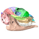
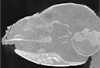
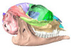
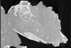
Skeletal embryonic development in the catshark
Bony connexions of the petrosal bone of extant hippos
bony labyrinth (14) , inner ear (11) , Eocene (11) , geometric morphometrics (10) , CT-scan (10) , Oligocene (9) , Micro-CT (9)
Maëva Judith Orliac (24) , Lionel Hautier (24) , Laurent Marivaux (18) , Renaud Lebrun (15) , Rodolphe Tabuce (14) , Pierre-Olivier Antoine (13) , Bastien Mennecart (13)
|
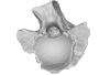
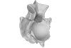
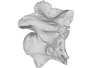
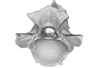
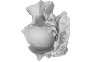
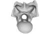
3D models related to the publication: Cranial anatomy of Hypisodus minimus (Artiodactyla: Ruminantia) from the Oligocene Brule Formation of North AmericaHannah Keppeler, Julia A. Schultz
Published online: 09/03/2023 |
|
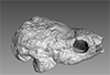
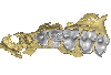
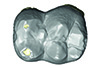
M3#1031CT image stack of a skull of Hypisodus minimus. Also includes a lumbar vertebra and a probable proximal phalanx of digit III or IV. Type: "3D_CT"doi: 10.18563/m3.sf.1031 state:published |
Download CT data |
|
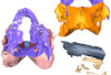
M3#10363D surface models of a skull of Hypisodus minimus (SMNK-PAL27212). The data includes a surface model for: basisphenoid, tympanic bullae, ethmoid (lamina perpendicularis), frontals, jugal (left), jugal (right), lacrimals, lower dentition, mandibles, mastoid processes, maxillaries, maxilloturbinals, nasals, occipital, palatine, parietals, petrosals, presphenoid, squamosals, turbinates, upper dentition, and the vomer. Type: "3D_surfaces"doi: 10.18563/m3.sf.1036 state:published |
Download 3D surface file |
Hypisodus minimus SMNK-PAL 27213 View specimen
|
M3#1033CT image stack of a skull of Hypisodus minimus. Also shows numerous postcranial material including an atlas articulated with the occipital bone, the distal part of a left humerus articulated to radius and ulna, a part of a femur, a part of a tibia and fibula, unidentifiable tarsal bones, parts of the metatarsals II, III, IV and V and their phalanges, a proximal phalanx of digit III or IV, a middle phalanx of digit III or IV, a possible patella and calcaneus, as well as numerous unidentifiable broken bony fragments. Type: "3D_CT"doi: 10.18563/m3.sf.1033 state:published |
Download CT data |
|
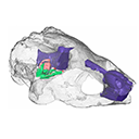
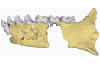
M3#10353D surface models of a skull of Hypisodus minimus (SMNK-PAL27213). The data includes a surface model for: atlas, basisphenoid, tympanic bullae, nasals, occipital, the petrosals, and the inner ear. Type: "3D_surfaces"doi: 10.18563/m3.sf.1035 state:published |
Download 3D surface file |
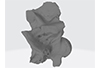
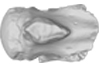
The present 3D Dataset contains the 3D model analyzed in the following publication: Paulina-Carabajal, A., Sterli, J., Werneburg, I., 2019. The endocranial anatomy of the stem turtle Naomichelys speciosa from the Early Cretaceous of North America. Acta Palaeontologica Polonica, https://doi.org/10.4202/app.00606.2019
Naomichelys speciosa FMNH PR273 View specimen
|
M3#428FMNH_PR273_1 - Naomichlys speciosa - skull Type: "3D_surfaces"doi: 10.18563/m3.sf.428 state:published |
Download 3D surface file |
This contribution contains the 3D models described and figured in the following publication: Georgalis, G.L., G. Guinot, K.E. Kassegne, Y.Z. Amoudji, A.K.C. Johnson, H. Cappetta and L. Hautier. 2021. An assemblage of giant aquatic snakes (Serpentes, Palaeophiidae) from the Eocene of Togo. Swiss Journal of Palaeontology 140, https://doi.org/10.1186/s13358-021-00236-w
Palaeophis africanus UM KPO 21 View specimen
|
M3#821Trunk vertebra UM KPO 21 of Palaeophis africanus Type: "3D_surfaces"doi: 10.18563/m3.sf.821 state:published |
Download 3D surface file |
Palaeophis africanus UM KPO 22 View specimen
|
M3#822Trunk vertebra UM KPO 22 of Palaeophis africanus from the Eocene of Togo Type: "3D_surfaces"doi: 10.18563/m3.sf.822 state:published |
Download 3D surface file |
Palaeophis africanus UM KPO 23 View specimen
|
M3#823Trunk vertebra UM KPO 23 of Palaeophis africanus Type: "3D_surfaces"doi: 10.18563/m3.sf.823 state:published |
Download 3D surface file |
Palaeophis africanus UM KPO 24 View specimen
|
M3#824Trunk vertebra UM KPO 24 of Palaeophis africanus Type: "3D_surfaces"doi: 10.18563/m3.sf.824 state:published |
Download 3D surface file |
Palaeophis africanus UM KPO 25 View specimen
|
M3#825Trunk vertebra UM KPO 25 of Palaeophis africanus Type: "3D_surfaces"doi: 10.18563/m3.sf.825 state:published |
Download 3D surface file |
Palaeophis africanus UM KPO 26 View specimen
|
M3#826Trunk vertebra UM KPO 26 of Palaeophis africanus Type: "3D_surfaces"doi: 10.18563/m3.sf.826 state:published |
Download 3D surface file |
Palaeophis africanus UM KPO 27 View specimen
|
M3#827Trunk vertebra UM KPO 27 of Palaeophis africanus Type: "3D_surfaces"doi: 10.18563/m3.sf.827 state:published |
Download 3D surface file |
Palaeophis africanus UM KPO 28 View specimen
|
M3#828Trunk vertebra UM KPO 28 of Palaeophis africanus Type: "3D_surfaces"doi: 10.18563/m3.sf.828 state:published |
Download 3D surface file |
Palaeophis africanus UM KPO 29 View specimen
|
M3#829Trunk vertebra UM KPO 29 of Palaeophis africanus Type: "3D_surfaces"doi: 10.18563/m3.sf.829 state:published |
Download 3D surface file |
Palaeophis africanus UM KPO 30 View specimen
|
M3#830Trunk vertebra UM KPO 30 of Palaeophis africanus Type: "3D_surfaces"doi: 10.18563/m3.sf.830 state:published |
Download 3D surface file |
Palaeophis africanus UM KPO 31 View specimen
|
M3#831Trunk vertebra UM KPO 28 of Palaeophis africanus Type: "3D_surfaces"doi: 10.18563/m3.sf.831 state:published |
Download 3D surface file |
Palaeophis africanus UM KPO 32 View specimen
|
M3#832Trunk vertebra UM KPO 32 of Palaeophis africanus Type: "3D_surfaces"doi: 10.18563/m3.sf.832 state:published |
Download 3D surface file |
Palaeophis africanus UM KPO 33 View specimen
|
M3#833Trunk vertebra UM KPO 33 of Palaeophis africanus Type: "3D_surfaces"doi: 10.18563/m3.sf.833 state:published |
Download 3D surface file |
Palaeophis africanus UM KPO 34 View specimen
|
M3#839Trunk vertebra UM KPO 34 of Palaeophis africanus Type: "3D_surfaces"doi: 10.18563/m3.sf.839 state:published |
Download 3D surface file |
Palaeophis africanus UM KPO 35 View specimen
|
M3#840Trunk vertebra UM KPO 35 of Palaeophis africanus Type: "3D_surfaces"doi: 10.18563/m3.sf.840 state:published |
Download 3D surface file |
Palaeophis africanus UM KPO 36 View specimen
|
M3#841Trunk vertebra UM KPO 36 of Palaeophis africanus Type: "3D_surfaces"doi: 10.18563/m3.sf.841 state:published |
Download 3D surface file |
Palaeophis africanus UM KPO 37 View specimen
|
M3#842Trunk vertebra UM KPO 37 of Palaeophis africanus Type: "3D_surfaces"doi: 10.18563/m3.sf.842 state:published |
Download 3D surface file |
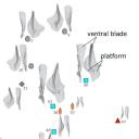
The present 3D dataset contains 15 specimens selected from the 69 3D models analyzed in the paper “3D topography as an indicator of change in food processing ability in the conodont genus Palmatolepis elements”. 3D topographic analysis of Palmatolepis P1 conodont elements from the Late Devonian period revealed an increase in blade sharpness together with a reduction in platform size. This indicates morphofunctional adaptation to more efficient prey processing.
Palmatolepis manticolepis UM CTB 144 View specimen

|
M3#1814Palmatolepis Manticolepis Type: "3D_surfaces"doi: 10.18563/m3.sf.1814 state:in_press |
Download 3D surface file |
Palmatolepis manticolepis UM CTB 151 View specimen

|
M3#1815Palmatolepis Manticolepis Type: "3D_surfaces"doi: 10.18563/m3.sf.1815 state:in_press |
Download 3D surface file |
Palmatolepis manticolepis UM CTB 078 View specimen

|
M3#1816Palmatolepis Manticolepis Type: "3D_surfaces"doi: 10.18563/m3.sf.1816 state:in_press |
Download 3D surface file |
Palmatolepis rhomboidea UM CTB 172 View specimen

|
M3#1817Palmatolepis rhomboidea Type: "3D_surfaces"doi: 10.18563/m3.sf.1817 state:in_press |
Download 3D surface file |
Palmatolepis glabra UM CTB 080 View specimen

|
M3#1818Palmatolepis glabra Type: "3D_surfaces"doi: 10.18563/m3.sf.1818 state:in_press |
Download 3D surface file |
Palmatolepis glabra UM CTB 177 View specimen

|
M3#1819Palmatolepis glabra Type: "3D_surfaces"doi: 10.18563/m3.sf.1819 state:in_press |
Download 3D surface file |
Palmatolepis glabra UM CTB 178 View specimen

|
M3#1820Palmatolepis glabra Type: "3D_surfaces"doi: 10.18563/m3.sf.1820 state:in_press |
Download 3D surface file |
Palmatolepis glabra UM CTB 179 View specimen

|
M3#1821Palmatolepis glabra Type: "3D_surfaces"doi: 10.18563/m3.sf.1821 state:in_press |
Download 3D surface file |
Palmatolepis gracilis UM CTB 186 View specimen

|
M3#1822Palmatolepis gracilis Type: "3D_surfaces"doi: 10.18563/m3.sf.1822 state:in_press |
Download 3D surface file |
Palmatolepis perlobata UM CTB 187 View specimen

|
M3#1823Palmatolepis perlobata Type: "3D_surfaces"doi: 10.18563/m3.sf.1823 state:in_press |
Download 3D surface file |
Palmatolepis perlobata UM CTB 189 View specimen

|
M3#1824Palmatolepis perlobata Type: "3D_surfaces"doi: 10.18563/m3.sf.1824 state:in_press |
Download 3D surface file |
Palmatolepis gracilis UM CTB 190 View specimen

|
M3#1825Palmatolepis gracilis Type: "3D_surfaces"doi: 10.18563/m3.sf.1825 state:in_press |
Download 3D surface file |
Palmatolepis perlobata UM CTB 191 View specimen

|
M3#1826Palmatolepis perlobata Type: "3D_surfaces"doi: 10.18563/m3.sf.1826 state:in_press |
Download 3D surface file |
Palmatolepis gracilis UM CTB 197 View specimen

|
M3#1827Palmatolepis gracilis Type: "3D_surfaces"doi: 10.18563/m3.sf.1827 state:in_press |
Download 3D surface file |
Palmatolepis gracilis UM CTB 200 View specimen

|
M3#1828Palmatolepis gracilis Type: "3D_surfaces"doi: 10.18563/m3.sf.1828 state:in_press |
Download 3D surface file |
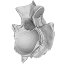
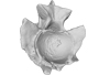
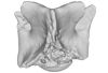
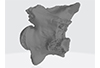
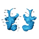
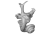
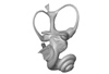
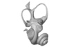
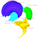
Our knowledge of the external brain morphology of the late Eocene artiodactyl ungulate Mixtotherium, relies on a plaster model realized on a specimen from the Victor Brun Museum in Montauban (France) and described by Dechaseaux (1973). Here, based on micro CT-scan data, we virtually reconstruct the 3D cast of the empty cavity of the partial cranium MA PHQ 716 from the Victor Brun Museum and compare it to the plaster model illustrated and described by Dechaseaux (1973). Indeed, the specimen from which the original plaster endocast originates was not identified by Dechaseaux by a specimen number. We confirm here that the studied specimen was indeed the one described and illustrated by Dechaseaux (1973). We also reconstruct a second, more detailed, model providing additional morphological and quantitative observations made available by micro CT scan investigation such as precisions on the neopallium folding and endocranial volumes.
Mixtotherium cuspidatum MA PHQ 716 View specimen
|
M3#857endocast of the brain cavity Type: "3D_surfaces"doi: 10.18563/m3.sf.857 state:published |
Download 3D surface file |
The present 3D Dataset contains the 3D models analyzed in Costeur L., Mennecart B., Müller B., Schulz G., 2016. Prenatal growth stages show the development of the ruminant bony labyrinth and petrosal bone. Journal of Anatomy. https://doi.org/10.1111/joa.12549
Bos taurus NMB3038 View specimen
|
M3#124Right bony labyrinth of a Bos taurus foetus (gestational age 115 days) Type: "3D_surfaces"doi: 10.18563/m3.sf.124 state:published |
Download 3D surface file |
Bos taurus NMB3367 View specimen
|
M3#125Right bony labyrinth of a Bos taurus foetus (gestational age 165 days) Type: "3D_surfaces"doi: 10.18563/m3.sf.125 state:published |
Download 3D surface file |
Bos taurus NMB3365 View specimen
|
M3#126Right bony labyrinth of a Bos taurus foetus (gestational age 210 days) Type: "3D_surfaces"doi: 10.18563/m3.sf.126 state:published |
Download 3D surface file |
Bos taurus NMB2855 View specimen
|
M3#127Right bony labyrinth of a Bos taurus foetus (gestational age 260 days) Type: "3D_surfaces"doi: 10.18563/m3.sf.127 state:published |
Download 3D surface file |
Bos taurus NMB1037 View specimen
|
M3#128Left bony labyrinth of an adult Bos taurus Type: "3D_surfaces"doi: 10.18563/m3.sf.128 state:published |
Download 3D surface file |
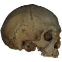
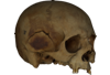
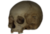
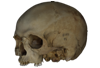
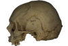
This contribution contains the 3D models described and figured in the following publications:
- Marini E., Lussu P., 2020. A virtual physical anthropology lab. Teaching in the time of coronavirus, in prep.;
- Lussu P., Bratzu D., Marini E., 2020. Cloud-based ultra close-range digital photogrammetry: validation of an approach for the effective virtual reconstruction of skeletal remains, in prep.
Homo sapiens MSAE 59 View specimen
|
M3#509MSAE 59 Type: "3D_surfaces"doi: 10.18563/m3.sf.509 state:published |
Download 3D surface file |
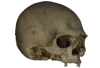
Homo sapiens MSAE 62 View specimen
|
M3#510MSAE 62 Type: "3D_surfaces"doi: 10.18563/m3.sf.510 state:published |
Download 3D surface file |
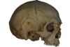
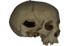
Homo sapiens MSAE 63 View specimen
|
M3#512MSAE 63 Type: "3D_surfaces"doi: 10.18563/m3.sf.512 state:published |
Download 3D surface file |
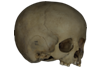
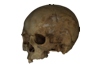
Homo sapiens MSAE 78 View specimen
|
M3#514MSAE 78 Type: "3D_surfaces"doi: 10.18563/m3.sf.514 state:published |
Download 3D surface file |
Homo sapiens MSAE 95 View specimen

|
M3#515MSAE 95 Type: "3D_surfaces"doi: 10.18563/m3.sf.515 state:published |
Download 3D surface file |
Homo sapiens MSAE 1852 View specimen
|
M3#516MSAE 1852 Type: "3D_surfaces"doi: 10.18563/m3.sf.516 state:published |
Download 3D surface file |
Homo sapiens MSAE 6426 View specimen
|
M3#517MSAE 6426 Type: "3D_surfaces"doi: 10.18563/m3.sf.517 state:published |
Download 3D surface file |
Homo sapiens MSAE 6428 View specimen
|
M3#518MSAE 6428 Type: "3D_surfaces"doi: 10.18563/m3.sf.518 state:published |
Download 3D surface file |
Homo sapiens MSAE 6992 View specimen
|
M3#519MSAE 6992 Type: "3D_surfaces"doi: 10.18563/m3.sf.519 state:published |
Download 3D surface file |
Homo sapiens MSAE 7688 View specimen
|
M3#520MSAE 7688 Type: "3D_surfaces"doi: 10.18563/m3.sf.520 state:published |
Download 3D surface file |
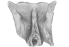
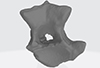
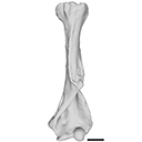
This contribution contains the 3D model described and figured in the following publication: Hautier L, Sarr R, Lihoreau F, Tabuce R, Marwan Hameh P. 2014. First record of the family Protocetidae in the Lutetian of Senegal (West Africa). Palaeovertebrata 38(2)-e2
indet. indet. SN103 View specimen

|
M3#5SN103, partial left innominate. Age and occurrence – Taïba Formation, Lutetian of the near Taïba Ndiaye, quarry of the Industries Chimiques du Sénégal (ICS) Type: "3D_surfaces"doi: 10.18563/m3.sf5 state:published |
Download 3D surface file |
This contribution contains the 3D models described and figured in the following publication: Shiraishi N et al. Morphology and morphometry of the human embryonic brain: A three-dimensional analysis NeuroImage 115, 2015, 96-103, DOI: 10.1016/j.neuroimage.2015.04.044.
Homo sapiens KC-CS13BRN50455 View specimen

|
M3#24Computationally reconstructed cerebral parenchyma and ventricle of the human embryo at Carnegie Stage 13. Type: "3D_surfaces"doi: 10.18563/m3.sf24 state:published |
Download 3D surface file |
Homo sapiens KC-CS14BRN18834 View specimen

|
M3#25Computationally reconstructed cerebral parenchyma and ventricle of the human embryo at Carnegie Stage 14. Type: "3D_surfaces"doi: 10.18563/m3.sf25 state:published |
Download 3D surface file |
Homo sapiens KC-CS15BRN19975 View specimen

|
M3#26Computationally reconstructed cerebral parenchyma and ventricle of the human embryo at Carnegie Stage 15. Type: "3D_surfaces"doi: 10.18563/m3.sf26 state:published |
Download 3D surface file |
Homo sapiens KC-CS16BRN7870 View specimen
|
M3#27Computationally reconstructed cerebral parenchyma and ventricle of the human embryo at Carnegie Stage 16. Type: "3D_surfaces"doi: 10.18563/m3.sf27 state:published |
Download 3D surface file |
Homo sapiens KC-CS17BRN26702 View specimen

|
M3#28Computationally reconstructed cerebral parenchyma and ventricle of the human embryo at Carnegie Stage 17. Type: "3D_surfaces"doi: 10.18563/m3.sf28 state:published |
Download 3D surface file |
Homo sapiens KC-CS18BRN25914 View specimen

|
M3#29Computationally reconstructed cerebral parenchyma and ventricle of the human embryo at Carnegie Stage 18. Type: "3D_surfaces"doi: 10.18563/m3.sf29 state:published |
Download 3D surface file |
Homo sapiens KC-CS19BRN16508 View specimen

|
M3#30Computationally reconstructed cerebral parenchyma and ventricle of the human embryo at Carnegie Stage 19. Type: "3D_surfaces"doi: 10.18563/m3.sf30 state:published |
Download 3D surface file |
Homo sapiens KC-CS20BRN26581 View specimen

|
M3#31Computationally reconstructed cerebral parenchyma and ventricle of the human embryo at Carnegie Stage 20. Type: "3D_surfaces"doi: 10.18563/m3.sf31 state:published |
Download 3D surface file |
Homo sapiens KC-CS21BRN33434 View specimen

|
M3#32Computationally reconstructed cerebral parenchyma and ventricle of the human embryo at Carnegie Stage 21. Type: "3D_surfaces"doi: 10.18563/m3.sf32 state:published |
Download 3D surface file |
Homo sapiens KC-CS22BRN27960 View specimen

|
M3#33Computationally reconstructed cerebral parenchyma and ventricle of the human embryo at Carnegie Stage 22. Type: "3D_surfaces"doi: 10.18563/m3.sf33 state:published |
Download 3D surface file |
Homo sapiens KC-CS23BRN28189 View specimen

|
M3#34Computationally reconstructed cerebral parenchyma and ventricle of the human embryo at Carnegie Stage 23. Type: "3D_surfaces"doi: 10.18563/m3.sf34 state:published |
Download 3D surface file |
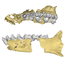
This contribution contains the 3D models of the fossil remains (maxilla, dentary, and talus) attributed to Djebelemur martinezi, a ca. 50 Ma primate from Tunisia (Djebel Chambi), described and figured in the following publication: Marivaux et al. (2013), Djebelemur, a tiny pre-tooth-combed primate from the Eocene of Tunisia: a glimpse into the origin of crown strepsirhines. PLoS ONE. https://doi.org/10.1371/journal.pone.0080778
Djebelemur martinezi CBI-1-544 View specimen
|
M3#365CBI-1-544, left maxilla preserving P3-M3 and alveoli for P2 and C1 Type: "3D_surfaces"doi: 10.18563/m3.sf.365 state:published |
Download 3D surface file |
Djebelemur martinezi CBI-1-567 View specimen
|
M3#363Isolated left upper P4 Type: "3D_surfaces"doi: 10.18563/m3.sf.363 state:published |
Download 3D surface file |
Djebelemur martinezi CBI-1-565-577-587-580 View specimen
|
M3#366- CBI-1-565, a damaged right mandible, which consists of three isolated pieces found together and reassembled here: the anterior part of the dentary bears the p3 and m1, and alveoli for p4, p2 and c, while the posterior part preserves m3 and a portion of the ascending ramus; the m2 was found isolated but in the same small calcareous block treated by acid processing. - CBI-1-577, isolated right lower p4. - CBI-1-587, isolated left lower p2 (reversed). - CBI-1-580, isolated left lower canine (reversed). Type: "3D_surfaces"doi: 10.18563/m3.sf.366 state:published |
Download 3D surface file |
Djebelemur martinezi CBI-1-545 View specimen
|
M3#364Right Talus Type: "3D_surfaces"doi: 10.18563/m3.sf.364 state:published |
Download 3D surface file |
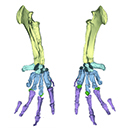
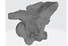
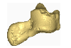
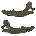
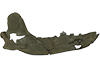
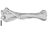
The present 3D Dataset contains the 3D model analyzed in Vautrin et al. (2019), Palaeontology, From limb to fin: an Eocene protocetid forelimb from Senegal sheds new light on the early locomotor evolution of early cetaceans.
?Carolinacetus indet. SNTB 2011-01 View specimen
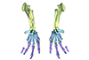
|
M3#3983D model of an articulated forelimb of a Carolinacetus-like protocetid from Senegal Type: "3D_surfaces"doi: 10.18563/m3.sf.398 state:published |
Download 3D surface file |
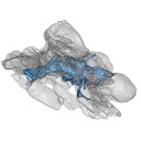
This contribution contains the 3D models described and figured in the following publication: Paulina-Carabajal, A. and Nieto, M. N. In press. Brief comment on the brain and inner ear of Giganotosaurus carolinii (Dinosauria: Theropoda) based on CT scans. Ameghiniana. https://doi.org/10.5710/AMGH.25.10.2019.3237
Giganotosaurus carolinii MUCPv-CH-1 View specimen
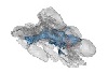
|
M3#504The current file contents 3D models of the braincase, brain, left and right inner ears Type: "3D_surfaces"doi: 10.18563/m3.sf.504 state:published |
Download 3D surface file |
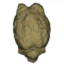
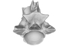
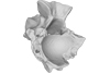
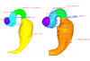
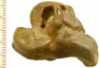
The present 3D Dataset contains the 3D model of the endocranial cast of Palaeolama sp. from the mid-Pleistocene (~1.2 Mya) of South America, analyzed in Balcarcel et al. 2023.
Palaeolama sp. PIMUZ A/V 4091 View specimen
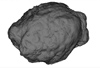
|
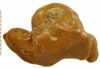
M3#11283D model of a natural endocast Type: "3D_surfaces"doi: 10.18563/m3.sf.1128 state:published |
Download 3D surface file |
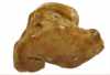
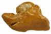
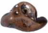
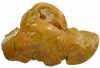
The present 3D Dataset contains the 3D model analyzed in Solé F., Lesport J.-F., Heitz A., and Mennecart B. minor revision. A new gigantic carnivore (Carnivora, Amphicyonidae) from the late middle Miocene of France. PeerJ.
Tartarocyon cazanavei MHNBx 2020.20.1 View specimen
|
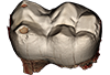
M3#903Surface scan (ply) and texture (png) of the holotype of Tartarocyon cazanavei (MHNBx 2020.20.1) Type: "3D_surfaces"doi: 10.18563/m3.sf.903 state:published |
Download 3D surface file |
This contribution contains the 3D model described and figured in the following publication: Billet G., Germain D., Ruf I., Muizon C. de, Hautier L. 2013. The inner ear of Megatherium and the evolution of the vestibular system in sloths. Journal of Anatomy 123:557-567, DOI: 10.1111/joa.12114.
Megatherium americanum MNHN.F.PAM276 View specimen

|
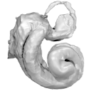
M3#14This model corresponds to a virtually reconstructed bony labyrinth of the right inner ear of the skull MNHN-F-PAM 276, attributed to the extinct giant ground sloth Megatherium americanum. The fossil comes from Pleistocene deposits at Rio Salado (Prov. Buenos Aires, Argentina). The bony labyrinth of Megatherium shows semicircular canals that are proportionally much larger than in the modern two-toed and three-toed sloths. The cochlea in Megatherium shows 2.5 turns, which is a rather high value within Xenarthra. Overall, the shape of the bony labyrinth of Megatherium resembles more that of extant armadillos than that of its extant sloth relatives. Type: "3D_surfaces"doi: 10.18563/m3.sf14 state:published |
Download 3D surface file |
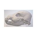
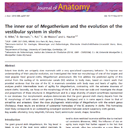
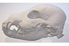
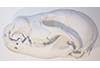
This contribution comprises the 3D models of three wolf pup skulls, which were used for the publication by Geiger et al. 2017 on Neomorphosis and heterochrony of skull shape in dog domestication.
Canis lupus CLL2 View specimen
|
M3#3123d model of a wolf pup skull Type: "3D_surfaces"doi: 10.18563/m3.sf.312 state:published |
Download 3D surface file |
Canis lupus CLL4 View specimen
|
M3#3133d model of a wolf pup skull Type: "3D_surfaces"doi: 10.18563/m3.sf.313 state:published |
Download 3D surface file |
Canis lupus CLL5 View specimen
|
M3#3143d model of a wolf pup skull Type: "3D_surfaces"doi: 10.18563/m3.sf.314 state:published |
Download 3D surface file |
The present 3D Dataset contains the 3D model analyzed in the following publication: occurrence of the ground sloth Nothrotheriops (Xenarthra, Folivora) in the Late Pleistocene of Uruguay: New information on its dietary and habitat preferences based on stable isotope analysis. Journal of Mammalian Evolution. https://doi.org/10.1007/s10914-023-09660-w
Nothrotheriops sp. CAV 1466 View specimen
|
M3#1129Left humerus Type: "3D_surfaces"doi: 10.18563/m3.sf.1129 state:published |
Download 3D surface file |
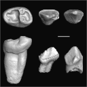
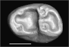
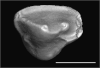
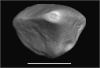
This contribution contains the 3D models of the fossil teeth of a small-bodied platyrrhine primate, Neosaimiri cf. fieldsi (Cebinae, Cebidae, Platyrrhini) discovered from Laventan deposits (late Middle Miocene) of Peruvian Amazonia, San Martín Department (TAR-31: Tarapoto/Juan Guerra vertebrate fossil-bearing locus n°31). These fossils were described and figured in the following publication: Marivaux et al. (2020), New record of Neosaimiri (Cebidae, Platyrrhini) from the late Middle Miocene of Peruvian Amazonia. Journal of Human Evolution. https://doi.org/10.1016/j.jhevol.2020.102835
Neosaimiri cf. fieldsi MUSM-3888 View specimen
|
M3#538MUSM-3888, right m3 of Neosaimiri cf. fieldsi. Type: "3D_surfaces"doi: 10.18563/m3.sf.538 state:published |
Download 3D surface file |
Neosaimiri cf. fieldsi MUSM-3890 View specimen
|
M3#540MUSM-3890, left dp2 of Neosaimiri cf. fieldsi. Type: "3D_surfaces"doi: 10.18563/m3.sf.540 state:published |
Download 3D surface file |
Neosaimiri cf. fieldsi MUSM-3895 View specimen
|
M3#541MUSM-3895, right DC1 of Neosaimiri cf. fieldsi. Type: "3D_surfaces"doi: 10.18563/m3.sf.541 state:published |
Download 3D surface file |
Neosaimiri cf. fieldsi MUSM-3891 View specimen
|
M3#542MUSM-3891, lingual part of a fragmentary right M1 or M2 of Neosaimiri cf. fieldsi. Type: "3D_surfaces"doi: 10.18563/m3.sf.542 state:published |
Download 3D surface file |
Neosaimiri cf. fieldsi MUSM-3892 View specimen
|
M3#543MUSM-3892, distobuccal part of a fragmentary right upper molar (metacone region) of Neosaimiri cf. fieldsi. Type: "3D_surfaces"doi: 10.18563/m3.sf.543 state:published |
Download 3D surface file |
Neosaimiri cf. fieldsi MUSM-3893 View specimen
|
M3#544MUSM-3893, buccal part of a fragmentary right P3 or P4 of Neosaimiri cf. fieldsi. Type: "3D_surfaces"doi: 10.18563/m3.sf.544 state:published |
Download 3D surface file |
Neosaimiri cf. fieldsi MUSM-3894 View specimen
|
M3#545MUSM-3894, lingual part of a fragmentary left P3 or P4 of Neosaimiri cf. fieldsi. Type: "3D_surfaces"doi: 10.18563/m3.sf.545 state:published |
Download 3D surface file |
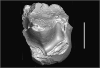
This contribution contains the 3D models described and figured in the following publication: Hautier L, Tabuce R, Kassegne KE, Amoudji YZ, Mourlam M, Orliac M, Quillévéré F, Charruault A-L, Johnson AKC, Guinot G. 2021. New middle Eocene proboscidean from Togo illuminates the early evolution of the elephantiform-like dental pattern.
Dagbatitherium tassyi ULDG-DAG1 View specimen
|
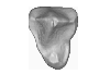
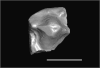
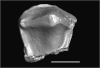
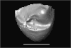
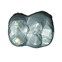
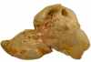
M3#7693D model of a molar of Dagbatitherium tassyi. Type: "3D_surfaces"doi: 10.18563/m3.sf.769 state:published |
Download 3D surface file |
|
M3#771µCT scan of a molar of Dagbatitherium tassyi. Type: "3D_CT"doi: 10.18563/m3.sf.771 state:published |
Download CT data |
This contribution contains the 3D models described and figured in the following publication: Aguirre-Fernández G, Jost J, and Hilfiker S. 2022. First records of extinct kentriodontid and squalodelphinid dolphins from the Upper Marine Molasse (Burdigalian age) of Switzerland and a reappraisal of the Swiss cetacean fauna.
Kentriodon sp. NMBE 5023944 View specimen
|
M3#8583D models of left periotic and bony labyrinth of NMBE 5023944 (Kentriodon sp.) Type: "3D_surfaces"doi: 10.18563/m3.sf.858 state:published |
Download 3D surface file |
Kentriodon sp. NMBE 5023945 View specimen
|
M3#8593D models of right periotic and bony labyrinth of NMBE 5023945 (Kentriodontidae indet.) Type: "3D_surfaces"doi: 10.18563/m3.sf.859 state:published |
Download 3D surface file |
Kentriodon sp. NMBE 5023946 View specimen
|
M3#8603D models of left periotic and bony labyrinth of NMBE 5023946 (Kentriodon sp.) Type: "3D_surfaces"doi: 10.18563/m3.sf.860 state:published |
Download 3D surface file |
Kentriodon sp. NMBE 5036436 View specimen
|
M3#8613D models of right periotic and bony labyrinth of NMBE 5036436 (Kentriodontidae indet.) Type: "3D_surfaces"doi: 10.18563/m3.sf.861 state:published |
Download 3D surface file |
indet. indet. NMBE 5023942 View specimen
|
M3#8623D models of right periotic and bony labyrinth of NMBE 5023942 (Squalodelphinidae indet.) Type: "3D_surfaces"doi: 10.18563/m3.sf.862 state:published |
Download 3D surface file |
indet. indet. NMBE 5023943 View specimen
|
M3#8633D models of left periotic and bony labyrinth of NMBE 5023943 (Squalodelphinidae indet.) Type: "3D_surfaces"doi: 10.18563/m3.sf.863 state:published |
Download 3D surface file |
indet. indet. NMBE 5036437 View specimen
|
M3#8643D models of left periotic and bony labyrinth of NMBE 5036437 (Physeteridae indet.) Type: "3D_surfaces"doi: 10.18563/m3.sf.864 state:published |
Download 3D surface file |


