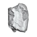
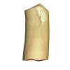
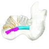
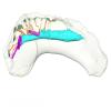
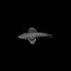
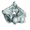
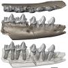
3D models of Kalakocetus, the earliest Cetacea
The specimens of Speothos pacivorus
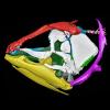
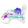
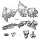
3D models related to the publication: Hidden diversity of Palaeogene metatherians: a new family of polydolopimorphian marsupials from Peruvian Amazonia
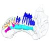
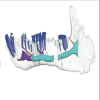
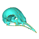
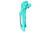
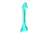
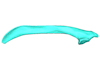
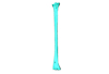
3D GM dataset of bird skeletal variation
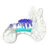
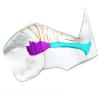
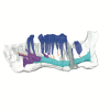
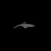
Skeletal embryonic development in the catshark
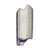
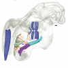
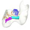
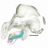
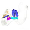
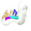
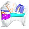
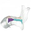
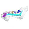
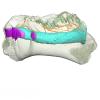
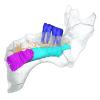
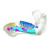
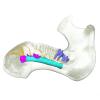
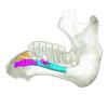
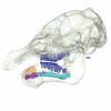
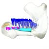
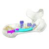
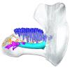
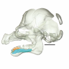
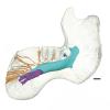
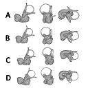
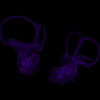
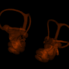
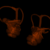
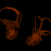
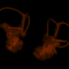
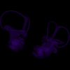
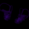
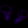
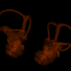
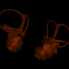
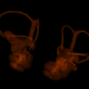
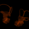
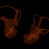
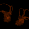
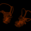
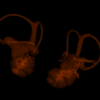
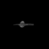
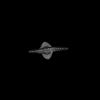
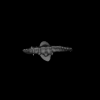
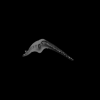
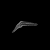
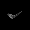
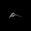
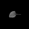
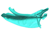
Bony connexions of the petrosal bone of extant hippos
bony labyrinth (14) , inner ear (11) , Eocene (11) , geometric morphometrics (10) , CT-scan (10) , Oligocene (9) , Micro-CT (9)
Maëva Judith Orliac (24) , Lionel Hautier (24) , Laurent Marivaux (18) , Renaud Lebrun (15) , Rodolphe Tabuce (14) , Pierre-Olivier Antoine (13) , Bastien Mennecart (13)
|
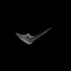
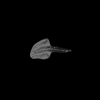
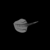
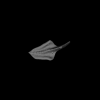
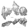
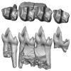
3D models related to the publication: Unexpected pampatheriid from the early Oligocene of Peruvian Amazonia: insights into the tropical differentiation of cingulate xenarthrans.François Pujos
Published online: 28/03/2025 |
|
M3#1600Molariform and associated dentinal microstructure Type: "3D_surfaces"doi: 10.18563/m3.sf.1600 state:published |
Download 3D surface file |
Choloepus didactylus UM-ZOOL-V12 View specimen
|
M3#1601Molariform and associated dentinal microstructure Type: "3D_surfaces"doi: 10.18563/m3.sf.1601 state:published |
Download 3D surface file |
Dasypus mexicanus UM-ZOOL-2787 View specimen

|
M3#1602Molariform and associated dentinal microstructure Type: "3D_surfaces"doi: 10.18563/m3.sf.1602 state:published |
Download 3D surface file |
Tolypeutes matacus UM-ZOOL-2789 View specimen
|
M3#1603Molariform and associated dentinal microstructure Type: "3D_surfaces"doi: 10.18563/m3.sf.1603 state:published |
Download 3D surface file |
Euphractus sexcinctus UM-ZOOL-2790 View specimen
|
M3#1604Molariform and associated dentinal microstructure Type: "3D_surfaces"doi: 10.18563/m3.sf.1604 state:published |
Download 3D surface file |
Holmesina septrionalis UM-FLD-1 View specimen
|
M3#1605Molariform and associated dentinal microstructure Type: "3D_surfaces"doi: 10.18563/m3.sf.1605 state:published |
Download 3D surface file |
Megatherium sp. UM-TAR-1 View specimen
|
M3#1607Molariform and associated dentinal microstructure Type: "3D_surfaces"doi: 10.18563/m3.sf.1607 state:published |
Download 3D surface file |
Indet indet MUSM-3965 View specimen
|
M3#1606Molariform and associated dentinal microstructure Type: "3D_surfaces"doi: 10.18563/m3.sf.1606 state:published |
Download 3D surface file |
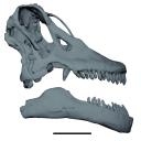
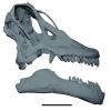
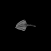
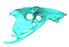
The study of titanosaur paleobiology has been severely hampered by the incomplete nature of their fossil record, particularly the scarcity of well-preserved and relatively complete cranial remains. Even the most complete titanosaur skulls are often fractured, incomplete, or deformed, which has resulted in a limited knowledge of the paleobiology related to cranial anatomy, especially functional morphology. In this context, we present the digital restoration of the skull of the Argentinean titanosaur Sarmientosaurus musacchioi, created using the open-source 3D modeling software Blender. The digitally restored model is freely accessible to other researchers, facilitating broader research and comparative studies.
Sarmientosaurus mussacchioi MDT-PV 02 View specimen
|
M3#1594Cranium and mandible of Sarmientosaurus mussacchioi Type: "3D_surfaces"doi: 10.18563/m3.sf.1594 state:published |
Download 3D surface file |
|
M3#1599Original object provided by Gabriel Casal (cranium) Type: "3D_surfaces"doi: 10.18563/m3.sf.1599 state:published |
Download 3D surface file |
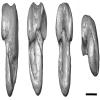
The present 3D Dataset contains sixteen 3D models of unornamented Polygnathus illustrating allometric variation and bilateral asymmetry within four “Operational Taxonomic Units” analyzed in the publication: Convergent allometric trajectories in Devonian-Carboniferous unornamented Polygnathus conodonts.
Polygnathus sp. UM-PSQ-010 View specimen
|
M3#1611Dextral P1 element Type: "3D_surfaces"doi: 10.18563/m3.sf.1611 state:published |
Download 3D surface file |
Polygnathus sp. UM-PSQ-011 View specimen

|
M3#1612Sinistral P1 element Type: "3D_surfaces"doi: 10.18563/m3.sf.1612 state:published |
Download 3D surface file |
Polygnathus sp. UM-PSQ-012 View specimen
|
M3#1613Sinistral P1 element Type: "3D_surfaces"doi: 10.18563/m3.sf.1613 state:published |
Download 3D surface file |
Polygnathus sp. UM-PSQ-013 View specimen

|
M3#1614Dextral P1 element Type: "3D_surfaces"doi: 10.18563/m3.sf.1614 state:published |
Download 3D surface file |
Polygnathus sp. UM-PSQ-014 View specimen
|
M3#1615Sinistral P1 element Type: "3D_surfaces"doi: 10.18563/m3.sf.1615 state:published |
Download 3D surface file |
Polygnathus sp. UM-PSQ-015 View specimen
|
M3#1616Sinistral P1 element Type: "3D_surfaces"doi: 10.18563/m3.sf.1616 state:published |
Download 3D surface file |
Polygnathus sp. UM-PSQ-016 View specimen

|
M3#1617Dextral P1 element Type: "3D_surfaces"doi: 10.18563/m3.sf.1617 state:published |
Download 3D surface file |
Polygnathus sp. UM-PSQ-017 View specimen
|
M3#1618Dextral P1 element Type: "3D_surfaces"doi: 10.18563/m3.sf.1618 state:published |
Download 3D surface file |
Polygnathus sp. UM-PSQ-018 View specimen
|
M3#1619Sinistral P1 element Type: "3D_surfaces"doi: 10.18563/m3.sf.1619 state:published |
Download 3D surface file |
Polygnathus sp. UM-PSQ-019 View specimen
|
M3#1620Sinistral P1 element Type: "3D_surfaces"doi: 10.18563/m3.sf.1620 state:published |
Download 3D surface file |
Polygnathus sp. UM-PSQ-020 View specimen

|
M3#1621Dextral P1 element Type: "3D_surfaces"doi: 10.18563/m3.sf.1621 state:published |
Download 3D surface file |
Polygnathus sp. UM-PSQ-021 View specimen
|
M3#1622Dextral P1 element Type: "3D_surfaces"doi: 10.18563/m3.sf.1622 state:published |
Download 3D surface file |
Polygnathus sp. UM-PSQ-022 View specimen
|
M3#1623Sinistral P1 element Type: "3D_surfaces"doi: 10.18563/m3.sf.1623 state:published |
Download 3D surface file |
Polygnathus sp. UM-PSQ-023 View specimen
|
M3#1624Sinistral P1 element Type: "3D_surfaces"doi: 10.18563/m3.sf.1624 state:published |
Download 3D surface file |
Polygnathus sp. UM-PSQ-024 View specimen

|
M3#1625Dextral P1 element Type: "3D_surfaces"doi: 10.18563/m3.sf.1625 state:published |
Download 3D surface file |
Polygnathus sp. UM-PSQ-025 View specimen
|
M3#1626Dextral P1 element Type: "3D_surfaces"doi: 10.18563/m3.sf.1626 state:published |
Download 3D surface file |
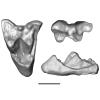
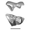
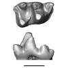
This contribution contains the three-dimensional models of the most informative fossil material attributed to both Peratherium musivum Gernelle, 2024, and Peratherium maximum (Crochet, 1979), respectively from early and middle early Eocene French localities. These specimens, which document the emergence of the relatively large peratheriines, were analyzed and discussed in: Gernelle et al. (2024), Dental morphology evolution in early peratheriines, including a new morphologically cryptic species and findings on the largest early Eocene European metatherian. https://doi.org/10.1080/08912963.2024.2403602
Peratherium musivum MNHN.F.SN122 View specimen
|
M3#16403D surface model of MNHN.F.SN122, right M3 Type: "3D_surfaces"doi: 10.18563/m3.sf.1640 state:published |
Download 3D surface file |
Peratherium musivum MNHN.F.RI220 View specimen
|
M3#16413D surface model of MNHN.F.RI220, left M2 (partial) Type: "3D_surfaces"doi: 10.18563/m3.sf.1641 state:published |
Download 3D surface file |
Peratherium musivum MNHN.F.RI296 View specimen
|
M3#16423D surface model of MNHN.F.RI296, right M1 (partial) Type: "3D_surfaces"doi: 10.18563/m3.sf.1642 state:published |
Download 3D surface file |
Peratherium musivum MNHN.F.RI368 View specimen
|
M3#16433D surface model of MNHN.F.RI368, right m2 Type: "3D_surfaces"doi: 10.18563/m3.sf.1643 state:published |
Download 3D surface file |
Peratherium musivum MNHN.F.RI385 View specimen
|
M3#16443D surface model of MNHN.F.RI385, left m1 Type: "3D_surfaces"doi: 10.18563/m3.sf.1644 state:published |
Download 3D surface file |
Peratherium maximum UM-BRI-17 View specimen
|
M3#16453D surface model of UM-BRI-17, right hemi-mandible with p1-p3, m1-m3 alveoli, and m4 Type: "3D_surfaces"doi: 10.18563/m3.sf.1645 state:published |
Download 3D surface file |
The present 3D Dataset contains the 3D models of the skull of the holotype of Miocaperea pulchra.
Miocaperea pulchra SMNS-P-46978 View specimen
|
M3#1656Blender file containing two models (the skull being preserved in two parts) Type: "3D_surfaces"doi: 10.18563/m3.sf.1656 state:published |
Download 3D surface file |
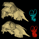
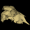
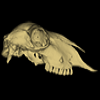
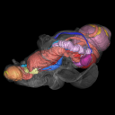
This contribution contains the 3D models described and figured in the following publication:Skull and Inner Ear Morphometrics in Sheep and Goats: Species and Breed Differentiation with Bioarchaeological Applications (Hemelsdael et al. submitted). The models include the external surface of a complete skull and inner ear of both a sheep (Ovis aries) and a goat (Capra hircus), generated from micro-CT scans. In the associated paper, we used 3D geometric morphometric data to assess inter and intra (i.e. between breeds) discrimination based on complete skulls, skull fragments and the semi-circular canals of the inner ear.
Capra hircus Amp_1 View specimen
|
M3#1806Skull of the goat Amp_1 Type: "3D_surfaces"doi: 10.18563/m3.sf.1806 state:published |
Download 3D surface file |

|
M3#1807Inner ear of the goat Amp_1 Type: "3D_surfaces"doi: 10.18563/m3.sf.1807 state:published |
Download 3D surface file |
Ovis aries UM_RR_2331 View specimen
|
M3#1808Skull of the sheep UM_RR_2331 Type: "3D_surfaces"doi: 10.18563/m3.sf.1808 state:published |
Download 3D surface file |

|
M3#1809Inner ear of the sheep UM_RR_2331 Type: "3D_surfaces"doi: 10.18563/m3.sf.1809 state:published |
Download 3D surface file |
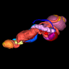
The present 3D dataset contains the 3D models analyzed in the publication: Rosa, R. M., Salvador, R. B., & Cavallari, D. C. (2025). The disappearing act of the magician tree snail: anatomy, distribution, and phylogenetic relationships of Drymaeus magus (Gastropoda: Bulimulidae), a long-lost species hidden in plain sight. Zoological Journal of the Linnean Society.
Drymaeus magus CMRP 1049 View specimen
|
M3#1597Internal organs of Drymaeus magus Type: "3D_surfaces"doi: 10.18563/m3.sf.1597 state:published |
Download 3D surface file |
|
M3#1598External surface of Drymaeus magus Type: "3D_surfaces"doi: 10.18563/m3.sf.1598 state:published |
Download 3D surface file |
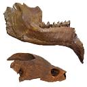
The present 3D Dataset contains the 3D models produced in the frame of the article Perthuis, A. de, Mennecart, B., Barrier, P., Chenot, É., Falconnet, J., Gagnaison, J.-C., Georgalis, G. L., Gilbert, C., Guevel, B., Langevin, D., Lapparent de Broin, F. de, Lemierre, A., Maubert, F., Ossó, À., Potel, S., Thivaiou, D., Tissier, J., Toullec, R., Xerri, S., Gagnaison, C. 2025. Révision des données sédimentologiques et biostratigraphiques des gisements à vertébrés des sables de l’Orléanais, à Beaugency, Tavers et Le Bardon (Miocène Moyen ; Loiret, France). Geodiversitas 47 (12): 2-76. https://doi.org/10.5252/geodiversitas2025v47a12
Bunolistriodon lockharti ULB-TAV-21 View specimen
|
M3#1837Left upper M3 Type: "3D_surfaces"doi: 10.18563/m3.sf.1837 state:published |
Download 3D surface file |
Megamphicyon giganteus ULB-TAV-13 View specimen
|
M3#1531Left first lower molar Type: "3D_surfaces"doi: 10.18563/m3.sf.1531 state:published |
Download 3D surface file |
Hispanotherium matritense ULB-TAV-17 View specimen
|
M3#1532Left first lower molar Type: "3D_surfaces"doi: 10.18563/m3.sf.1532 state:published |
Download 3D surface file |
Plesiaceratherium lumiarense ULB-TAV-18 View specimen
|
M3#1533Left third upper molar Type: "3D_surfaces"doi: 10.18563/m3.sf.1533 state:published |
Download 3D surface file |
Chelydropsis aff. sansaniensis ULB-TAV-23 View specimen
|
M3#1535Cast of a skull Type: "3D_surfaces"doi: 10.18563/m3.sf.1535 state:published |
Download 3D surface file |
Ronzotherium romani ULB-TAV-4 View specimen
|
M3#1556Right fourth upper premolar Type: "3D_surfaces"doi: 10.18563/m3.sf.1556 state:published |
Download 3D surface file |
Prodeinotherium bavaricum ULB-TAV-24 View specimen
|
M3#1557left hemimandibule Type: "3D_surfaces"doi: 10.18563/m3.sf.1557 state:published |
Download 3D surface file |

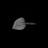
We provide a 3D reconstruction of the skull of Latimeria chalumnae that can be easily accessed and visualized for a better understanding of its cranial anatomy. Different skeletal elements are saved as separate PLY files that can be combined to visualize the entire skull or isolated to virtually dissect the skull. We included some guidelines for a fast and easy visualization of the 3D skull.
Latimeria chalumnae MHNG 1080.070 View specimen
|
M3#1254the skeletal elements of the skull of Latimeria chalumnae included in 26 different PLY files Type: "3D_surfaces"doi: 10.18563/m3.sf.1254 state:published |
Download 3D surface file |

This contribution contains the 3D models described and figured in the following publication: Hautier L, Gomes Rodrigues H, Ferreira-Cardoso S, Emerling CA, Porcher M-L, Asher R, Portela Miguez R, Delsuc F. 2023. From teeth to pad: tooth loss and development of keratinous structures in sirenians. Proceedings of the Royal Society B. https://doi.org/10.1098/rspb.2023.1932
Dugong dugon 2005.51 View specimen
|
M3#1275Internal mandibular morphology. Orange = dorsal canaliculi; purple = mental branches; cyan = mandibular canal; dark blue = teeth; green = tooth alveoli. Type: "3D_surfaces"doi: 10.18563/m3.sf.1275 state:published |
Download 3D surface file |
Dugong dugon 2023.66 View specimen
|
M3#1274Internal mandibular morphology. Orange = dorsal canaliculi; purple = mental branches; cyan = mandibular canal; dark blue = teeth; green = tooth alveoli. Type: "3D_surfaces"doi: 10.18563/m3.sf.1274 state:published |
Download 3D surface file |
Dugong dugon 5386 View specimen
|
M3#1276Internal mandibular morphology. Orange = dorsal canaliculi; purple = mental branches; cyan = mandibular canal; dark blue = teeth; green = tooth alveoli. Type: "3D_surfaces"doi: 10.18563/m3.sf.1276 state:published |
Download 3D surface file |
Dugong dugon 1848.8.29.7/GERM 1027g View specimen
|
M3#1277Internal mandibular morphology. Orange = dorsal canaliculi; purple = mental branches; cyan = mandibular canal; dark blue = teeth. Type: "3D_surfaces"doi: 10.18563/m3.sf.1277 state:published |
Download 3D surface file |
Dugong dugon 1991.413 View specimen
|
M3#1278Internal mandibular morphology. Orange = dorsal canaliculi; purple = mental branches; cyan = mandibular canal; dark blue = teeth. Type: "3D_surfaces"doi: 10.18563/m3.sf.1278 state:published |
Download 3D surface file |
Dugong dugon 1991.427 View specimen
|
M3#1279Internal mandibular morphology. Orange = dorsal canaliculi; purple = mental branches; cyan = mandibular canal; dark blue = teeth. Type: "3D_surfaces"doi: 10.18563/m3.sf.1279 state:published |
Download 3D surface file |
Dugong dugon 2017-3-9 View specimen
|
M3#1280Internal mandibular morphology. Orange = dorsal canaliculi; purple = mental branches; cyan = mandibular canal; dark blue = teeth. Type: "3D_surfaces"doi: 10.18563/m3.sf.1280 state:published |
Download 3D surface file |
Eosiren lybica 1913-22 View specimen
|
M3#1281Internal mandibular morphology. Orange = dorsal canaliculi; purple = mental branches; cyan = mandibular canal; dark blue = teeth; green = tooth alveoli. Type: "3D_surfaces"doi: 10.18563/m3.sf.1281 state:published |
Download 3D surface file |
Halitherium taulannense RGHP C001 View specimen
|
M3#1282Internal mandibular morphology. Cyan = mandibular canal; dark blue = teeth. Type: "3D_surfaces"doi: 10.18563/m3.sf.1282 state:published |
Download 3D surface file |
Halitherium taulannense RGHP C009 View specimen
|
M3#1283Internal mandibular morphology. Orange = dorsal canaliculi; purple = mental branches; cyan = mandibular canal; dark blue = teeth; green = tooth alveoli. Type: "3D_surfaces"doi: 10.18563/m3.sf.1283 state:published |
Download 3D surface file |
Hydrodamalis gigas 1947.10.21.1 View specimen
|
M3#1284Internal mandibular morphology. Orange = dorsal canaliculi; purple = mental branches; cyan = mandibular canal Type: "3D_surfaces"doi: 10.18563/m3.sf.1284 state:published |
Download 3D surface file |
Hydrodamalis gigas C1021 View specimen

|
M3#1285Anterior part of the mandible Type: "3D_surfaces"doi: 10.18563/m3.sf.1285 state:published |
Download 3D surface file |
|
M3#1286Posterior part of the mandible Type: "3D_surfaces"doi: 10.18563/m3.sf.1286 state:published |
Download 3D surface file |
Hydrodamalis gigas 2023.67 View specimen
|
M3#1287Internal mandibular morphology. Orange = dorsal canaliculi; purple = mental branches; cyan = mandibular canal Type: "3D_surfaces"doi: 10.18563/m3.sf.1287 state:published |
Download 3D surface file |
Libysiren sickenbergi M.82429 View specimen
|
M3#1288Internal mandibular morphology. Orange = dorsal canaliculi; purple = mental branches; cyan = mandibular canal; dark blue = teeth; green = tooth alveoli. Type: "3D_surfaces"doi: 10.18563/m3.sf.1288 state:published |
Download 3D surface file |
Libysiren sickenbergi M.45675 View specimen
|
M3#1289Internal mandibular morphology. Orange = dorsal canaliculi; purple = mental branches; cyan = mandibular canal; dark blue = teeth; green = tooth alveoli. Type: "3D_surfaces"doi: 10.18563/m3.sf.1289 state:published |
Download 3D surface file |
Prorastomus sirenoides OR.448976 View specimen
|
M3#1290Internal morphology of the left mandible. Orange = dorsal canaliculi; purple = mental branches; cyan = mandibular canal; dark blue = teeth; green = tooth alveoli. Type: "3D_surfaces"doi: 10.18563/m3.sf.1290 state:published |
Download 3D surface file |
|
M3#1304Internal morphology of the right mandible. Orange = dorsal canaliculi; purple = mental branches; cyan = mandibular canal; dark blue = teeth. Type: "3D_surfaces"doi: 10.18563/m3.sf.1304 state:published |
Download 3D surface file |
Ribodon limbatus M.7073 View specimen
|
M3#1292Internal mandibular morphology. Orange = dorsal canaliculi; purple = mental branches; cyan = mandibular canal; dark blue = teeth; green = tooth alveoli. Type: "3D_surfaces"doi: 10.18563/m3.sf.1292 state:published |
Download 3D surface file |
Rytiodus capgrandi PAL2017-8-1 View specimen
|
M3#1293Internal mandibular morphology. Orange = dorsal canaliculi; purple = mental branches; cyan = mandibular canal; dark blue = teeth; green = tooth alveoli. Type: "3D_surfaces"doi: 10.18563/m3.sf.1293 state:published |
Download 3D surface file |
Trichechus inunguis 1868.12.19.2 View specimen
|
M3#1294Internal mandibular morphology. Orange = dorsal canaliculi; purple = mental branches; cyan = mandibular canal; dark blue = teeth; green = tooth alveoli. Type: "3D_surfaces"doi: 10.18563/m3.sf.1294 state:published |
Download 3D surface file |
Trichechus manatus 1843.3.10.12 View specimen
|
M3#1295Internal mandibular morphology. Orange = dorsal canaliculi; purple = mental branches; cyan = mandibular canal; dark blue = teeth. Type: "3D_surfaces"doi: 10.18563/m3.sf.1295 state:published |
Download 3D surface file |
Trichechus manatus 1864.6.5.1 View specimen
|
M3#1296Internal mandibular morphology. Orange = dorsal canaliculi; purple = mental branches; cyan = mandibular canal; dark blue = teeth; green = tooth alveoli. Type: "3D_surfaces"doi: 10.18563/m3.sf.1296 state:published |
Download 3D surface file |
Trichechus manatus 1950.1.23.1 View specimen
|
M3#1297Internal mandibular morphology. Orange = dorsal canaliculi; purple = mental branches; cyan = mandibular canal; dark blue = teeth; green = tooth alveoli. Type: "3D_surfaces"doi: 10.18563/m3.sf.1297 state:published |
Download 3D surface file |
Trichechus senegalensis 1885.6.30.2 View specimen
|
M3#1298Internal mandibular morphology. Orange = dorsal canaliculi; purple = mental branches; cyan = mandibular canal; dark blue = teeth; green = tooth alveoli. Type: "3D_surfaces"doi: 10.18563/m3.sf.1298 state:published |
Download 3D surface file |
Trichechus senegalensis 1894.7.25.8 View specimen
|
M3#1299Internal mandibular morphology. Orange = dorsal canaliculi; purple = mental branches; cyan = mandibular canal; dark blue = teeth. Type: "3D_surfaces"doi: 10.18563/m3.sf.1299 state:published |
Download 3D surface file |
Trichechus senegalensis V97 View specimen
|
M3#1302Mandibular internal morphology. Orange = dorsal canaliculi; purple = mental branches; cyan = mandibular canal; dark blue = teeth. Type: "3D_surfaces"doi: 10.18563/m3.sf.1302 state:published |
Download 3D surface file |
Trichechus sp. 65.4.28.9 View specimen
|
M3#1300Internal mandibular morphology. Orange = dorsal canaliculi; purple = mental branches; cyan = mandibular canal; dark blue = teeth; green = tooth alveoli. Type: "3D_surfaces"doi: 10.18563/m3.sf.1300 state:published |
Download 3D surface file |
Dugong dugon 1946.8.6.2 View specimen
|
M3#1301Mandibular internal morphology. Orange = dorsal canaliculi; purple = mental branches; cyan = mandibular canal; dark blue = teeth. Type: "3D_surfaces"doi: 10.18563/m3.sf.1301 state:published |
Download 3D surface file |

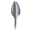
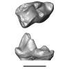
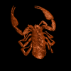
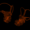
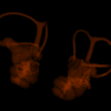
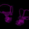
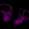
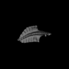
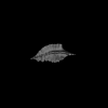
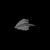
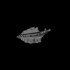
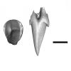
In this contribution a third new species of the rare genus Burmesescorpiops Lourenço, 2016 is described. The discovery of this new element belonging to the family Palaeoeuscorpiidae Lourenço, 2003 and to the subfamily Archaeoscorpiopinae Lourenço, 2015 brings further elements to support the validity of the genus Burmesescorpiops. This generic group remains however, poorly speciose. This is the latest discovery of Burmesescorpiops wunpawng, the name is derived from the Kachin Hilltribe peoples who are indigenous to the area. The data provided here is a 3D surface.
Burmesescorpiops wunpawng ps-gyi-01-25 View specimen
|
M3#18463d Surface Volume Type: "3D_surfaces"doi: 10.18563/m3.sf.1846 state:published |
Download 3D surface file |
The present 3D Dataset contains the 3D models analyzed in the following publication: Size variation under domestication: Conservatism in the inner ear shape of wolves, dogs and dingoes. Scientific Reports 7, Article number: 13330, https://doi.org/10.1038/s41598-017-13523-9.
Canis lupus familiaris NMBE 16 View specimen

|
M3#2293D virtual endocast of the left inner ear Type: "3D_surfaces"doi: 10.18563/m3.sf.229 state:published |
Download 3D surface file |
Canis lupus familiaris NMBE-LAT-1136 View specimen

|
M3#2423D virtual endocast of the left inner ear Type: "3D_surfaces"doi: 10.18563/m3.sf.242 state:published |
Download 3D surface file |
Canis lupus familiaris NMBE-LAT-1119 View specimen

|
M3#2433D virtual endocast of the left inner ear Type: "3D_surfaces"doi: 10.18563/m3.sf.243 state:published |
Download 3D surface file |
Canis lupus familiaris NMBE-BUR-1057 View specimen

|
M3#2443D virtual endocast of the left inner ear Type: "3D_surfaces"doi: 10.18563/m3.sf.244 state:published |
Download 3D surface file |
Canis lupus familiaris NMBE-LUS-1102 View specimen

|
M3#2453D virtual endocast of the left inner ear Type: "3D_surfaces"doi: 10.18563/m3.sf.245 state:published |
Download 3D surface file |
Canis lupus familiaris NMBE-LUS-1095 View specimen

|
M3#2463D virtual endocast of the left inner ear Type: "3D_surfaces"doi: 10.18563/m3.sf.246 state:published |
Download 3D surface file |
Canis lupus familiaris NMBE-DUR-1124 View specimen

|
M3#2473D virtual endocast of the left inner ear Type: "3D_surfaces"doi: 10.18563/m3.sf.247 state:published |
Download 3D surface file |
Canis lupus chanco ZMUZH 17603 View specimen

|
M3#2483D virtual endocast of the left inner ear Type: "3D_surfaces"doi: 10.18563/m3.sf.248 state:published |
Download 3D surface file |
Canis lupus chanco ZMUZH 20201 View specimen

|
M3#2493D virtual endocast of the left inner ear Type: "3D_surfaces"doi: 10.18563/m3.sf.249 state:published |
Download 3D surface file |
Canis lupus chanco ZMUZH 17602 View specimen

|
M3#2503D virtual endocast of the left inner ear Type: "3D_surfaces"doi: 10.18563/m3.sf.250 state:published |
Download 3D surface file |
Canis lupus ZMUZH 13854 View specimen

|
M3#2403D virtual endocast of the left inner ear Type: "3D_surfaces"doi: 10.18563/m3.sf.240 state:published |
Download 3D surface file |
Canis lupus chanco ZMUZH 20202 View specimen

|
M3#2393D virtual endocast of the left inner ear Type: "3D_surfaces"doi: 10.18563/m3.sf.239 state:published |
Download 3D surface file |
Canis lupus chanco ZMUZH 17612 View specimen

|
M3#2303D virtual endocast of the left inner ear Type: "3D_surfaces"doi: 10.18563/m3.sf.230 state:published |
Download 3D surface file |
Canis lupus chanco ZMUZH 18082 View specimen

|
M3#2313D virtual endocast of the left inner ear Type: "3D_surfaces"doi: 10.18563/m3.sf.231 state:published |
Download 3D surface file |
Canis lupus ZMUZH 17118 View specimen

|
M3#2323D virtual endocast of the left inner ear Type: "3D_surfaces"doi: 10.18563/m3.sf.232 state:published |
Download 3D surface file |
Canis lupus ZMUZH 15858 View specimen

|
M3#2333D virtual endocast of the left inner ear Type: "3D_surfaces"doi: 10.18563/m3.sf.233 state:published |
Download 3D surface file |
Canis lupus familiaris ZMUZH 17712 View specimen

|
M3#2343D virtual endocast of the left inner ear Type: "3D_surfaces"doi: 10.18563/m3.sf.234 state:published |
Download 3D surface file |
Canis lupus familiaris ZMUZH 17713 View specimen

|
M3#2353D virtual endocast of the left inner ear Type: "3D_surfaces"doi: 10.18563/m3.sf.235 state:published |
Download 3D surface file |
Canis lupus familiaris ZMUZH 10166 View specimen

|
M3#2363D virtual endocast of the left inner ear Type: "3D_surfaces"doi: 10.18563/m3.sf.236 state:published |
Download 3D surface file |
Canis lupus familiaris ZMUZH 10175 View specimen

|
M3#2373D virtual endocast of the left inner ear Type: "3D_surfaces"doi: 10.18563/m3.sf.237 state:published |
Download 3D surface file |
Canis lupus familiaris ZMUZH 14842 View specimen

|
M3#2383D virtual endocast of the left inner ear Type: "3D_surfaces"doi: 10.18563/m3.sf.238 state:published |
Download 3D surface file |
Canis lupus familiaris ZMUZH 10342 View specimen

|
M3#2513D virtual endocast of the left inner ear Type: "3D_surfaces"doi: 10.18563/m3.sf.251 state:published |
Download 3D surface file |
Canis lupus familiaris ZMUZH 10343 View specimen

|
M3#2523D virtual endocast of the left inner ear Type: "3D_surfaces"doi: 10.18563/m3.sf.252 state:published |
Download 3D surface file |
Canis lupus familiaris ZMUZH 13766 View specimen

|
M3#2533D virtual endocast of the left inner ear Type: "3D_surfaces"doi: 10.18563/m3.sf.253 state:published |
Download 3D surface file |
Canis lupus familiaris ZMUZH 17717 View specimen

|
M3#2653D virtual endocast of the left inner ear Type: "3D_surfaces"doi: 10.18563/m3.sf.265 state:published |
Download 3D surface file |
Canis lupus familiaris ZMUZH 17711 View specimen

|
M3#2663D virtual endocast of the left inner ear Type: "3D_surfaces"doi: 10.18563/m3.sf.266 state:published |
Download 3D surface file |
Canis lupus familiaris ZMUZH 17714 View specimen

|
M3#2673D virtual endocast of the left inner ear Type: "3D_surfaces"doi: 10.18563/m3.sf.267 state:published |
Download 3D surface file |
Canis lupus familiaris ZMUZH 17715 View specimen

|
M3#2683D virtual endocast of the left inner ear Type: "3D_surfaces"doi: 10.18563/m3.sf.268 state:published |
Download 3D surface file |
Canis lupus familiaris PIMUZ A/V 2835 View specimen

|
M3#2693D virtual endocast of the left inner ear Type: "3D_surfaces"doi: 10.18563/m3.sf.269 state:published |
Download 3D surface file |
Canis lupus familiaris PIMUZ A/V 2834 View specimen

|
M3#2703D virtual endocast of the left inner ear Type: "3D_surfaces"doi: 10.18563/m3.sf.270 state:published |
Download 3D surface file |
Canis lupus familiaris PIMUZ A/V 2837 View specimen

|
M3#2713D virtual endocast of the left inner ear Type: "3D_surfaces"doi: 10.18563/m3.sf.271 state:published |
Download 3D surface file |
Canis lupus familiaris PIMUZ A/V 2831 View specimen

|
M3#2723D virtual endocast of the left inner ear Type: "3D_surfaces"doi: 10.18563/m3.sf.272 state:published |
Download 3D surface file |
Canis lupus familiaris PIMUZ A/V 2845 View specimen

|
M3#2733D virtual endocast of the left inner ear Type: "3D_surfaces"doi: 10.18563/m3.sf.273 state:published |
Download 3D surface file |
Canis lupus familiaris PIMUZ A/V 3001 View specimen

|
M3#2643D virtual endocast of the left inner ear Type: "3D_surfaces"doi: 10.18563/m3.sf.264 state:published |
Download 3D surface file |
Canis lupus familiaris PIMUZ A/V 2832 View specimen

|
M3#2633D virtual endocast of the left inner ear Type: "3D_surfaces"doi: 10.18563/m3.sf.263 state:published |
Download 3D surface file |
Canis lupus familiaris PIMUZ A/V 3000 View specimen

|
M3#2543D virtual endocast of the left inner ear Type: "3D_surfaces"doi: 10.18563/m3.sf.254 state:published |
Download 3D surface file |
Canis lupus familiaris PIMUZ A/V 2847 View specimen

|
M3#2553D virtual endocast of the left inner ear Type: "3D_surfaces"doi: 10.18563/m3.sf.255 state:published |
Download 3D surface file |
Canis lupus familiaris PIMUZ A/V 2846 View specimen

|
M3#2563D virtual endocast of the left inner ear Type: "3D_surfaces"doi: 10.18563/m3.sf.256 state:published |
Download 3D surface file |
Canis lupus familiaris PIMUZ A/V 2836 View specimen

|
M3#2573D virtual endocast of the left inner ear Type: "3D_surfaces"doi: 10.18563/m3.sf.257 state:published |
Download 3D surface file |
Canis lupus familiaris NMB 12080 View specimen

|
M3#2583D virtual endocast of the left inner ear Type: "3D_surfaces"doi: 10.18563/m3.sf.258 state:published |
Download 3D surface file |
Canis lupus familiaris NMB 12081 View specimen

|
M3#2593D virtual endocast of the left inner ear Type: "3D_surfaces"doi: 10.18563/m3.sf.259 state:published |
Download 3D surface file |
Canis lupus familiaris NMB 12079 View specimen

|
M3#2603D virtual endocast of the left inner ear Type: "3D_surfaces"doi: 10.18563/m3.sf.260 state:published |
Download 3D surface file |
Canis lupus familiaris NMB 12078 View specimen

|
M3#2613D virtual endocast of the left inner ear Type: "3D_surfaces"doi: 10.18563/m3.sf.261 state:published |
Download 3D surface file |
Canis lupus familiaris NMBE 1051209 View specimen

|
M3#2623D virtual endocast of the left inner ear Type: "3D_surfaces"doi: 10.18563/m3.sf.262 state:published |
Download 3D surface file |
Canis lupus familiaris NMBE 1051226 View specimen

|
M3#2283D virtual endocast of the left inner ear Type: "3D_surfaces"doi: 10.18563/m3.sf.228 state:published |
Download 3D surface file |
Canis lupus familiaris NMBE 1051381 View specimen

|
M3#2213D virtual endocast of the left inner ear Type: "3D_surfaces"doi: 10.18563/m3.sf.221 state:published |
Download 3D surface file |
Canis lupus familiaris NMBE 1051418 View specimen

|
M3#1843D virtual endocast of the left inner ear Type: "3D_surfaces"doi: 10.18563/m3.sf.184 state:published |
Download 3D surface file |
Canis lupus familiaris ZMUZH A.II. View specimen

|
M3#1973D virtual endocast of the left inner ear Type: "3D_surfaces"doi: 10.18563/m3.sf.197 state:published |
Download 3D surface file |
Canis lupus familiaris ZMUZH A.VII. View specimen

|
M3#1983D virtual endocast of the left inner ear Type: "3D_surfaces"doi: 10.18563/m3.sf.198 state:published |
Download 3D surface file |
Canis lupus familiaris ZMUZH We.6. View specimen

|
M3#1993D virtual endocast of the left inner ear Type: "3D_surfaces"doi: 10.18563/m3.sf.199 state:published |
Download 3D surface file |
Canis lupus familiaris ZMUZH Ez.2. View specimen

|
M3#2003D virtual endocast of the left inner ear Type: "3D_surfaces"doi: 10.18563/m3.sf.200 state:published |
Download 3D surface file |
Canis lupus familiaris ZMUZH Ez.E. View specimen

|
M3#2013D virtual endocast of the left inner ear Type: "3D_surfaces"doi: 10.18563/m3.sf.201 state:published |
Download 3D surface file |
Canis lupus familiaris ZMUZH A.6. View specimen

|
M3#2023D virtual endocast of the left inner ear Type: "3D_surfaces"doi: 10.18563/m3.sf.202 state:published |
Download 3D surface file |
Canis lupus familiaris ZMUZH Wyn.9. View specimen

|
M3#2033D virtual endocast of the left inner ear Type: "3D_surfaces"doi: 10.18563/m3.sf.203 state:published |
Download 3D surface file |
Canis lupus familiaris ZMUZH F.48. View specimen

|
M3#2043D virtual endocast of the left inner ear Type: "3D_surfaces"doi: 10.18563/m3.sf.204 state:published |
Download 3D surface file |
Canis lupus familiaris ZMUZH Terp.1. View specimen

|
M3#2053D virtual endocast of the left inner ear Type: "3D_surfaces"doi: 10.18563/m3.sf.205 state:published |
Download 3D surface file |
Canis lupus familiaris ZMUZH A.VIII. View specimen

|
M3#1963D virtual endocast of the left inner ear Type: "3D_surfaces"doi: 10.18563/m3.sf.196 state:published |
Download 3D surface file |
Canis lupus familiaris ZMUZH A.VI. View specimen

|
M3#1953D virtual endocast of the left inner ear Type: "3D_surfaces"doi: 10.18563/m3.sf.195 state:published |
Download 3D surface file |
Canis lupus familiaris ZMUZH A.IV. View specimen

|
M3#1853D virtual endocast of the left inner ear Type: "3D_surfaces"doi: 10.18563/m3.sf.185 state:published |
Download 3D surface file |
Canis lupus familiaris NMBE A.403. View specimen

|
M3#1873D virtual endocast of the left inner ear Type: "3D_surfaces"doi: 10.18563/m3.sf.187 state:published |
Download 3D surface file |
Canis lupus familiaris NMBE A.5.a. View specimen

|
M3#1883D virtual endocast of the left inner ear Type: "3D_surfaces"doi: 10.18563/m3.sf.188 state:published |
Download 3D surface file |
Canis lupus NMB 8381 View specimen

|
M3#1893D virtual endocast of the left inner ear Type: "3D_surfaces"doi: 10.18563/m3.sf.189 state:published |
Download 3D surface file |
Canis lupus lycaon NMB C.1362 View specimen

|
M3#1903D virtual endocast of the left inner ear Type: "3D_surfaces"doi: 10.18563/m3.sf.190 state:published |
Download 3D surface file |
Canis lupus NMB Z309 View specimen

|
M3#1913D virtual endocast of the left inner ear Type: "3D_surfaces"doi: 10.18563/m3.sf.191 state:published |
Download 3D surface file |
Canis lupus NMB 2761 View specimen

|
M3#1923D virtual endocast of the left inner ear Type: "3D_surfaces"doi: 10.18563/m3.sf.192 state:published |
Download 3D surface file |
Canis lupus occidentalis NMB No Nb View specimen

|
M3#1933D virtual endocast of the left inner ear Type: "3D_surfaces"doi: 10.18563/m3.sf.193 state:published |
Download 3D surface file |
Canis lupus NMB 5258 View specimen

|
M3#1943D virtual endocast of the left inner ear Type: "3D_surfaces"doi: 10.18563/m3.sf.194 state:published |
Download 3D surface file |
Canis lupus NMB SCM320 View specimen

|
M3#2063D virtual endocast of the left inner ear Type: "3D_surfaces"doi: 10.18563/m3.sf.206 state:published |
Download 3D surface file |
Canis lupus arabs NMB 11019 View specimen

|
M3#2073D virtual endocast of the left inner ear Type: "3D_surfaces"doi: 10.18563/m3.sf.207 state:published |
Download 3D surface file |
Canis lupus UMZC K.3141 View specimen

|
M3#2083D virtual endocast of the left inner ear Type: "3D_surfaces"doi: 10.18563/m3.sf.208 state:published |
Download 3D surface file |
Canis lupus UMZC K.3150.1 View specimen

|
M3#2193D virtual endocast of the left inner ear Type: "3D_surfaces"doi: 10.18563/m3.sf.219 state:published |
Download 3D surface file |
Canis lupus UMZC K.3152 View specimen

|
M3#2203D virtual endocast of the left inner ear Type: "3D_surfaces"doi: 10.18563/m3.sf.220 state:published |
Download 3D surface file |
Canis lupus UMZC K.3149 View specimen

|
M3#2223D virtual endocast of the left inner ear Type: "3D_surfaces"doi: 10.18563/m3.sf.222 state:published |
Download 3D surface file |
Canis lupus familiaris UMZC K.3016 View specimen

|
M3#2233D virtual endocast of the left inner ear Type: "3D_surfaces"doi: 10.18563/m3.sf.223 state:published |
Download 3D surface file |
Canis lupus occidentalis ZMUZH 17210 View specimen

|
M3#2243D virtual endocast of the left inner ear Type: "3D_surfaces"doi: 10.18563/m3.sf.224 state:published |
Download 3D surface file |
Canis lupus familiaris SZ 7961 View specimen

|
M3#2253D virtual endocast of the left inner ear Type: "3D_surfaces"doi: 10.18563/m3.sf.225 state:published |
Download 3D surface file |
Canis lupus familiaris SZ 7959 View specimen

|
M3#2263D virtual endocast of the left inner ear Type: "3D_surfaces"doi: 10.18563/m3.sf.226 state:published |
Download 3D surface file |
Canis lupus familiaris SZ 7958 View specimen

|
M3#2173D virtual endocast of the left inner ear Type: "3D_surfaces"doi: 10.18563/m3.sf.217 state:published |
Download 3D surface file |
Canis lupus familiaris SZ 7930 View specimen

|
M3#2163D virtual endocast of the left inner ear Type: "3D_surfaces"doi: 10.18563/m3.sf.216 state:published |
Download 3D surface file |
Canis lupus familiaris SZ 7926 View specimen

|
M3#2183D virtual endocast of the left inner ear Type: "3D_surfaces"doi: 10.18563/m3.sf.218 state:published |
Download 3D surface file |
Canis lupus familiaris SZ 7929 View specimen

|
M3#2093D virtual endocast of the left inner ear Type: "3D_surfaces"doi: 10.18563/m3.sf.209 state:published |
Download 3D surface file |
Canis lupus dingo M6297 View specimen

|
M3#1863D virtual endocast of the left inner ear Type: "3D_surfaces"doi: 10.18563/m3.sf.186 state:published |
Download 3D surface file |
Canis lupus dingo M24153 View specimen

|
M3#2103D virtual endocast of the left inner ear Type: "3D_surfaces"doi: 10.18563/m3.sf.210 state:published |
Download 3D surface file |
Canis lupus dingo M33608 View specimen

|
M3#2113D virtual endocast of the left inner ear Type: "3D_surfaces"doi: 10.18563/m3.sf.211 state:published |
Download 3D surface file |
Canis lupus dingo M38587 View specimen

|
M3#2123D virtual endocast of the left inner ear Type: "3D_surfaces"doi: 10.18563/m3.sf.212 state:published |
Download 3D surface file |
Canis lupus dingo Blumenbach UMZC K.3221 View specimen

|
M3#2133D virtual endocast of the left inner ear Type: "3D_surfaces"doi: 10.18563/m3.sf.213 state:published |
Download 3D surface file |
Canis lupus dingo Blumenbach UMZC K.3223 View specimen

|
M3#2143D virtual endocast of the left inner ear Type: "3D_surfaces"doi: 10.18563/m3.sf.214 state:published |
Download 3D surface file |
Canis lupus dingo UniSyd FVS 45 View specimen

|
M3#2153D virtual endocast of the left inner ear Type: "3D_surfaces"doi: 10.18563/m3.sf.215 state:published |
Download 3D surface file |
Canis lupus dingo UNSW Z354 View specimen

|
M3#2273D virtual endocast of the left inner ear Type: "3D_surfaces"doi: 10.18563/m3.sf.227 state:published |
Download 3D surface file |
Canis lupus familiaris TMM M-150 View specimen

|
M3#2413D virtual endocast of the left inner ear Type: "3D_surfaces"doi: 10.18563/m3.sf.241 state:published |
Download 3D surface file |
Canis lupus M39960 View specimen

|
M3#2743D virtual endocast of the left inner ear Type: "3D_surfaces"doi: 10.18563/m3.sf.274 state:published |
Download 3D surface file |
Canis lupus NMB 8635 View specimen

|
M3#2753D virtual endocast of the left inner ear Type: "3D_surfaces"doi: 10.18563/m3.sf.275 state:published |
Download 3D surface file |
This contribution contains 3D models of left and right house mouse (Mus musculus domesticus) inner ears analyzed in Renaud et al. (2024). The studied mice belong to four groups: wild-trapped mice, wild-derived lab offspring, a typical laboratory strain (Swiss) and hybrids between wild-derived and Swiss mice. They have been analyzed to assess the impact of mobility reduction on inner ear morphology, including patterns of divergence, levels of inter-individual variance (disparity) and intra-individual variance (fluctuating asymmetry)
Mus musculus Tourch_7819 View specimen
|
M3#1366Two bony labyrinths Type: "3D_surfaces"doi: 10.18563/m3.sf.1366 state:published |
Download 3D surface file |
Mus musculus Tourch_7821 View specimen

|
M3#1367Two bony labyrinths Type: "3D_surfaces"doi: 10.18563/m3.sf.1367 state:published |
Download 3D surface file |
Mus musculus Tourch_7839 View specimen
|
M3#1368Two bony labyrinths Type: "3D_surfaces"doi: 10.18563/m3.sf.1368 state:published |
Download 3D surface file |
Mus musculus Tourch_7873 View specimen
|
M3#1369Two bony labyrinths Type: "3D_surfaces"doi: 10.18563/m3.sf.1369 state:published |
Download 3D surface file |
Mus musculus Tourch_7877 View specimen
|
M3#1370Two bony labyrinths Type: "3D_surfaces"doi: 10.18563/m3.sf.1370 state:published |
Download 3D surface file |
Mus musculus Tourch_7922 View specimen
|
M3#1371Two bony labyrinths Type: "3D_surfaces"doi: 10.18563/m3.sf.1371 state:published |
Download 3D surface file |
Mus musculus Tourch_7923 View specimen
|
M3#1372Two bony labyrinths Type: "3D_surfaces"doi: 10.18563/m3.sf.1372 state:published |
Download 3D surface file |
Mus musculus Tourch_7925 View specimen
|
M3#1373Two bony labyrinths Type: "3D_surfaces"doi: 10.18563/m3.sf.1373 state:published |
Download 3D surface file |
Mus musculus Tourch_7927 View specimen
|
M3#1374Two bony labyrinths Type: "3D_surfaces"doi: 10.18563/m3.sf.1374 state:published |
Download 3D surface file |
Mus musculus Tourch_7932 View specimen
|
M3#1375Two bony labyrinths Type: "3D_surfaces"doi: 10.18563/m3.sf.1375 state:published |
Download 3D surface file |
Mus musculus Bal02 View specimen
|
M3#1320Two bony labyrinths Type: "3D_surfaces"doi: 10.18563/m3.sf.1320 state:published |
Download 3D surface file |
Mus musculus Bal04 View specimen
|
M3#1321Two bony labyrinths Type: "3D_surfaces"doi: 10.18563/m3.sf.1321 state:published |
Download 3D surface file |
Mus musculus Bal06 View specimen
|
M3#1322Two bony labyrinths Type: "3D_surfaces"doi: 10.18563/m3.sf.1322 state:published |
Download 3D surface file |
Mus musculus Bal08 View specimen
|
M3#1323Two bony labyrinths Type: "3D_surfaces"doi: 10.18563/m3.sf.1323 state:published |
Download 3D surface file |
Mus musculus Bal11 View specimen
|
M3#1324Two bony labyrinths Type: "3D_surfaces"doi: 10.18563/m3.sf.1324 state:published |
Download 3D surface file |
Mus musculus Bal12 View specimen
|
M3#1325Two bony labyrinths Type: "3D_surfaces"doi: 10.18563/m3.sf.1325 state:published |
Download 3D surface file |
Mus musculus Bal15 View specimen
|
M3#1326Two bony labyrinths Type: "3D_surfaces"doi: 10.18563/m3.sf.1326 state:published |
Download 3D surface file |
Mus musculus Bal16 View specimen
|
M3#1327Two bony labyrinths Type: "3D_surfaces"doi: 10.18563/m3.sf.1327 state:published |
Download 3D surface file |
Mus musculus Bal17 View specimen
|
M3#1328Two bony labyrinths Type: "3D_surfaces"doi: 10.18563/m3.sf.1328 state:published |
Download 3D surface file |
Mus musculus Bal18 View specimen
|
M3#1329Two bony labyrinths Type: "3D_surfaces"doi: 10.18563/m3.sf.1329 state:published |
Download 3D surface file |
Mus musculus Bal19 View specimen
|
M3#1330Two bony labyrinths Type: "3D_surfaces"doi: 10.18563/m3.sf.1330 state:published |
Download 3D surface file |
Mus musculus Bal20 View specimen
|
M3#1331Two bony labyrinths Type: "3D_surfaces"doi: 10.18563/m3.sf.1331 state:published |
Download 3D surface file |
Mus musculus Bal21 View specimen
|
M3#1332Two bony labyrinths Type: "3D_surfaces"doi: 10.18563/m3.sf.1332 state:published |
Download 3D surface file |
Mus musculus Bal22 View specimen

|
M3#1333Two bony labyrinths Type: "3D_surfaces"doi: 10.18563/m3.sf.1333 state:published |
Download 3D surface file |
Mus musculus Bal23 View specimen
|
M3#1334Two bony labyrinths Type: "3D_surfaces"doi: 10.18563/m3.sf.1334 state:published |
Download 3D surface file |
Mus musculus Bal24 View specimen
|
M3#1335Two bony labyrinths Type: "3D_surfaces"doi: 10.18563/m3.sf.1335 state:published |
Download 3D surface file |
Mus musculus Bal25 View specimen
|
M3#1336Two bony labyrinths Type: "3D_surfaces"doi: 10.18563/m3.sf.1336 state:published |
Download 3D surface file |
Mus musculus Balan_LAB_035 View specimen

|
M3#1337Two bony labyrinths Type: "3D_surfaces"doi: 10.18563/m3.sf.1337 state:published |
Download 3D surface file |
Mus musculus Balan_LAB_046 View specimen
|
M3#1338Two bony labyrinths Type: "3D_surfaces"doi: 10.18563/m3.sf.1338 state:published |
Download 3D surface file |
Mus musculus Balan_LAB_054 View specimen
|
M3#1339Two bony labyrinths Type: "3D_surfaces"doi: 10.18563/m3.sf.1339 state:published |
Download 3D surface file |
Mus musculus Balan_LAB_056 View specimen
|
M3#1340Two bony labyrinths Type: "3D_surfaces"doi: 10.18563/m3.sf.1340 state:published |
Download 3D surface file |
Mus musculus Balan_LAB_082 View specimen
|
M3#1341Two bony labyrinths Type: "3D_surfaces"doi: 10.18563/m3.sf.1341 state:published |
Download 3D surface file |
Mus musculus Balan_LAB_086 View specimen

|
M3#1342Two bony labyrinths Type: "3D_surfaces"doi: 10.18563/m3.sf.1342 state:published |
Download 3D surface file |
Mus musculus Balan_LAB_092 View specimen

|
M3#1343Two bony labyrinths Type: "3D_surfaces"doi: 10.18563/m3.sf.1343 state:published |
Download 3D surface file |
Mus musculus Balan_LAB_319 View specimen
|
M3#1344Two bony labyrinths Type: "3D_surfaces"doi: 10.18563/m3.sf.1344 state:published |
Download 3D surface file |
Mus musculus Balan_LAB_325 View specimen
|
M3#1345Two bony labyrinths Type: "3D_surfaces"doi: 10.18563/m3.sf.1345 state:published |
Download 3D surface file |
Mus musculus Balan_LAB_329 View specimen
|
M3#1346Two bony labyrinths Type: "3D_surfaces"doi: 10.18563/m3.sf.1346 state:published |
Download 3D surface file |
Mus musculus Balan_LAB_330 View specimen
|
M3#1347Two bony labyrinths Type: "3D_surfaces"doi: 10.18563/m3.sf.1347 state:published |
Download 3D surface file |
Mus musculus Balan_LAB_F2b View specimen
|
M3#1348Two bony labyrinths Type: "3D_surfaces"doi: 10.18563/m3.sf.1348 state:published |
Download 3D surface file |
Mus musculus Balan_LAB_BB3weeks View specimen
|
M3#1349Two bony labyrinths Type: "3D_surfaces"doi: 10.18563/m3.sf.1349 state:published |
Download 3D surface file |
Mus musculus SW0ter View specimen
|
M3#1376Two bony labyrinths Type: "3D_surfaces"doi: 10.18563/m3.sf.1376 state:published |
Download 3D surface file |
Mus musculus SW343 View specimen
|
M3#1377Two bony labyrinths Type: "3D_surfaces"doi: 10.18563/m3.sf.1377 state:published |
Download 3D surface file |
Mus musculus SW1 View specimen
|
M3#1378Two bony labyrinths Type: "3D_surfaces"doi: 10.18563/m3.sf.1378 state:published |
Download 3D surface file |
Mus musculus SW2 View specimen
|
M3#1379Two bony labyrinths Type: "3D_surfaces"doi: 10.18563/m3.sf.1379 state:published |
Download 3D surface file |
Mus musculus BAL_F1_30x17_27j View specimen
|
M3#1350Two bony labyrinths Type: "3D_surfaces"doi: 10.18563/m3.sf.1350 state:published |
Download 3D surface file |
Mus musculus BAL_F1_167_48j View specimen
|
M3#1351Two bony labyrinths Type: "3D_surfaces"doi: 10.18563/m3.sf.1351 state:published |
Download 3D surface file |
Mus musculus BAL_F1_188_32j View specimen
|
M3#1352Two bony labyrinths Type: "3D_surfaces"doi: 10.18563/m3.sf.1352 state:published |
Download 3D surface file |
Mus musculus BAL_F1_192_28j View specimen
|
M3#1353Two bony labyrinths Type: "3D_surfaces"doi: 10.18563/m3.sf.1353 state:published |
Download 3D surface file |
Mus musculus BAL_F1_194_46j View specimen
|
M3#1354Two bony labyrinths Type: "3D_surfaces"doi: 10.18563/m3.sf.1354 state:published |
Download 3D surface file |
Mus musculus BAL_F1_196_44j View specimen
|
M3#1355Two bony labyrinths Type: "3D_surfaces"doi: 10.18563/m3.sf.1355 state:published |
Download 3D surface file |
Mus musculus BAL_F2_40x56_24j View specimen

|
M3#1356Two bony labyrinths Type: "3D_surfaces"doi: 10.18563/m3.sf.1356 state:published |
Download 3D surface file |
Mus musculus BAL_F2_47x61_22j View specimen
|
M3#1357Two bony labyrinths Type: "3D_surfaces"doi: 10.18563/m3.sf.1357 state:published |
Download 3D surface file |
Mus musculus Gardouch_3419 View specimen
|
M3#1358Two bony labyrinths Type: "3D_surfaces"doi: 10.18563/m3.sf.1358 state:published |
Download 3D surface file |
Mus musculus Gardouch_3432 View specimen
|
M3#1359Two bony labyrinths Type: "3D_surfaces"doi: 10.18563/m3.sf.1359 state:published |
Download 3D surface file |
Mus musculus Gardouch_3437 View specimen
|
M3#1360Two bony labyrinths Type: "3D_surfaces"doi: 10.18563/m3.sf.1360 state:published |
Download 3D surface file |
Mus musculus Gardouch_3439 View specimen
|
M3#1361Two bony labyrinths Type: "3D_surfaces"doi: 10.18563/m3.sf.1361 state:published |
Download 3D surface file |
Mus musculus Gardouch_3450 View specimen
|
M3#1362Two bony labyrinths Type: "3D_surfaces"doi: 10.18563/m3.sf.1362 state:published |
Download 3D surface file |
Mus musculus Gardouch_3453 View specimen
|
M3#1363Two bony labyrinths Type: "3D_surfaces"doi: 10.18563/m3.sf.1363 state:published |
Download 3D surface file |
Mus musculus Gardouch_3459 View specimen
|
M3#1364Two bony labyrinths Type: "3D_surfaces"doi: 10.18563/m3.sf.1364 state:published |
Download 3D surface file |
Mus musculus Gardouch_3462 View specimen
|
M3#1365Two bony labyrinths Type: "3D_surfaces"doi: 10.18563/m3.sf.1365 state:published |
Download 3D surface file |
Mus musculus SW5 View specimen
|
M3#1380Two bony labyrinths Type: "3D_surfaces"doi: 10.18563/m3.sf.1380 state:published |
Download 3D surface file |
Mus musculus SWF3 View specimen
|
M3#1381Two bony labyrinths Type: "3D_surfaces"doi: 10.18563/m3.sf.1381 state:published |
Download 3D surface file |
Mus musculus SW342 View specimen
|
M3#1382Two bony labyrinths Type: "3D_surfaces"doi: 10.18563/m3.sf.1382 state:published |
Download 3D surface file |
Mus musculus SW341 View specimen
|
M3#1383Two bony labyrinths Type: "3D_surfaces"doi: 10.18563/m3.sf.1383 state:published |
Download 3D surface file |
Mus musculus SW339 View specimen
|
M3#1384Two bony labyrinths Type: "3D_surfaces"doi: 10.18563/m3.sf.1384 state:published |
Download 3D surface file |
Mus musculus SWF4 View specimen
|
M3#1385Two bony labyrinths Type: "3D_surfaces"doi: 10.18563/m3.sf.1385 state:published |
Download 3D surface file |
Mus musculus SW0bis_350 View specimen
|
M3#1386Two bony labyrinths Type: "3D_surfaces"doi: 10.18563/m3.sf.1386 state:published |
Download 3D surface file |
Mus musculus SW0_348 View specimen
|
M3#1387Two bony labyrinths Type: "3D_surfaces"doi: 10.18563/m3.sf.1387 state:published |
Download 3D surface file |
Mus musculus SW347 View specimen
|
M3#1388Two bony labyrinths Type: "3D_surfaces"doi: 10.18563/m3.sf.1388 state:published |
Download 3D surface file |
Mus musculus SW345 View specimen
|
M3#1389Two bony labyrinths Type: "3D_surfaces"doi: 10.18563/m3.sf.1389 state:published |
Download 3D surface file |
Mus musculus hyb_125xSW_01 View specimen
|
M3#1390Two bony labyrinths Type: "3D_surfaces"doi: 10.18563/m3.sf.1390 state:published |
Download 3D surface file |
Mus musculus hyb_125xSW_02 View specimen
|
M3#1391Two bony labyrinths Type: "3D_surfaces"doi: 10.18563/m3.sf.1391 state:published |
Download 3D surface file |
Mus musculus hyb_SWx126_01 View specimen
|
M3#1392Two bony labyrinths Type: "3D_surfaces"doi: 10.18563/m3.sf.1392 state:published |
Download 3D surface file |
Mus musculus hyb_SWx126_02 View specimen
|
M3#1393Two bony labyrinths Type: "3D_surfaces"doi: 10.18563/m3.sf.1393 state:published |
Download 3D surface file |

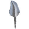
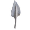
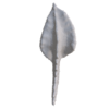
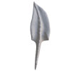
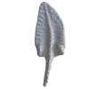
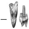
This contribution contains the three-dimensional digital model of one isolated fossil tooth of an anthropoid primate (Ashaninkacebus simpsoni), discovered in sedimentary deposits located on the upper Rio Juruá in State of Acre, Brazil (Western Amazonia). This fossil was described, figured and discussed in the following publication: Marivaux et al. (2023), An eosimiid primate of South Asian affinities in the Paleogene of Western Amazonia and the origin of New World monkeys. Proceedings of the National Academy of Sciences USA. https://doi.org/10.1073/pnas.2301338120
Ashaninkacebus simpsoni UFAC-CS 066 View specimen
|
M3#1114Right first upper molar (rM1), pristine. Type: "3D_surfaces"doi: 10.18563/m3.sf.1114 state:published |
Download 3D surface file |
The present 3D Dataset contains the 3D models analyzed in Assemat et al. 2023: Shape diversity in conodont elements, a quantitative study using 3D topography. Marine Micropaleontology 184. https://doi.org/10.1016/j.marmicro.2023.102292
P1 elements represent dental components of the conodont apparatus that perform the final stage of food processing before ingestion. Consequently, quantifying the shape of P1 elements across the topographic indices of different conodont species becomes crucial for deciphering the diversity in feeding behavior within this group.
Bispathodus aculeatus UM CTB 082 View specimen
|
M3#1404P element Type: "3D_surfaces"doi: 10.18563/m3.sf.1404 state:published |
Download 3D surface file |
Bispathodus aculeatus UM CTB 083 View specimen

|
M3#1405P element Type: "3D_surfaces"doi: 10.18563/m3.sf.1405 state:published |
Download 3D surface file |
Bispathodus aculeatus UM CTB 086 View specimen
|
M3#1406P element Type: "3D_surfaces"doi: 10.18563/m3.sf.1406 state:published |
Download 3D surface file |
Bispathodus ultimus UM CTB 088 View specimen

|
M3#1407P element Type: "3D_surfaces"doi: 10.18563/m3.sf.1407 state:published |
Download 3D surface file |
Bispathodus aculeatus UM CTB 089 View specimen
|
M3#1408P element Type: "3D_surfaces"doi: 10.18563/m3.sf.1408 state:published |
Download 3D surface file |
Bispathodus costatus UM CTB 090 View specimen
|
M3#1409P element Type: "3D_surfaces"doi: 10.18563/m3.sf.1409 state:published |
Download 3D surface file |
Bispathodus ultimus UM CTB 092 View specimen
|
M3#1410P element Type: "3D_surfaces"doi: 10.18563/m3.sf.1410 state:published |
Download 3D surface file |
Bispathodus costatus UM CTB 093 View specimen
|
M3#1411P element Type: "3D_surfaces"doi: 10.18563/m3.sf.1411 state:published |
Download 3D surface file |
Bispathodus spinulicostatus UM CTB 094 View specimen
|
M3#1412P element Type: "3D_surfaces"doi: 10.18563/m3.sf.1412 state:published |
Download 3D surface file |
Bispathodus aculeatus UM CTB 096 View specimen
|
M3#1413P element Type: "3D_surfaces"doi: 10.18563/m3.sf.1413 state:published |
Download 3D surface file |
Bispathodus ultimus UM CTB 098 View specimen
|
M3#1414P element Type: "3D_surfaces"doi: 10.18563/m3.sf.1414 state:published |
Download 3D surface file |
Bispathodus costatus UM CTB 060 View specimen
|
M3#1415P element Type: "3D_surfaces"doi: 10.18563/m3.sf.1415 state:published |
Download 3D surface file |
Bispathodus spinulicostatus UM CTB 073 View specimen
|
M3#1416P element Type: "3D_surfaces"doi: 10.18563/m3.sf.1416 state:published |
Download 3D surface file |
Branmehla suprema UM CTB 049 View specimen

|
M3#1417P element Type: "3D_surfaces"doi: 10.18563/m3.sf.1417 state:published |
Download 3D surface file |
Branmehla inornata UM CTB 100 View specimen
|
M3#1418P element Type: "3D_surfaces"doi: 10.18563/m3.sf.1418 state:published |
Download 3D surface file |
Bispathodus stabilis (morphe 1) UM CTB 101 View specimen

|
M3#1419P element Type: "3D_surfaces"doi: 10.18563/m3.sf.1419 state:published |
Download 3D surface file |
Branmehla suprema UM CTB 102 View specimen

|
M3#1420P element Type: "3D_surfaces"doi: 10.18563/m3.sf.1420 state:published |
Download 3D surface file |
Branmehla suprema UM CTB 103 View specimen

|
M3#1421P element Type: "3D_surfaces"doi: 10.18563/m3.sf.1421 state:published |
Download 3D surface file |
Branmehla suprema UM CTB 104 View specimen
|
M3#1422P element Type: "3D_surfaces"doi: 10.18563/m3.sf.1422 state:published |
Download 3D surface file |
Branmehla suprema UM CTB 105 View specimen

|
M3#1423P element Type: "3D_surfaces"doi: 10.18563/m3.sf.1423 state:published |
Download 3D surface file |
Branmehla suprema UM CTB 106 View specimen
|
M3#1424P element Type: "3D_surfaces"doi: 10.18563/m3.sf.1424 state:published |
Download 3D surface file |
Branmehla suprema UM CTB 072 View specimen

|
M3#1425P element Type: "3D_surfaces"doi: 10.18563/m3.sf.1425 state:published |
Download 3D surface file |
Branmehla suprema UM CTB 107 View specimen
|
M3#1426P element Type: "3D_surfaces"doi: 10.18563/m3.sf.1426 state:published |
Download 3D surface file |
Branmehla suprema UM CTB 108 View specimen

|
M3#1427P element Type: "3D_surfaces"doi: 10.18563/m3.sf.1427 state:published |
Download 3D surface file |
Branmehla suprema UM CTB 109 View specimen
|
M3#1428P element Type: "3D_surfaces"doi: 10.18563/m3.sf.1428 state:published |
Download 3D surface file |
Bispathodus stabilis (morphe 1) UM CTB 110 View specimen

|
M3#1429P element Type: "3D_surfaces"doi: 10.18563/m3.sf.1429 state:published |
Download 3D surface file |
Palmatolepis gracilis UM CTB 112 View specimen

|
M3#1430P element Type: "3D_surfaces"doi: 10.18563/m3.sf.1430 state:published |
Download 3D surface file |
Palmatolepis gracilis UM CTB 061 View specimen

|
M3#1431P element Type: "3D_surfaces"doi: 10.18563/m3.sf.1431 state:published |
Download 3D surface file |
Palmatolepis gracilis UM CTB 115 View specimen
|
M3#1432P element Type: "3D_surfaces"doi: 10.18563/m3.sf.1432 state:published |
Download 3D surface file |
Palmatolepis gracilis UM CTB 116 View specimen

|
M3#1433P element Type: "3D_surfaces"doi: 10.18563/m3.sf.1433 state:published |
Download 3D surface file |
Palmatolepis gracilis UM CTB 117 View specimen
|
M3#1434P element Type: "3D_surfaces"doi: 10.18563/m3.sf.1434 state:published |
Download 3D surface file |
Palmatolepis gracilis UM CTB 062 View specimen
|
M3#1435P element Type: "3D_surfaces"doi: 10.18563/m3.sf.1435 state:published |
Download 3D surface file |
Palmatolepis gracilis UM CTB 118 View specimen
|
M3#1436P element Type: "3D_surfaces"doi: 10.18563/m3.sf.1436 state:published |
Download 3D surface file |
Palmatolepis gracilis UM CTB 119 View specimen

|
M3#1437P element Type: "3D_surfaces"doi: 10.18563/m3.sf.1437 state:published |
Download 3D surface file |
Palmatolepis gracilis UM CTB 120 View specimen
|
M3#1438P element Type: "3D_surfaces"doi: 10.18563/m3.sf.1438 state:published |
Download 3D surface file |
Polygnathus communis UM CTB 075 View specimen
|
M3#1439P element Type: "3D_surfaces"doi: 10.18563/m3.sf.1439 state:published |
Download 3D surface file |
Polygnathus communis UM CTB 121 View specimen
|
M3#1440P element Type: "3D_surfaces"doi: 10.18563/m3.sf.1440 state:published |
Download 3D surface file |
Polygnathus communis UM CTB 122 View specimen
|
M3#1441P element Type: "3D_surfaces"doi: 10.18563/m3.sf.1441 state:published |
Download 3D surface file |
Polygnathus communis UM CTB 123 View specimen
|
M3#1442P element Type: "3D_surfaces"doi: 10.18563/m3.sf.1442 state:published |
Download 3D surface file |
Polygnathus communis UM CTB 125 View specimen
|
M3#1443P element Type: "3D_surfaces"doi: 10.18563/m3.sf.1443 state:published |
Download 3D surface file |
Polygnathus communis UM CTB 126 View specimen
|
M3#1444P element Type: "3D_surfaces"doi: 10.18563/m3.sf.1444 state:published |
Download 3D surface file |
Polygnathus communis UM CTB 128 View specimen
|
M3#1445P element Type: "3D_surfaces"doi: 10.18563/m3.sf.1445 state:published |
Download 3D surface file |
Polygnathus communis UM CTB 130 View specimen
|
M3#1446P element Type: "3D_surfaces"doi: 10.18563/m3.sf.1446 state:published |
Download 3D surface file |
Polygnathus communis UM CTB 131 View specimen
|
M3#1447P element Type: "3D_surfaces"doi: 10.18563/m3.sf.1447 state:published |
Download 3D surface file |
Polygnathus communis UM CTB 132 View specimen
|
M3#1448P element Type: "3D_surfaces"doi: 10.18563/m3.sf.1448 state:published |
Download 3D surface file |
Polygnathus communis UM CTB 133 View specimen
|
M3#1449P element Type: "3D_surfaces"doi: 10.18563/m3.sf.1449 state:published |
Download 3D surface file |
Polygnathus symmetricus UM CTB 139 View specimen
|
M3#1450P element Type: "3D_surfaces"doi: 10.18563/m3.sf.1450 state:published |
Download 3D surface file |
Polygnathus symmetricus UM CTB 140 View specimen
|
M3#1451P element Type: "3D_surfaces"doi: 10.18563/m3.sf.1451 state:published |
Download 3D surface file |
Polygnathus symmetricus UM CTB 141 View specimen
|
M3#1452P element Type: "3D_surfaces"doi: 10.18563/m3.sf.1452 state:published |
Download 3D surface file |
Polygnathus symmetricus UM CTB 142 View specimen
|
M3#1453P element Type: "3D_surfaces"doi: 10.18563/m3.sf.1453 state:published |
Download 3D surface file |

The present 3D Dataset contains the 3D model analyzed in Wazir, W. A., Sehgal, R. K., Čerňanský, A., Patnaik, R., Kumar, N., Singh, A. P. and Singh, N. P. 2022. A find from the Ladakh Himalaya reveals a survival of madtsoiid snakes (Serpentes, Madtsoiidae) in India through the late Oligocene. Journal of Vertebrate Paleontology, 41(6), e2058401. https://doi.org/10.1080/02724634.2021.2058401
indet. indet. WIMF/A 4816 View specimen
|
M3#1754Vertebra Type: "3D_surfaces"doi: 10.18563/m3.sf.1754 state:published |
Download 3D surface file |

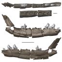
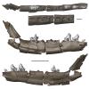
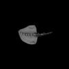
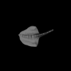
Turtles are one of the most impressive vertebrates. Much of the body is either hidden in a shell or can be drawn into it. Turtles impress with their individual longevity and their often peaceful disposition. Also, with their resilience, they have survived all extinction events since their emergence in the Late Triassic. Today's diversity of shapes is impressive and ranges from the large and high domed Galapagos turtles to the hamster-sized flat pancake turtles. The holotype of one of the oldest fossil turtles, Proganochelys quenstedtii, is housed in the paleontological collection in Tübingen/Germany. Since its discovery some years before 1873, P. quenstedtii has represented the 'prototype' of the turtle and has had an eventful scientific history. It was found in Neuenhaus (Häfner-Neuhausen in Schönbuch forest), Baden-Württemberg, Germany, and stems from Löwenstein-Formation (Weißer Keupersandstein), Late Triassic. The current catalogue number is GPIT-PV-30000. The specimen is listed in the historical inventory “Tübinger Petrefaktenverzeichnis 1841 bis 1896, [folio 326v.]“, as “[catalogue number: PV]16549, Schildkröte Weiser Keupersandstein Hafnerhausen” [turtle from White Keuper Sandstone]. Another, more recent synonym is “GPIT/RE/9396”. The same specimen was presented as uncatalogued by Gaffney (1990). Here we provide a surface scan of the steinkern for easier access of this famous specimen to the scientific community.
Proganochelys quenstedtii GPIT-PV-30000 View specimen
|
M3#967This the surface model of the steinkern of the shell of Proganochelys quenstedtii. Type: "3D_surfaces"doi: 10.18563/m3.sf.967 state:published |
Download 3D surface file |
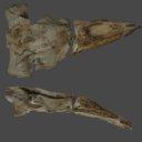
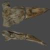
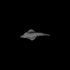
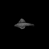
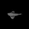
The present 3D Dataset contains the 3D model analyzed in The largest freshwater odontocete: a South Asian river dolphin relative from the Proto-Amazonia.
Pebanista yacuruna MUSM 4017 View specimen
|
M3#1394Holotype skull of Pebanista yacuruna MUSM 4017 Type: "3D_surfaces"doi: 10.18563/m3.sf.1394 state:published |
Download 3D surface file |
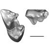
This contribution contains the three-dimensional models of the most complete and/or informative fossil materials attributed to Peradectes crocheti Gernelle, 2024, the earliest peradectid metatherian species of Europe, from its type locality (Palette, Provence, ~55 Ma). These specimens were analyzed and discussed in: Gernelle et al. (2024), Taxonomy and evolutionary history of peradectids (Metatheria): new data from the early Eocene of France. https://doi.org/10.1007/s10914-024-09724-5
Peradectes crocheti MHN.AIX.PV.2018.26.14 View specimen
|
M3#14993D surface model of MHN.AIX.PV.2018.26.14, fragmentary left maxilla with C-P1, anterior root of P2, and M1-M3 Type: "3D_surfaces"doi: 10.18563/m3.sf.1499 state:published |
Download 3D surface file |
Peradectes crocheti MHN.AIX.PV.2017.6.6 View specimen
|
M3#15003D surface model of MHN.AIX.PV.2017.6.6, left P2 Type: "3D_surfaces"doi: 10.18563/m3.sf.1500 state:published |
Download 3D surface file |
Peradectes crocheti MHN.AIX.PV.2017.6.7 View specimen
|
M3#15013D surface model of MHN.AIX.PV.2017.6.7, left M3 Type: "3D_surfaces"doi: 10.18563/m3.sf.1501 state:published |
Download 3D surface file |
Peradectes crocheti MHN.AIX.PV.2017.6.8 View specimen
|
M3#15023D surface model of MHN.AIX.PV.2017.6.8, right hemi-mandible fragment with canine alveolus, posterior root of p1, partial p2, p3, partial m1, and m2-m3 Type: "3D_surfaces"doi: 10.18563/m3.sf.1502 state:published |
Download 3D surface file |
Peradectes crocheti MHN.AIX.PV.2017.6.9 View specimen
|
M3#15033D surface model of MHN.AIX.PV.2017.6.9, leftm1-m4 row with fragments of dentary Type: "3D_surfaces"doi: 10.18563/m3.sf.1503 state:published |
Download 3D surface file |
Peradectes crocheti MHN.AIX.PV.2017.6.14 View specimen
|
M3#15043D surface model of MHN.AIX.PV.2017.6.14, right astragalus Type: "3D_surfaces"doi: 10.18563/m3.sf.1504 state:published |
Download 3D surface file |
Macroevolution is integral to understanding the patterns of the diversification of life. As the life sciences increasingly use big data approaches, large multivariate datasets are required to test fundamental macroevolutionary hypotheses. In vertebrate evolution, large datasets have been created to quantify morphological variation, largely focusing on particular areas of the skeleton. We provide a landmarking protocol to quantify morphological variation in skeletal elements across the head, trunk, hindlimb and forelimb using 3-dimensional landmarks and semilandmarks, and present a large pan-skeletal database of bird morphology for 149 taxa across avian phylogeny using CT scan data. This large collection of 3D models and geometric morphometric data is open access and can be used in the future for new research, teaching and outreach. The 3D models and CT scans of the 149 specimens related to this project can be downloaded at MorphoSource (https://www.morphosource.org/projects/00000C420)
Menura novaehollandiae FMNH 336751 View specimen
|
M3#5613D model of the left carpometacarpus of the superb lyrebird, Menura novaehollandia (displayed as a mirror image in the 3DHOP viewer). Type: "3D_surfaces"doi: 10.18563/m3.sf.561 state:published |
Download 3D surface file |

|
M3#5623D model of the mandible of the superb lyrebird, Menura novaehollandiae. Type: "3D_surfaces"doi: 10.18563/m3.sf.562 state:published |
Download 3D surface file |
|
M3#5633D model of the right coracoid of the superb lyrebird, Menura novaehollandiae. Type: "3D_surfaces"doi: 10.18563/m3.sf.563 state:published |
Download 3D surface file |
|
M3#5643D model of the right scapula of the superb lyrebird, Menura novaehollandiae. Type: "3D_surfaces"doi: 10.18563/m3.sf.564 state:published |
Download 3D surface file |
|
M3#5653D model of the right tarsometatarsus of the superb lyrebird, Menura novaehollandiae. Type: "3D_surfaces"doi: 10.18563/m3.sf.565 state:published |
Download 3D surface file |
|
M3#5663D model of the sternum of the superb lyrebird, Menura novaehollandiae. Type: "3D_surfaces"doi: 10.18563/m3.sf.566 state:published |
Download 3D surface file |
|
M3#5673D model of the left femur of the superb lyrebird, Menura novaehollandiae (displayed as a mirror image in the 3DHOP viewer). Type: "3D_surfaces"doi: 10.18563/m3.sf.567 state:published |
Download 3D surface file |
|
M3#5683D model of the skull of the superb lyrebird, Menura novaehollandiae. Type: "3D_surfaces"doi: 10.18563/m3.sf.568 state:published |
Download 3D surface file |
|
M3#5693D model of the left humerus of the superb lyrebird, Menura novaehollandiae (displayed as a mirror image in the 3DHOP viewer). Type: "3D_surfaces"doi: 10.18563/m3.sf.569 state:published |
Download 3D surface file |
|
M3#5703D model of the synsacrum of the superb lyrebird, Menura novaehollandiae. Type: "3D_surfaces"doi: 10.18563/m3.sf.570 state:published |
Download 3D surface file |
|
M3#5713D model of the left radius of the superb lyrebird, Menura novaehollandiae (displayed as a mirror image in the 3DHOP viewer). Type: "3D_surfaces"doi: 10.18563/m3.sf.571 state:published |
Download 3D surface file |

|
M3#5723D model of the left tibiotarsus of the superb lyrebird, Menura novaehollandiae (displayed as a mirror image in the 3DHOP viewer). Type: "3D_surfaces"doi: 10.18563/m3.sf.572 state:published |
Download 3D surface file |
|
M3#5733D model of the left ulna of the superb lyrebird, Menura novaehollandiae (displayed as a mirror image in the 3DHOP viewer). Type: "3D_surfaces"doi: 10.18563/m3.sf.573 state:published |
Download 3D surface file |


