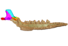
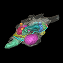
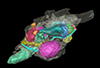
3D models of Kalakocetus, the earliest Cetacea
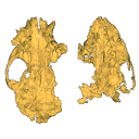
The specimens of Speothos pacivorus
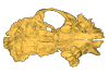
3D models related to the publication: Hidden diversity of Palaeogene metatherians: a new family of polydolopimorphian marsupials from Peruvian Amazonia
3D GM dataset of bird skeletal variation
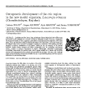
Skeletal embryonic development in the catshark
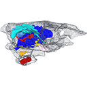
Bony connexions of the petrosal bone of extant hippos
bony labyrinth (14) , inner ear (11) , Eocene (11) , geometric morphometrics (10) , CT-scan (10) , Oligocene (9) , Micro-CT (9)
Maëva Judith Orliac (24) , Lionel Hautier (24) , Laurent Marivaux (18) , Renaud Lebrun (15) , Rodolphe Tabuce (14) , Pierre-Olivier Antoine (13) , Bastien Mennecart (13)
|
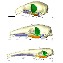
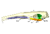
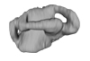
3D models related to the publication: “Comparative masticatory myology in anteaters and its implications for interpreting morphological convergence in myrmecophagous placentals”Sérgio Ferreira-Cardoso, Pierre-Henri Fabre
Published online: 29/07/2020 |
|
M3#522Skull, mandible, and muscles of Cyclopes didactylus Type: "3D_surfaces"doi: 10.18563/m3.sf.522 state:published |
Download 3D surface file |
Tamandua tetradactyla M3075_JAG View specimen

|
M3#524Skull, left mandibles, and muscles of Tamandua tetradactyla. Type: "3D_surfaces"doi: 10.18563/m3.sf.524 state:published |
Download 3D surface file |
Myrmecophaga tridactyla M3023_JAG View specimen
|
M3#523Skull, left mandible and muscles of Myrmecophaga tridactyla. Type: "3D_surfaces"doi: 10.18563/m3.sf.523 state:published |
Download 3D surface file |
The present 3D Dataset contains the 3D models analyzed in the following publication: Georgalis, G. L., and T. M. Scheyer. A new species of Palaeopython (Serpentes) and other extinct squamates from the Eocene of Dielsdorf (Zurich, Switzerland). Swiss Journal of Geosciences (in press). https://doi.org/10.1007/s00015-019-00341-6
Palaeopython helveticus PIMUZ A/III 631 View specimen
|
M3#399ZIP file containing .ply of vertebra PIMUZ A/III 631 from Palaeopython helveticus n. sp. Type: "3D_surfaces"doi: 10.18563/m3.sf.399 state:published |
Download 3D surface file |
|
M3#403dataset of snake vertebra PIMUZ A/III 631 Type: "3D_CT"doi: 10.18563/m3.sf.403 state:published |
Download CT data |
Palaeopython helveticus PIMUZ A/III 634 View specimen
|
M3#400ZIP file containing .ply of vertebra PIMUZ A/III 634 from Palaeopython helveticus n. sp. (holotype) Type: "3D_surfaces"doi: 10.18563/m3.sf.400 state:published |
Download 3D surface file |
|
M3#404dataset of snake vertbra PIMUZ A/III 634 (holotype) Type: "3D_CT"doi: 10.18563/m3.sf.404 state:published |
Download CT data |
Palaeopython helveticus PIMUZ A/III 636 View specimen
|
M3#401ZIP file containing .ply of vertebra PIMUZ A/III 636 from Palaeopython helveticus n. sp. Type: "3D_surfaces"doi: 10.18563/m3.sf.401 state:published |
Download 3D surface file |
|
M3#406dataset of snake vertebra PIMUZ A/III 636 Type: "3D_CT"doi: 10.18563/m3.sf.406 state:published |
Download CT data |
Palaeovaranus sp. PIMUZ A/III 234 View specimen
|
M3#402ZIP file containing .ply of dentary PIMUZ A/III 234 of Palaeovaranus sp. Type: "3D_surfaces"doi: 10.18563/m3.sf.402 state:published |
Download 3D surface file |

|
M3#405dataset of dentary of Palaeovaranus sp. (PIMUZ A/III 234) Type: "3D_CT"doi: 10.18563/m3.sf.405 state:published |
Download CT data |
The present 3D Dataset contains the 3D models analyzed in the publication ‘Ontogenetic development of the otic region in the new model organism, Leucoraja erinacea (Chondrichthyes; Rajidae)’, https://doi.org/10.1017/S1755691018000993
Leucoraja erinacea 2018.9.26.1 View specimen
|
M3#3673D model of the right skeletal labyrinth of the adult specimen of Leucoraja erincea. T Type: "3D_surfaces"doi: 10.18563/m3.sf.367 state:published |
Download 3D surface file |
Leucoraja erinacea 2018.9.25.2 View specimen
|
M3#3683D model of the right skeletal labyrinth of the stage 34 specimen of Leucoraja erincea. Type: "3D_surfaces"doi: 10.18563/m3.sf.368 state:published |
Download 3D surface file |
Leucoraja erinacea 2018.9.25.3 View specimen
|
M3#3693D model of the right skeletal labyrinth of the stage 32 specimen of Leucoraja erinacea. Type: "3D_surfaces"doi: 10.18563/m3.sf.369 state:published |
Download 3D surface file |
|
M3#3723D model of the right membranous system of stage 32 of Leucoraja erincea. Type: "3D_surfaces"doi: 10.18563/m3.sf.372 state:published |
Download 3D surface file |
Leucoraja erinacea 2018.9.25.4 View specimen
|
M3#3703D model of the right skeletal labyrinth of the stage 31 specimen of Leucoraja erinacea. Type: "3D_surfaces"doi: 10.18563/m3.sf.370 state:published |
Download 3D surface file |
Leucoraja erinacea 2018.9.26.5 View specimen

|
M3#3763D model of the right skeletal labyrinth of the stage 29 specimen of Leucoraja erinacea. Type: "3D_surfaces"doi: 10.18563/m3.sf.376 state:published |
Download 3D surface file |
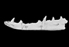
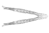
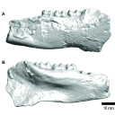
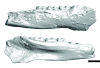
This note presents the 3D model of the hemi-mandible UM-PAT 159 of the MP7 Diacodexis species D. cf. gigasei and 3D models corresponding to the restoration of the ascending ramus, broken on the original specimen, and to a restoration of a complete mandible based on the preserved left hemi-mandible.
Diacodexis cf. gigasei UMPAT159 View specimen
|
M3#3153D models of UM PAT 159 after the restoration of the ascending ramus Type: "3D_surfaces"doi: 10.18563/m3.sf.315 state:published |
Download 3D surface file |
|
M3#316restoration of a complete mandible based on the preserved left hemi-mandible UM PAT 159 Type: "3D_surfaces"doi: 10.18563/m3.sf.316 state:published |
Download 3D surface file |
|
M3#3173D model of the hemi-mandible UM PAT 159 Type: "3D_surfaces"doi: 10.18563/m3.sf.317 state:published |
Download 3D surface file |
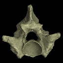
This contribution contains the 3D model described and figured in the following publication: Albino, A., Carrillo-Briceño, J. D. & Neenan, J. M. 2016. An enigmatic aquatic snake from the Cenomanian of northern South America. PeerJ 4:e2027 http://dx.doi.org/10.7717/peerj.2027
Lunaophis aquaticus MCNC-1827-F View specimen

|
M3#116Articulated precloacal vertebrae of Lunaophis aquaticus Type: "3D_surfaces"doi: 10.18563/m3.sf.116 state:published |
Download 3D surface file |
This contribution contains the 3D models described and figured in the following publication: Orliac M.J., Karadenizli L., Antoine P.-O., Sen S. 2015. Small suids (Mammalia, Artiodactyla) from the late Early Miocene of Turkey and a short overview of Early Miocene small suoids in the Old World. Paleontologia electronica 18(2): 18.2.30A: 1-48. https://doi.org/10.26879/547
?Nguruwe galaticum SMT-1 View specimen
|
M3#16fragment of palate with left broken M1-M3 Type: "3D_surfaces"doi: 10.18563/m3.sf16 state:published |
Download 3D surface file |
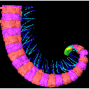
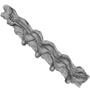
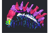
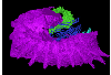
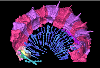
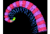
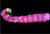
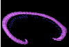
The present 3D Dataset contains the 3D models analyzed in the publication Fossils from the Montceau-les-Mines Lagerstätte (305 Ma) shed light on the anatomy, ecology and phylogeny of Carboniferous millipedes. Authors: Lheritier Mickael, Perroux Maëva, Vannier Jean, Escarguel Gilles, Wesener Thomas, Moritz Leif, Chabard Dominique, Adrien Jerome and Perrier Vincent. Journal of Systematics Palaeontology. https://doi.org/10.1080/14772019.2023.2169891
Amynilyspes fatimae MNHN.F.SOT.2134 View specimen
|
M3#1073Nearly complete specimen. Type: "3D_surfaces"doi: 10.18563/m3.sf.1073 state:published |
Download 3D surface file |
Amynilyspes fatimae MNHN.F.SOT.14983 View specimen
|
M3#1074Nearly complete specimen. Type: "3D_surfaces"doi: 10.18563/m3.sf.1074 state:published |
Download 3D surface file |
Amynilyspes fatimae MNHN.F.SOT.2129 View specimen
|
M3#1075Nearly complete specimen. Type: "3D_surfaces"doi: 10.18563/m3.sf.1075 state:published |
Download 3D surface file |
Blanzilius parriati MNHN.F.SOT.2114A View specimen
|
M3#1076Front part. Type: "3D_surfaces"doi: 10.18563/m3.sf.1076 state:published |
Download 3D surface file |
Blanzilius parriati MNHN.F.SOT.5148 View specimen
|
M3#1077Front part. Type: "3D_surfaces"doi: 10.18563/m3.sf.1077 state:published |
Download 3D surface file |
Blanzilius parriati MNHN.F.SOT.2113 View specimen
|
M3#1078Fragment with legs, sternites and possible tracheal openings. Type: "3D_surfaces"doi: 10.18563/m3.sf.1078 state:published |
Download 3D surface file |
Blanzilius parriati MNHN.F.SOT.81522 View specimen
|
M3#1079Nealry complete specimen. Type: "3D_surfaces"doi: 10.18563/m3.sf.1079 state:published |
Download 3D surface file |
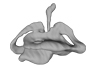
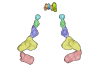
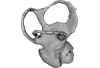
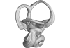
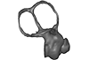
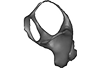
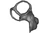
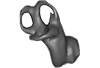
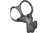
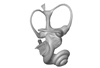
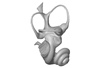
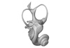
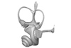
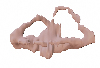
The present Dataset contains the 3D model of the male genital organs of greater horseshoe bat, Rhinolophus ferrumequinum. This is the first detailed 3D structure of the soft-tissue genital organs of bats. The 3D model was generated using microCT and techniques of virtual reconstruction.
Rhinolophus ferrumequinum JP18-006 View specimen
|
M3#521The genital organs of male greater horseshoe bat. Type: "3D_surfaces"doi: 10.18563/m3.sf.521 state:published |
Download 3D surface file |
The present 3D Dataset contains the 3D models described and figured in the following publication: Grohé C., Bonis L. de, Chaimanee Y., Chavasseau O., Rugbumrung M., Yamee C., Suraprasit K., Gibert C., Surault J., Blondel C., Jaeger J.-J. 2020. The late middle Miocene Mae Moh Basin of northern Thailand: the richest Neogene assemblage of Carnivora from Southeast Asia and a paleobiogeographic analysis of Miocene Asian carnivorans. American Museum Novitates. http://digitallibrary.amnh.org/handle/2246/7223
Siamogale bounosa MM-54 View specimen
|
M3#5053D model of the skull of Siamogale bounosa The zip file contains: - the 3D surface in PLY - the orientation files in .pos and .ori - the project in .ntw Type: "3D_surfaces"doi: 10.18563/m3.sf.505 state:published |
Download 3D surface file |
Vishnuonyx maemohensis MM-78 View specimen
|
M3#5063D model of the skull of Vishnuonyx maemohensis The zip file contains: - the 3D surface in PLY - the orientation files in .pos and .ori - the project in .ntw Type: "3D_surfaces"doi: 10.18563/m3.sf.506 state:published |
Download 3D surface file |
|
M3#5073D model of the reconstructed upper teeth of Vishnuonyx maemohensis The zip file contains: - the 3D surface in PLY - the orientation files in .pos and .ori - the project in .ntw Type: "3D_surfaces"doi: 10.18563/m3.sf.507 state:published |
Download 3D surface file |
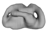
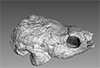
The present 3D Dataset contains the 3D model of a left dentary with m1-m3 analyzed in “A new fossil of Tayassuidae (Mammalia: Certartiodactyla) from the Pleistocene of northern Brazil”. The 3D model was generated using a laser scanning.
cf. Pecari tajacu UFSM 11606 View specimen
|
M3#498Left dentary with m1-m3 Type: "3D_surfaces"doi: 10.18563/m3.sf.498 state:published |
Download 3D surface file |
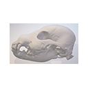
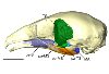
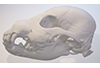
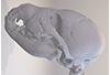
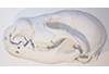
This contribution comprises the 3D models of three wolf pup skulls, which were used for the publication by Geiger et al. 2017 on Neomorphosis and heterochrony of skull shape in dog domestication.
Canis lupus CLL2 View specimen
|
M3#3123d model of a wolf pup skull Type: "3D_surfaces"doi: 10.18563/m3.sf.312 state:published |
Download 3D surface file |
Canis lupus CLL4 View specimen
|
M3#3133d model of a wolf pup skull Type: "3D_surfaces"doi: 10.18563/m3.sf.313 state:published |
Download 3D surface file |
Canis lupus CLL5 View specimen
|
M3#3143d model of a wolf pup skull Type: "3D_surfaces"doi: 10.18563/m3.sf.314 state:published |
Download 3D surface file |
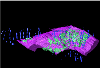
The present 3D Dataset contains the 3D models analyzed in: "a giant dapediid from the Late Triassic of Switzerland and insights into neopterygian phylogeny", Royal Society Open Science, https://doi.org/10.1098/rsos.180497
Scopulipiscis saxciput PIMUZ A/I 3026 View specimen
|
M3#1773D surfaces of the skull and endocranial spaces inside neurocranium, including the aortic canal, braincase, fossa bridgei, lateral cranial canal, nerves and other passageways, notochord, posterior myodome, and right semicircular canals. Type: "3D_surfaces"doi: 10.18563/m3.sf.177 state:published |
Download 3D surface file |

|
M3#178Scan of the neurocranium of PIMUZ A/I 3026 Type: "3D_CT"doi: 10.18563/m3.sf.178 state:published |
Download CT data |
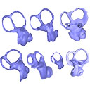
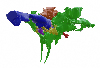
Here, the semicircular canals of the most aquatic seal, the rare Antarctic Ross Seal (Ommatophoca rossii), are presented for the first time, along with representatives of every species in the Lobodontini: the leopard seal (Hydrurga leptonyx), Weddell seal (Leptonychotes weddellii), and crabeater seal (Lobodon carcinophagus). Because encounters with wild Ross seal are rare, and few specimens are available in collections worldwide, this dataset increases accessibility to a rare species. For further comparison, we present the bony labyrinths of other carnivorans, the elephant seal (Mirounga leonina), harbor seal (Phoca vitulina), walrus (Odobenus rosmarus), South American sea lion (Otaria byronia).
Odobenus rosmarus MVZ 125566 View specimen
|
M3#173Surface of the semicircular canals and cochlea of the walrus, Odobenus rosmarus Type: "3D_surfaces"doi: 10.18563/m3.sf.173 state:published |
Download 3D surface file |
Phoca vitulina UZNH 17973 View specimen
|
M3#174Endocast surface of the semicircular canals and cochlea of the harbor seal, Phoca vitulina. Type: "3D_surfaces"doi: 10.18563/m3.sf.174 state:published |
Download 3D surface file |
Hydrurga leptonyx MLP 14.IV.48.11 View specimen
|
M3#285Endocast surface of the semicircular canals and cochlea of the leopard seal, Hydrurga leptonyx. Type: "3D_surfaces"doi: 10.18563/m3.sf.285 state:published |
Download 3D surface file |
Leptonychotes weddellii IAA 02-13 View specimen
|
M3#288Endocast surface of the semicircular canals and cochlea of the Weddell seal Leptonychotes weddellii. Type: "3D_surfaces"doi: 10.18563/m3.sf.288 state:published |
Download 3D surface file |
Lobodon carcinophagus IAA 530 View specimen
|
M3#286Endocast surface of the semicircular canals and cochlea of the crabeater seal, Lobodon carcinophagus. Type: "3D_surfaces"doi: 10.18563/m3.sf.286 state:published |
Download 3D surface file |
Ommatophoca rossii MACN 48259 View specimen
|
M3#176Endocast surface of the semicircular canals and cochlea of the Ross seal Ommatophoca rossii. Type: "3D_surfaces"doi: 10.18563/m3.sf.176 state:published |
Download 3D surface file |
Mirounga leonina IAA 03-5 View specimen
|
M3#287Right endocast surface of the semicircular canals and cochlea of the elephant seal, Mirounga leonina. Type: "3D_surfaces"doi: 10.18563/m3.sf.287 state:published |
Download 3D surface file |
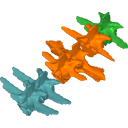
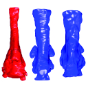
The present 3D Dataset contains the 3D models analyzed in Costeur L., Mennecart B., Müller B., Schulz G., 2016. Prenatal growth stages show the development of the ruminant bony labyrinth and petrosal bone. Journal of Anatomy. https://doi.org/10.1111/joa.12549
Bos taurus NMB3038 View specimen
|
M3#124Right bony labyrinth of a Bos taurus foetus (gestational age 115 days) Type: "3D_surfaces"doi: 10.18563/m3.sf.124 state:published |
Download 3D surface file |
Bos taurus NMB3367 View specimen
|
M3#125Right bony labyrinth of a Bos taurus foetus (gestational age 165 days) Type: "3D_surfaces"doi: 10.18563/m3.sf.125 state:published |
Download 3D surface file |
Bos taurus NMB3365 View specimen
|
M3#126Right bony labyrinth of a Bos taurus foetus (gestational age 210 days) Type: "3D_surfaces"doi: 10.18563/m3.sf.126 state:published |
Download 3D surface file |
Bos taurus NMB2855 View specimen
|
M3#127Right bony labyrinth of a Bos taurus foetus (gestational age 260 days) Type: "3D_surfaces"doi: 10.18563/m3.sf.127 state:published |
Download 3D surface file |
Bos taurus NMB1037 View specimen
|
M3#128Left bony labyrinth of an adult Bos taurus Type: "3D_surfaces"doi: 10.18563/m3.sf.128 state:published |
Download 3D surface file |
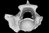
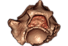
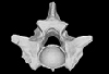
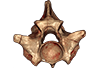
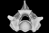
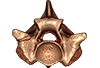
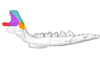
This contribution contains the 3D models described and figured in the following publication: Molnar, JL, Pierce, SE, Bhullar, B-A, Turner, AH, Hutchinson, JR (accepted). Morphological and functional changes in the crocodylomorph vertebral column with increasing aquatic adaptation. Royal Society Open Science.
Protosuchus richardsoni AMNH-VP 3024 View specimen

|
M3#448th and 9th dorsal vertebrae, 1st and 2nd lumbar vertebrae, and 5th lumbar and sacral vertebrae. Type: "3D_surfaces"doi: 10.18563/m3.sf44 state:published |
Download 3D surface file |
Terrestrisuchus gracilis NHM-PV R 7562 View specimen

|
M3#451st and 2nd lumbar vertebrae, and 5th lumbar and sacral vertebrae Type: "3D_surfaces"doi: 10.18563/m3.sf45 state:published |
Download 3D surface file |
Pelagosaurus typus NHM-PV OR 32598 View specimen

|
M3#467th and 8th dorsal vertebrae, 11th and 12th dorsal vertebrae, 15th dorsal vertebra and sacral vertebra. Type: "3D_surfaces"doi: 10.18563/m3.sf46 state:published |
Download 3D surface file |
Metriorhynchus superciliosus NHM-PV R 2054 View specimen

|
M3#476th and 7th dorsal vertebrae, 10th and 11th dorsal vertebrae, 17th dorsal vertebra and sacral vertebra Type: "3D_surfaces"doi: 10.18563/m3.sf47 state:published |
Download 3D surface file |
Crocodylus niloticus FNC0 View specimen

|
M3#487th and 8th dorsal vertebrae, 1st and 2nd lumbar vertebrae, 5th lumbar vertebra and sacral vertebra. Type: "3D_surfaces"doi: 10.18563/m3.sf48 state:published |
Download 3D surface file |
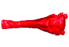
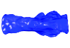
The present 3D Dataset contains the 3D model analyzed in Presence of the ground sloth Valgipes bucklandi (Xenarthra, Folivora, Scelidotheriinae) in southern Uruguay during the Late Pleistocene: Ecological and biogeographical implications. Quaternary International. https://doi.org/10.1016/j.quaint.2021.06.011
Valgipes bucklandi CAV 1573 View specimen
|
M3#797Left tibia-fibula Type: "3D_surfaces"doi: 10.18563/m3.sf.797 state:published |
Download 3D surface file |
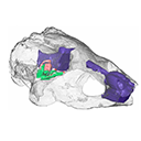
The present 3D Dataset contains the 3D model analyzed in the following publication: Paulina-Carabajal, A., Sterli, J., Werneburg, I., 2019. The endocranial anatomy of the stem turtle Naomichelys speciosa from the Early Cretaceous of North America. Acta Palaeontologica Polonica, https://doi.org/10.4202/app.00606.2019
Naomichelys speciosa FMNH PR273 View specimen
|
M3#428FMNH_PR273_1 - Naomichlys speciosa - skull Type: "3D_surfaces"doi: 10.18563/m3.sf.428 state:published |
Download 3D surface file |
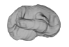
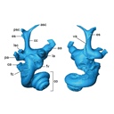
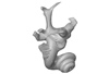
The present 3D Dataset contains the 3D models of brain endocast of traversodontid cynodonts studied in: Pavanatto et al. 2019. Virtual reconstruction of cranial endocasts of traversodontid cynodonts (Eucynodontia: Gomphodontia) from the upper Triassic of Southern Brazil. Journal of Morphology. https://doi.org/10.1002/jmor.21029
Siriusgnathus niemeyerorum CAPPA/UFSM 0032 View specimen
|
M3#4253D model of the brain endocast Type: "3D_surfaces"doi: 10.18563/m3.sf.425 state:published |
Download 3D surface file |
Exaeretodon riograndensis CAPPA/UFSM 0030 View specimen

|
M3#4263D model of the brain endocast Type: "3D_surfaces"doi: 10.18563/m3.sf.426 state:published |
Download 3D surface file |
Exaeretodon riograndensis CAPPA/UFSM 0227 View specimen
|
M3#4273D model of the brain endocast Type: "3D_surfaces"doi: 10.18563/m3.sf.427 state:published |
Download 3D surface file |
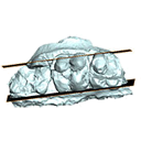
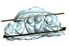
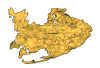
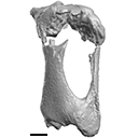
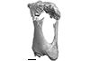
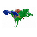
This contribution contains the 3D model described and figured in the following publication: Hautier L, Sarr R, Lihoreau F, Tabuce R, Marwan Hameh P. 2014. First record of the family Protocetidae in the Lutetian of Senegal (West Africa). Palaeovertebrata 38(2)-e2
indet. indet. SN103 View specimen

|
M3#5SN103, partial left innominate. Age and occurrence – Taïba Formation, Lutetian of the near Taïba Ndiaye, quarry of the Industries Chimiques du Sénégal (ICS) Type: "3D_surfaces"doi: 10.18563/m3.sf5 state:published |
Download 3D surface file |
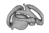
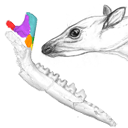
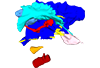
The present 3D Dataset contains the 3D models analyzed in Pochat-Cottilloux Y., Rinder N., Perrichon G., Adrien J., Amiot R., Hua S. & Martin J. E. (2023). The neuroanatomy and pneumaticity of Hamadasuchus from the Cretaceous of Morocco and its significance for the paleoecology of Peirosauridae and other altirostral crocodylomorphs. Journal of Anatomy, https://doi.org/10.1111/joa.13887
Hamadasuchus sp. UCBL-FSL 532408 View specimen
|
M3#10943D volume reconstruction of the braincase osteology Type: "3D_surfaces"doi: 10.18563/m3.sf.1094 state:published |
Download 3D surface file |
|
M3#10963D volume reconstruction of the endocast Type: "3D_surfaces"doi: 10.18563/m3.sf.1096 state:published |
Download 3D surface file |
|
M3#10973D volume reconstruction of the labyrinths Type: "3D_surfaces"doi: 10.18563/m3.sf.1097 state:published |
Download 3D surface file |
|
M3#10983D volume reconstruction of the pneumatic cavities Type: "3D_surfaces"doi: 10.18563/m3.sf.1098 state:published |
Download 3D surface file |


