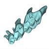
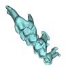
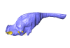
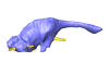
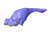
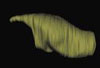
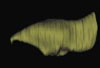
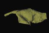
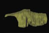
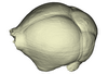
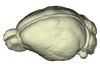
3D models of Kalakocetus, the earliest Cetacea
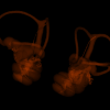
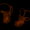
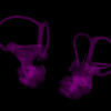
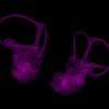
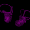
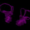
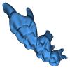
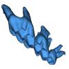
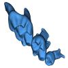
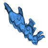
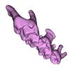
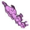
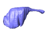
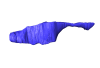
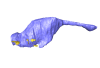
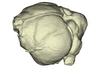
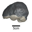
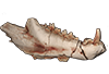
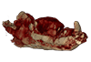
The specimens of Speothos pacivorus
3D models related to the publication: Hidden diversity of Palaeogene metatherians: a new family of polydolopimorphian marsupials from Peruvian Amazonia
3D GM dataset of bird skeletal variation
Skeletal embryonic development in the catshark
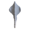
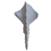
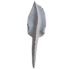
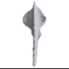
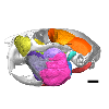
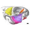
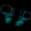
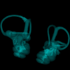
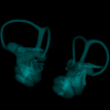
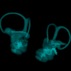
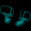
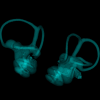
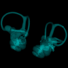
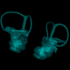
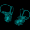
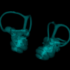
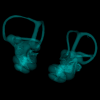
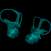
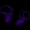
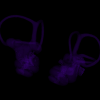
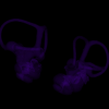
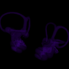
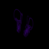
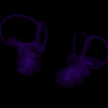
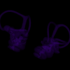
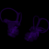
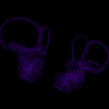
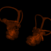
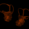
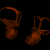
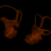
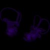
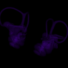
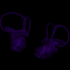
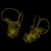
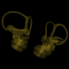
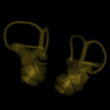
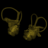
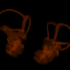
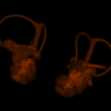
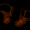
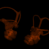
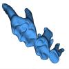
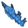
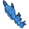
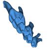
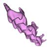
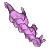
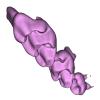
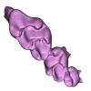
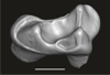
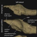
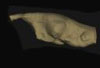
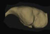
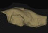
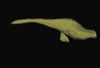
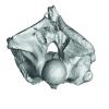
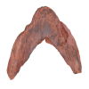
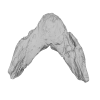
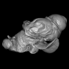
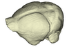
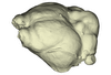
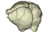
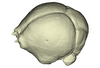
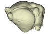
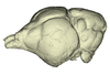
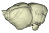
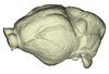
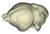
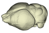
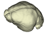
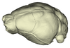
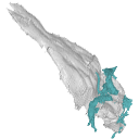
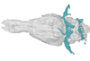
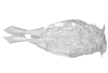
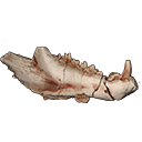
Bony connexions of the petrosal bone of extant hippos
bony labyrinth (14) , inner ear (11) , Eocene (11) , geometric morphometrics (10) , CT-scan (10) , Oligocene (9) , Micro-CT (9)
Maëva Judith Orliac (24) , Lionel Hautier (24) , Laurent Marivaux (18) , Renaud Lebrun (15) , Rodolphe Tabuce (14) , Pierre-Olivier Antoine (13) , Bastien Mennecart (13)
|
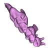
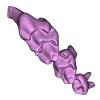
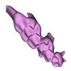
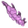
3D models related to the publication: Convergent allometric trajectories in Devonian-Carboniferous unornamented Polygnathus conodontsFélix Nesme
Published online: 25/04/2025 |
|
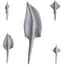
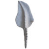
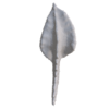
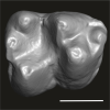
M3#1611Dextral P1 element Type: "3D_surfaces"doi: 10.18563/m3.sf.1611 state:published |
Download 3D surface file |
Polygnathus sp. UM-PSQ-011 View specimen
|
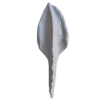
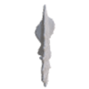
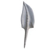
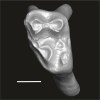
M3#1612Sinistral P1 element Type: "3D_surfaces"doi: 10.18563/m3.sf.1612 state:published |
Download 3D surface file |
Polygnathus sp. UM-PSQ-012 View specimen
|
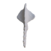
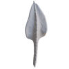
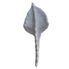
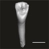
M3#1613Sinistral P1 element Type: "3D_surfaces"doi: 10.18563/m3.sf.1613 state:published |
Download 3D surface file |
Polygnathus sp. UM-PSQ-013 View specimen
|
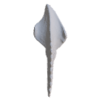
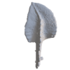
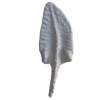
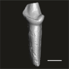
M3#1614Dextral P1 element Type: "3D_surfaces"doi: 10.18563/m3.sf.1614 state:published |
Download 3D surface file |
Polygnathus sp. UM-PSQ-014 View specimen
|
M3#1615Sinistral P1 element Type: "3D_surfaces"doi: 10.18563/m3.sf.1615 state:published |
Download 3D surface file |
Polygnathus sp. UM-PSQ-015 View specimen
|
M3#1616Sinistral P1 element Type: "3D_surfaces"doi: 10.18563/m3.sf.1616 state:published |
Download 3D surface file |
Polygnathus sp. UM-PSQ-016 View specimen
|
M3#1617Dextral P1 element Type: "3D_surfaces"doi: 10.18563/m3.sf.1617 state:published |
Download 3D surface file |
Polygnathus sp. UM-PSQ-017 View specimen
|
M3#1618Dextral P1 element Type: "3D_surfaces"doi: 10.18563/m3.sf.1618 state:published |
Download 3D surface file |
Polygnathus sp. UM-PSQ-018 View specimen
|
M3#1619Sinistral P1 element Type: "3D_surfaces"doi: 10.18563/m3.sf.1619 state:published |
Download 3D surface file |
Polygnathus sp. UM-PSQ-019 View specimen
|
M3#1620Sinistral P1 element Type: "3D_surfaces"doi: 10.18563/m3.sf.1620 state:published |
Download 3D surface file |
Polygnathus sp. UM-PSQ-020 View specimen

|
M3#1621Dextral P1 element Type: "3D_surfaces"doi: 10.18563/m3.sf.1621 state:published |
Download 3D surface file |
Polygnathus sp. UM-PSQ-021 View specimen
|
M3#1622Dextral P1 element Type: "3D_surfaces"doi: 10.18563/m3.sf.1622 state:published |
Download 3D surface file |
Polygnathus sp. UM-PSQ-022 View specimen
|
M3#1623Sinistral P1 element Type: "3D_surfaces"doi: 10.18563/m3.sf.1623 state:published |
Download 3D surface file |
Polygnathus sp. UM-PSQ-023 View specimen
|
M3#1624Sinistral P1 element Type: "3D_surfaces"doi: 10.18563/m3.sf.1624 state:published |
Download 3D surface file |
Polygnathus sp. UM-PSQ-024 View specimen
|
M3#1625Dextral P1 element Type: "3D_surfaces"doi: 10.18563/m3.sf.1625 state:published |
Download 3D surface file |
Polygnathus sp. UM-PSQ-025 View specimen
|
M3#1626Dextral P1 element Type: "3D_surfaces"doi: 10.18563/m3.sf.1626 state:published |
Download 3D surface file |
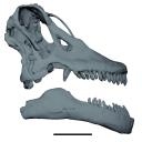
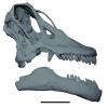
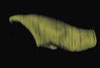
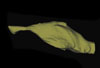
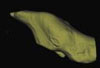
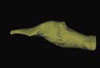
The study of titanosaur paleobiology has been severely hampered by the incomplete nature of their fossil record, particularly the scarcity of well-preserved and relatively complete cranial remains. Even the most complete titanosaur skulls are often fractured, incomplete, or deformed, which has resulted in a limited knowledge of the paleobiology related to cranial anatomy, especially functional morphology. In this context, we present the digital restoration of the skull of the Argentinean titanosaur Sarmientosaurus musacchioi, created using the open-source 3D modeling software Blender. The digitally restored model is freely accessible to other researchers, facilitating broader research and comparative studies.
Sarmientosaurus mussacchioi MDT-PV 02 View specimen
|
M3#1594Cranium and mandible of Sarmientosaurus mussacchioi Type: "3D_surfaces"doi: 10.18563/m3.sf.1594 state:published |
Download 3D surface file |
|
M3#1599Original object provided by Gabriel Casal (cranium) Type: "3D_surfaces"doi: 10.18563/m3.sf.1599 state:published |
Download 3D surface file |

This contribution contains the 3D model described and figured in the following publication: Martin, T., Averianov, A. O., Schultz, J. A., & Schwermann, A. H. (2023). A stem therian mammal from the Lower Cretaceous of Germany. Journal of Vertebrate Paleontology, e2224848.
Spelaeomolitor speratus WMNM P99101 View specimen
|
M3#12573D_model_Spelaeomolitor_lower_molar Type: "3D_surfaces"doi: 10.18563/m3.sf.1257 state:published |
Download 3D surface file |
|
M3#1258CT imagestack (jpgs) and info data sheet (pca file) in one zip folder Type: "3D_CT"doi: 10.18563/m3.sf.1258 state:published |
Download CT data |
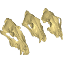
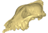
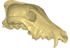
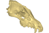
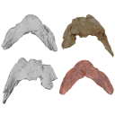
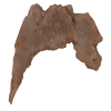
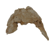
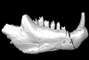
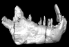
Archaeozoological studies are increasingly using new methods and approaches to explore questions about domestication. Here, we provide 3D models of three archaeological Canis lupus skulls from Belgium originating from the sites of Goyet (31,680±250BP; 31,890+240/-220BP), Trou des Nutons (21,810±90BP) and Trou Balleux (postglacial). Since their identification as either wolves or early dogs is still debated, we present these models as additional tools for further investigating their evolutionary history and the history of dog domestication.
Canis lupus Goyet 2860 View specimen
|
M3#213D surface model of the cranium of the Late Pleistocene Canis lupus "Goyet 2860" from the Royal Belgian Institute of Natural Sciences. Type: "3D_surfaces"doi: 10.18563/m3.sf21 state:published |
Download 3D surface file |
Canis lupus Trou Balleux no-nr View specimen
|
M3#223D surface model of the cranium of the Late Pleistocene Canis lupus "Trou Balleux no-nr" from the University of Liège, Belgium Type: "3D_surfaces"doi: 10.18563/m3.sf22 state:published |
Download 3D surface file |
Canis lupus Trou des Nutons 2559-1 View specimen
|
M3#233D surface model of the cranium of the Late Pleistocene Canis lupus "Trou des Nutons 2559-1" from the Royal Belgian Institute of Natural Sciences. Type: "3D_surfaces"doi: 10.18563/m3.sf23 state:published |
Download 3D surface file |
This contribution contains the 3D model(s) described and figured in the following publication: Da Cunha, L., Fabre, P.-H. & Hautier, L. (2024) Springhares, flying and flightless scaly-tailed squirrels (Anomaluromorpha, Rodentia) are the squirrely mouse: comparative anatomy of the masticatory musculature and its implications on the evolution of hystricomorphy in rodents. Journal of Anatomy, 244, 900–928.
Anomalurus derbianus 21804 View specimen
|
M3#1493Masticatory apparatus of Anomalurus Type: "3D_surfaces"doi: 10.18563/m3.sf.1493 state:published |
Download 3D surface file |
Idiurus macrotis 29335 View specimen
|
M3#1492Masticatory apparatus of Idiurus Type: "3D_surfaces"doi: 10.18563/m3.sf.1492 state:published |
Download 3D surface file |
Zenkerella insignis 5.5.23.27 View specimen
|
M3#1490Masticatory apparatus of Zenkerella Type: "3D_surfaces"doi: 10.18563/m3.sf.1490 state:published |
Download 3D surface file |
Pedetes capensis NA View specimen
|
M3#1491Masticatory apparatus of Pedetes Type: "3D_surfaces"doi: 10.18563/m3.sf.1491 state:published |
Download 3D surface file |
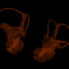
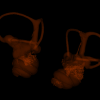
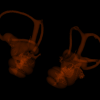
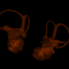
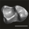
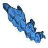
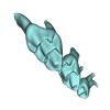
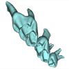
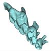
This contribution contains 3D models of left and right house mouse (Mus musculus domesticus) inner ears analyzed in Renaud et al. (2024). The studied mice belong to four groups: wild-trapped mice, wild-derived lab offspring, a typical laboratory strain (Swiss) and hybrids between wild-derived and Swiss mice. They have been analyzed to assess the impact of mobility reduction on inner ear morphology, including patterns of divergence, levels of inter-individual variance (disparity) and intra-individual variance (fluctuating asymmetry)
Mus musculus Tourch_7819 View specimen
|
M3#1366Two bony labyrinths Type: "3D_surfaces"doi: 10.18563/m3.sf.1366 state:published |
Download 3D surface file |
Mus musculus Tourch_7821 View specimen
|
M3#1367Two bony labyrinths Type: "3D_surfaces"doi: 10.18563/m3.sf.1367 state:published |
Download 3D surface file |
Mus musculus Tourch_7839 View specimen
|
M3#1368Two bony labyrinths Type: "3D_surfaces"doi: 10.18563/m3.sf.1368 state:published |
Download 3D surface file |
Mus musculus Tourch_7873 View specimen
|
M3#1369Two bony labyrinths Type: "3D_surfaces"doi: 10.18563/m3.sf.1369 state:published |
Download 3D surface file |
Mus musculus Tourch_7877 View specimen
|
M3#1370Two bony labyrinths Type: "3D_surfaces"doi: 10.18563/m3.sf.1370 state:published |
Download 3D surface file |
Mus musculus Tourch_7922 View specimen
|
M3#1371Two bony labyrinths Type: "3D_surfaces"doi: 10.18563/m3.sf.1371 state:published |
Download 3D surface file |
Mus musculus Tourch_7923 View specimen
|
M3#1372Two bony labyrinths Type: "3D_surfaces"doi: 10.18563/m3.sf.1372 state:published |
Download 3D surface file |
Mus musculus Tourch_7925 View specimen
|
M3#1373Two bony labyrinths Type: "3D_surfaces"doi: 10.18563/m3.sf.1373 state:published |
Download 3D surface file |
Mus musculus Tourch_7927 View specimen
|
M3#1374Two bony labyrinths Type: "3D_surfaces"doi: 10.18563/m3.sf.1374 state:published |
Download 3D surface file |
Mus musculus Tourch_7932 View specimen
|
M3#1375Two bony labyrinths Type: "3D_surfaces"doi: 10.18563/m3.sf.1375 state:published |
Download 3D surface file |
Mus musculus Bal02 View specimen
|
M3#1320Two bony labyrinths Type: "3D_surfaces"doi: 10.18563/m3.sf.1320 state:published |
Download 3D surface file |
Mus musculus Bal04 View specimen
|
M3#1321Two bony labyrinths Type: "3D_surfaces"doi: 10.18563/m3.sf.1321 state:published |
Download 3D surface file |
Mus musculus Bal06 View specimen
|
M3#1322Two bony labyrinths Type: "3D_surfaces"doi: 10.18563/m3.sf.1322 state:published |
Download 3D surface file |
Mus musculus Bal08 View specimen
|
M3#1323Two bony labyrinths Type: "3D_surfaces"doi: 10.18563/m3.sf.1323 state:published |
Download 3D surface file |
Mus musculus Bal11 View specimen
|
M3#1324Two bony labyrinths Type: "3D_surfaces"doi: 10.18563/m3.sf.1324 state:published |
Download 3D surface file |
Mus musculus Bal12 View specimen
|
M3#1325Two bony labyrinths Type: "3D_surfaces"doi: 10.18563/m3.sf.1325 state:published |
Download 3D surface file |
Mus musculus Bal15 View specimen
|
M3#1326Two bony labyrinths Type: "3D_surfaces"doi: 10.18563/m3.sf.1326 state:published |
Download 3D surface file |
Mus musculus Bal16 View specimen
|
M3#1327Two bony labyrinths Type: "3D_surfaces"doi: 10.18563/m3.sf.1327 state:published |
Download 3D surface file |
Mus musculus Bal17 View specimen
|
M3#1328Two bony labyrinths Type: "3D_surfaces"doi: 10.18563/m3.sf.1328 state:published |
Download 3D surface file |
Mus musculus Bal18 View specimen
|
M3#1329Two bony labyrinths Type: "3D_surfaces"doi: 10.18563/m3.sf.1329 state:published |
Download 3D surface file |
Mus musculus Bal19 View specimen
|
M3#1330Two bony labyrinths Type: "3D_surfaces"doi: 10.18563/m3.sf.1330 state:published |
Download 3D surface file |
Mus musculus Bal20 View specimen
|
M3#1331Two bony labyrinths Type: "3D_surfaces"doi: 10.18563/m3.sf.1331 state:published |
Download 3D surface file |
Mus musculus Bal21 View specimen
|
M3#1332Two bony labyrinths Type: "3D_surfaces"doi: 10.18563/m3.sf.1332 state:published |
Download 3D surface file |
Mus musculus Bal22 View specimen

|
M3#1333Two bony labyrinths Type: "3D_surfaces"doi: 10.18563/m3.sf.1333 state:published |
Download 3D surface file |
Mus musculus Bal23 View specimen
|
M3#1334Two bony labyrinths Type: "3D_surfaces"doi: 10.18563/m3.sf.1334 state:published |
Download 3D surface file |
Mus musculus Bal24 View specimen
|
M3#1335Two bony labyrinths Type: "3D_surfaces"doi: 10.18563/m3.sf.1335 state:published |
Download 3D surface file |
Mus musculus Bal25 View specimen
|
M3#1336Two bony labyrinths Type: "3D_surfaces"doi: 10.18563/m3.sf.1336 state:published |
Download 3D surface file |
Mus musculus Balan_LAB_035 View specimen
|
M3#1337Two bony labyrinths Type: "3D_surfaces"doi: 10.18563/m3.sf.1337 state:published |
Download 3D surface file |
Mus musculus Balan_LAB_046 View specimen
|
M3#1338Two bony labyrinths Type: "3D_surfaces"doi: 10.18563/m3.sf.1338 state:published |
Download 3D surface file |
Mus musculus Balan_LAB_054 View specimen
|
M3#1339Two bony labyrinths Type: "3D_surfaces"doi: 10.18563/m3.sf.1339 state:published |
Download 3D surface file |
Mus musculus Balan_LAB_056 View specimen
|
M3#1340Two bony labyrinths Type: "3D_surfaces"doi: 10.18563/m3.sf.1340 state:published |
Download 3D surface file |
Mus musculus Balan_LAB_082 View specimen
|
M3#1341Two bony labyrinths Type: "3D_surfaces"doi: 10.18563/m3.sf.1341 state:published |
Download 3D surface file |
Mus musculus Balan_LAB_086 View specimen

|
M3#1342Two bony labyrinths Type: "3D_surfaces"doi: 10.18563/m3.sf.1342 state:published |
Download 3D surface file |
Mus musculus Balan_LAB_092 View specimen
|
M3#1343Two bony labyrinths Type: "3D_surfaces"doi: 10.18563/m3.sf.1343 state:published |
Download 3D surface file |
Mus musculus Balan_LAB_319 View specimen
|
M3#1344Two bony labyrinths Type: "3D_surfaces"doi: 10.18563/m3.sf.1344 state:published |
Download 3D surface file |
Mus musculus Balan_LAB_325 View specimen
|
M3#1345Two bony labyrinths Type: "3D_surfaces"doi: 10.18563/m3.sf.1345 state:published |
Download 3D surface file |
Mus musculus Balan_LAB_329 View specimen
|
M3#1346Two bony labyrinths Type: "3D_surfaces"doi: 10.18563/m3.sf.1346 state:published |
Download 3D surface file |
Mus musculus Balan_LAB_330 View specimen
|
M3#1347Two bony labyrinths Type: "3D_surfaces"doi: 10.18563/m3.sf.1347 state:published |
Download 3D surface file |
Mus musculus Balan_LAB_F2b View specimen
|
M3#1348Two bony labyrinths Type: "3D_surfaces"doi: 10.18563/m3.sf.1348 state:published |
Download 3D surface file |
Mus musculus Balan_LAB_BB3weeks View specimen
|
M3#1349Two bony labyrinths Type: "3D_surfaces"doi: 10.18563/m3.sf.1349 state:published |
Download 3D surface file |
Mus musculus SW0ter View specimen
|
M3#1376Two bony labyrinths Type: "3D_surfaces"doi: 10.18563/m3.sf.1376 state:published |
Download 3D surface file |
Mus musculus SW343 View specimen
|
M3#1377Two bony labyrinths Type: "3D_surfaces"doi: 10.18563/m3.sf.1377 state:published |
Download 3D surface file |
Mus musculus SW1 View specimen
|
M3#1378Two bony labyrinths Type: "3D_surfaces"doi: 10.18563/m3.sf.1378 state:published |
Download 3D surface file |
Mus musculus SW2 View specimen
|
M3#1379Two bony labyrinths Type: "3D_surfaces"doi: 10.18563/m3.sf.1379 state:published |
Download 3D surface file |
Mus musculus BAL_F1_30x17_27j View specimen
|
M3#1350Two bony labyrinths Type: "3D_surfaces"doi: 10.18563/m3.sf.1350 state:published |
Download 3D surface file |
Mus musculus BAL_F1_167_48j View specimen
|
M3#1351Two bony labyrinths Type: "3D_surfaces"doi: 10.18563/m3.sf.1351 state:published |
Download 3D surface file |
Mus musculus BAL_F1_188_32j View specimen
|
M3#1352Two bony labyrinths Type: "3D_surfaces"doi: 10.18563/m3.sf.1352 state:published |
Download 3D surface file |
Mus musculus BAL_F1_192_28j View specimen
|
M3#1353Two bony labyrinths Type: "3D_surfaces"doi: 10.18563/m3.sf.1353 state:published |
Download 3D surface file |
Mus musculus BAL_F1_194_46j View specimen
|
M3#1354Two bony labyrinths Type: "3D_surfaces"doi: 10.18563/m3.sf.1354 state:published |
Download 3D surface file |
Mus musculus BAL_F1_196_44j View specimen
|
M3#1355Two bony labyrinths Type: "3D_surfaces"doi: 10.18563/m3.sf.1355 state:published |
Download 3D surface file |
Mus musculus BAL_F2_40x56_24j View specimen

|
M3#1356Two bony labyrinths Type: "3D_surfaces"doi: 10.18563/m3.sf.1356 state:published |
Download 3D surface file |
Mus musculus BAL_F2_47x61_22j View specimen
|
M3#1357Two bony labyrinths Type: "3D_surfaces"doi: 10.18563/m3.sf.1357 state:published |
Download 3D surface file |
Mus musculus Gardouch_3419 View specimen
|
M3#1358Two bony labyrinths Type: "3D_surfaces"doi: 10.18563/m3.sf.1358 state:published |
Download 3D surface file |
Mus musculus Gardouch_3432 View specimen
|
M3#1359Two bony labyrinths Type: "3D_surfaces"doi: 10.18563/m3.sf.1359 state:published |
Download 3D surface file |
Mus musculus Gardouch_3437 View specimen
|
M3#1360Two bony labyrinths Type: "3D_surfaces"doi: 10.18563/m3.sf.1360 state:published |
Download 3D surface file |
Mus musculus Gardouch_3439 View specimen
|
M3#1361Two bony labyrinths Type: "3D_surfaces"doi: 10.18563/m3.sf.1361 state:published |
Download 3D surface file |
Mus musculus Gardouch_3450 View specimen
|
M3#1362Two bony labyrinths Type: "3D_surfaces"doi: 10.18563/m3.sf.1362 state:published |
Download 3D surface file |
Mus musculus Gardouch_3453 View specimen
|
M3#1363Two bony labyrinths Type: "3D_surfaces"doi: 10.18563/m3.sf.1363 state:published |
Download 3D surface file |
Mus musculus Gardouch_3459 View specimen
|
M3#1364Two bony labyrinths Type: "3D_surfaces"doi: 10.18563/m3.sf.1364 state:published |
Download 3D surface file |
Mus musculus Gardouch_3462 View specimen
|
M3#1365Two bony labyrinths Type: "3D_surfaces"doi: 10.18563/m3.sf.1365 state:published |
Download 3D surface file |
Mus musculus SW5 View specimen
|
M3#1380Two bony labyrinths Type: "3D_surfaces"doi: 10.18563/m3.sf.1380 state:published |
Download 3D surface file |
Mus musculus SWF3 View specimen
|
M3#1381Two bony labyrinths Type: "3D_surfaces"doi: 10.18563/m3.sf.1381 state:published |
Download 3D surface file |
Mus musculus SW342 View specimen
|
M3#1382Two bony labyrinths Type: "3D_surfaces"doi: 10.18563/m3.sf.1382 state:published |
Download 3D surface file |
Mus musculus SW341 View specimen
|
M3#1383Two bony labyrinths Type: "3D_surfaces"doi: 10.18563/m3.sf.1383 state:published |
Download 3D surface file |
Mus musculus SW339 View specimen
|
M3#1384Two bony labyrinths Type: "3D_surfaces"doi: 10.18563/m3.sf.1384 state:published |
Download 3D surface file |
Mus musculus SWF4 View specimen
|
M3#1385Two bony labyrinths Type: "3D_surfaces"doi: 10.18563/m3.sf.1385 state:published |
Download 3D surface file |
Mus musculus SW0bis_350 View specimen
|
M3#1386Two bony labyrinths Type: "3D_surfaces"doi: 10.18563/m3.sf.1386 state:published |
Download 3D surface file |
Mus musculus SW0_348 View specimen
|
M3#1387Two bony labyrinths Type: "3D_surfaces"doi: 10.18563/m3.sf.1387 state:published |
Download 3D surface file |
Mus musculus SW347 View specimen
|
M3#1388Two bony labyrinths Type: "3D_surfaces"doi: 10.18563/m3.sf.1388 state:published |
Download 3D surface file |
Mus musculus SW345 View specimen
|
M3#1389Two bony labyrinths Type: "3D_surfaces"doi: 10.18563/m3.sf.1389 state:published |
Download 3D surface file |
Mus musculus hyb_125xSW_01 View specimen
|
M3#1390Two bony labyrinths Type: "3D_surfaces"doi: 10.18563/m3.sf.1390 state:published |
Download 3D surface file |
Mus musculus hyb_125xSW_02 View specimen
|
M3#1391Two bony labyrinths Type: "3D_surfaces"doi: 10.18563/m3.sf.1391 state:published |
Download 3D surface file |
Mus musculus hyb_SWx126_01 View specimen
|
M3#1392Two bony labyrinths Type: "3D_surfaces"doi: 10.18563/m3.sf.1392 state:published |
Download 3D surface file |
Mus musculus hyb_SWx126_02 View specimen
|
M3#1393Two bony labyrinths Type: "3D_surfaces"doi: 10.18563/m3.sf.1393 state:published |
Download 3D surface file |
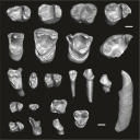
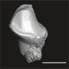
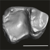
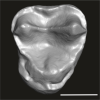
This contribution contains the three-dimensional digital models of the dental fossil material of anthropoid and strepsirrhine primates, discovered in Lower Oligocene detrital deposits outcropping in the Porto Rico and El Argoub areas, east of the Dakhla peninsula region (Atlantic Sahara; in the south of Morocco, near the northern border of Mauritania). These fossils were described, figured and discussed in the following publication: Marivaux et al. (2024), A new primate community from the earliest Oligocene of the Atlantic margin of Northwest Africa: Systematic, paleobiogeographic and paleoenvironmental implications. Journal of Human Evolution. https://doi.org/10.1016/j.jhevol.2024.103548
Catopithecus aff. browni DAK-Arg-087 View specimen
|
M3#1211Isolated right lower m3 (worn) Type: "3D_surfaces"doi: 10.18563/m3.sf.1211 state:published |
Download 3D surface file |
Catopithecus aff. browni DAK-Arg-088 View specimen
|
M3#1212Isolated right lower m2 (abraded/corroded) Type: "3D_surfaces"doi: 10.18563/m3.sf.1212 state:published |
Download 3D surface file |
Catopithecus aff. browni DAK-Arg-089 View specimen
|
M3#1213Isolated left lower m1 (worn) Type: "3D_surfaces"doi: 10.18563/m3.sf.1213 state:published |
Download 3D surface file |
Catopithecus aff. browni DAK-Pto-052 View specimen
|
M3#1214Isolated right lower m1 (pristine but lacking the mesiobuccal region) Type: "3D_surfaces"doi: 10.18563/m3.sf.1214 state:published |
Download 3D surface file |
Catopithecus aff. browni DAK-Arg-090 View specimen
|
M3#1215Isolated left upper P4 Type: "3D_surfaces"doi: 10.18563/m3.sf.1215 state:published |
Download 3D surface file |
Catopithecus aff. browni DAK-Arg-091 View specimen
|
M3#1216Isolated left upper M2 (worn and corroded) Type: "3D_surfaces"doi: 10.18563/m3.sf.1216 state:published |
Download 3D surface file |
Catopithecus aff. browni DAK-Pto-053 View specimen
|
M3#1217Isolated right upper M1 (lacking the buccal region) Type: "3D_surfaces"doi: 10.18563/m3.sf.1217 state:published |
Download 3D surface file |
Abuqatrania cf. basiodontos DAK-Arg-092 View specimen
|
M3#1218Isolated left lower c1 Type: "3D_surfaces"doi: 10.18563/m3.sf.1218 state:published |
Download 3D surface file |
?Propliopithecus sp. DAK-Pto-056 View specimen
|
M3#1219Isolated right lower m3 (fragment of talonid of a germ) Type: "3D_surfaces"doi: 10.18563/m3.sf.1219 state:published |
Download 3D surface file |
Abuqatrania cf. basiodontos DAK-Arg-093 View specimen
|
M3#1469Isolated right lower m1 Type: "3D_surfaces"doi: 10.18563/m3.sf.1469 state:published |
Download 3D surface file |
Abuqatrania cf. basiodontos DAK-Arg-094 View specimen
|
M3#1221Isolated left upper M1 or M2 (corroded, lacking the enamel cap [exposed dentine]) Type: "3D_surfaces"doi: 10.18563/m3.sf.1221 state:published |
Download 3D surface file |
Abuqatrania cf. basiodontos DAK-Arg-095 View specimen
|
M3#1222Isolated right lower i1 or i2 Type: "3D_surfaces"doi: 10.18563/m3.sf.1222 state:published |
Download 3D surface file |
Abuqatrania cf. basiodontos DAK-Arg-096 View specimen
|
M3#1223Isolated right lower p2 (worn apex) Type: "3D_surfaces"doi: 10.18563/m3.sf.1223 state:published |
Download 3D surface file |
Abuqatrania cf. basiodontos DAK-Arg-097 View specimen
|
M3#1224Isolated right lower p2 (worn apex and broken root) Type: "3D_surfaces"doi: 10.18563/m3.sf.1224 state:published |
Download 3D surface file |
Afrotarsius sp. DAK-Arg-098 View specimen
|
M3#1225Isolated left lower p3 Type: "3D_surfaces"doi: 10.18563/m3.sf.1225 state:published |
Download 3D surface file |
Afrotarsius sp. DAK-Pto-054 View specimen
|
M3#1226Isolated right lower m1 (abraded/corroded) Type: "3D_surfaces"doi: 10.18563/m3.sf.1226 state:published |
Download 3D surface file |
Orolemur mermozi DAK-Pto-055 View specimen
|
M3#1227Isolated right upper M1 or M2 (pristine, Holotype) Type: "3D_surfaces"doi: 10.18563/m3.sf.1227 state:published |
Download 3D surface file |
Wadilemur cf. elegans DAK-Arg-099 View specimen
|
M3#1228Isolated right lower m2 Type: "3D_surfaces"doi: 10.18563/m3.sf.1228 state:published |
Download 3D surface file |
cf. 'Anchomomys' milleri DAK-Arg-100 View specimen
|
M3#1229Isolated right lower c1 Type: "3D_surfaces"doi: 10.18563/m3.sf.1229 state:published |
Download 3D surface file |
Abuqatrania cf. basiodontos DAK-Arg-101 View specimen
|
M3#1396Isolated left upper M3 (abraded) Type: "3D_surfaces"doi: 10.18563/m3.sf.1396 state:published |
Download 3D surface file |
Orogalago saintexuperyi DAK-Arg-102 View specimen
|
M3#1397Isolated left lower m2 Type: "3D_surfaces"doi: 10.18563/m3.sf.1397 state:published |
Download 3D surface file |
Wadilemur cf. elegans DAK-Arg-103 View specimen
|
M3#1473Isolated right upper M1 or M2 (lacking the mesial and buccal regions) Type: "3D_surfaces"doi: 10.18563/m3.sf.1473 state:published |
Download 3D surface file |
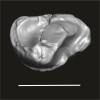
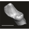
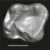
This contribution contains 3D models of upper molar rows of house mice (Mus musculus domesticus) belonging to Western European commensal and Sub-Antarctic feral populations. These two groups are characterized by different patterns of wear and alignment of the three molars along the row, related to contrasted masticatory demand in relation with their diet. These models are analyzed in the following publication: Renaud et al 2023, “Molar wear in house mice, insight into diet preferences at an ecological time scale?”, https://doi.org/10.1093/biolinnean/blad091
Mus musculus G09_06 View specimen
|
M3#1166right upper molar row Type: "3D_surfaces"doi: 10.18563/m3.sf.1166 state:published |
Download 3D surface file |
Mus musculus G09_10 View specimen
|
M3#1168right upper molar row Type: "3D_surfaces"doi: 10.18563/m3.sf.1168 state:published |
Download 3D surface file |
Mus musculus G09_15 View specimen
|
M3#1169right upper molar row Type: "3D_surfaces"doi: 10.18563/m3.sf.1169 state:published |
Download 3D surface file |
Mus musculus G09_16 View specimen
|
M3#1170right upper molar row Type: "3D_surfaces"doi: 10.18563/m3.sf.1170 state:published |
Download 3D surface file |
Mus musculus G09_17 View specimen
|
M3#1171right upper molar row Type: "3D_surfaces"doi: 10.18563/m3.sf.1171 state:published |
Download 3D surface file |
Mus musculus G09_21 View specimen
|
M3#1172right upper molar row Type: "3D_surfaces"doi: 10.18563/m3.sf.1172 state:published |
Download 3D surface file |
Mus musculus G09_26 View specimen
|
M3#1173right upper molar row Type: "3D_surfaces"doi: 10.18563/m3.sf.1173 state:published |
Download 3D surface file |
Mus musculus G09_27 View specimen
|
M3#1174right upper molar row Type: "3D_surfaces"doi: 10.18563/m3.sf.1174 state:published |
Download 3D surface file |
Mus musculus G09_29 View specimen
|
M3#1175right upper molar row Type: "3D_surfaces"doi: 10.18563/m3.sf.1175 state:published |
Download 3D surface file |
Mus musculus G09_65 View specimen
|
M3#1176right upper molar row Type: "3D_surfaces"doi: 10.18563/m3.sf.1176 state:published |
Download 3D surface file |
Mus musculus G09_66 View specimen
|
M3#1177right upper molar row Type: "3D_surfaces"doi: 10.18563/m3.sf.1177 state:published |
Download 3D surface file |
Mus musculus G93_03 View specimen
|
M3#1178right upper molar row Type: "3D_surfaces"doi: 10.18563/m3.sf.1178 state:published |
Download 3D surface file |
Mus musculus G93_04 View specimen
|
M3#1179right upper molar row Type: "3D_surfaces"doi: 10.18563/m3.sf.1179 state:published |
Download 3D surface file |
Mus musculus G93_10 View specimen
|
M3#1180right upper molar row Type: "3D_surfaces"doi: 10.18563/m3.sf.1180 state:published |
Download 3D surface file |
Mus musculus G93_11 View specimen
|
M3#1181right upper molar row Type: "3D_surfaces"doi: 10.18563/m3.sf.1181 state:published |
Download 3D surface file |
Mus musculus G93_13 View specimen
|
M3#1182right upper molar row Type: "3D_surfaces"doi: 10.18563/m3.sf.1182 state:published |
Download 3D surface file |
Mus musculus G93_14 View specimen
|
M3#1183right upper molar row Type: "3D_surfaces"doi: 10.18563/m3.sf.1183 state:published |
Download 3D surface file |
Mus musculus G93_15 View specimen
|
M3#1184right upper molar row Type: "3D_surfaces"doi: 10.18563/m3.sf.1184 state:published |
Download 3D surface file |
Mus musculus G93_24 View specimen
|
M3#1185left molar row Type: "3D_surfaces"doi: 10.18563/m3.sf.1185 state:published |
Download 3D surface file |
Mus musculus Tourch_7819 View specimen
|
M3#1186right upper molar row Type: "3D_surfaces"doi: 10.18563/m3.sf.1186 state:published |
Download 3D surface file |
Mus musculus G93_25 View specimen
|
M3#1187right upper molar row Type: "3D_surfaces"doi: 10.18563/m3.sf.1187 state:published |
Download 3D surface file |
Mus musculus Tourch_7821 View specimen
|
M3#1188right upper molar row Type: "3D_surfaces"doi: 10.18563/m3.sf.1188 state:published |
Download 3D surface file |
Mus musculus Tourch_7839 View specimen
|
M3#1189right upper molar row Type: "3D_surfaces"doi: 10.18563/m3.sf.1189 state:published |
Download 3D surface file |
Mus musculus Tourch_7873 View specimen
|
M3#1190right upper molar row Type: "3D_surfaces"doi: 10.18563/m3.sf.1190 state:published |
Download 3D surface file |
Mus musculus Tourch_7877 View specimen
|
M3#1196right upper molar row Type: "3D_surfaces"doi: 10.18563/m3.sf.1196 state:published |
Download 3D surface file |
Mus musculus Tourch_7922 View specimen
|
M3#1191right upper molar row Type: "3D_surfaces"doi: 10.18563/m3.sf.1191 state:published |
Download 3D surface file |
Mus musculus Tourch_7923 View specimen
|
M3#1192right upper molar row Type: "3D_surfaces"doi: 10.18563/m3.sf.1192 state:published |
Download 3D surface file |
Mus musculus Tourch_7925 View specimen
|
M3#1193right upper molar row Type: "3D_surfaces"doi: 10.18563/m3.sf.1193 state:published |
Download 3D surface file |
Mus musculus Tourch_7927 View specimen
|
M3#1194right upper molar row Type: "3D_surfaces"doi: 10.18563/m3.sf.1194 state:published |
Download 3D surface file |
Mus musculus Tourch_7932 View specimen
|
M3#1195right upper molar row Type: "3D_surfaces"doi: 10.18563/m3.sf.1195 state:published |
Download 3D surface file |

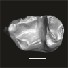
This contribution contains the three-dimensional digital model of one isolated fossil tooth of an anthropoid primate (Ashaninkacebus simpsoni), discovered in sedimentary deposits located on the upper Rio Juruá in State of Acre, Brazil (Western Amazonia). This fossil was described, figured and discussed in the following publication: Marivaux et al. (2023), An eosimiid primate of South Asian affinities in the Paleogene of Western Amazonia and the origin of New World monkeys. Proceedings of the National Academy of Sciences USA. https://doi.org/10.1073/pnas.2301338120
Ashaninkacebus simpsoni UFAC-CS 066 View specimen
|
M3#1114Right first upper molar (rM1), pristine. Type: "3D_surfaces"doi: 10.18563/m3.sf.1114 state:published |
Download 3D surface file |
The present 3D Dataset contains the 3D models analyzed in the following manuscript: L. Roese-Miron, M.E.H. Jones, J.D. Ferreira and A.S. Hsiou., 2023. Virtual endocasts of Clevosaurus brasiliensis and the tuatara: Rhynchocephalian neuroanatomy and the oldest endocranial record for Lepidosauria.
Sphenodon punctatus CM 30660 View specimen
|
M3#10993D surface model of the cranial endocast of specimen CM 30660 (Sphenodon punctatus). Type: "3D_surfaces"doi: 10.18563/m3.sf.1099 state:published |
Download 3D surface file |
Sphenodon punctatus KCLZJ 001 View specimen
|
M3#11003D surface models of the cranial endocast and the initial trunks of the cranial nerves of specimen KCLZJ 001 (Sphenodon punctatus). Type: "3D_surfaces"doi: 10.18563/m3.sf.1100 state:published |
Download 3D surface file |
Sphenodon punctatus LDUCZ x0036 View specimen
|
M3#11013D surface models of the cranial endocast and the initial trunks of the cranial nerves of specimen LDUCZ x0036 (Sphenodon punctatus). Type: "3D_surfaces"doi: 10.18563/m3.sf.1101 state:published |
Download 3D surface file |
Sphenodon punctatus LDUCZ x1126 View specimen
|
M3#11023D surface model of the cranial endocast of specimen LDUCZ x1126 (Sphenodon punctatus). Type: "3D_surfaces"doi: 10.18563/m3.sf.1102 state:published |
Download 3D surface file |
Clevosaurus brasiliensis MCN PV 2852 View specimen
|
M3#11033D surface model of the cranial endocast of specimen MCN PV 2852 (Clevosaurus brasiliensis). Type: "3D_surfaces"doi: 10.18563/m3.sf.1103 state:published |
Download 3D surface file |
Sphenodon punctatus SAMA 70524 View specimen
|
M3#11043D surface models of the cranial endocast, brain, endosseous labyrinth and initial trunks of the cranial nerves of specimen SAMA 70524 (Sphenodon punctatus). Type: "3D_surfaces"doi: 10.18563/m3.sf.1104 state:published |
Download 3D surface file |
Sphenodon punctatus SU1 View specimen
|
M3#11053D surface models of the cranial endocast and the initial trunks of the cranial nerves of specimen SU1 (Sphenodon punctatus). Type: "3D_surfaces"doi: 10.18563/m3.sf.1105 state:published |
Download 3D surface file |
Sphenodon punctatus YPM HERR 009194 View specimen
|
M3#11063D surface models of the cranial endocast and the initial trunks of the cranial nerves of specimen YPM HERR 009194 (Sphenodon punctatus). Type: "3D_surfaces"doi: 10.18563/m3.sf.1106 state:published |
Download 3D surface file |
The present 3D Dataset contains the 3D model analyzed in Hendrickx, C. and Bell, P. R. 2021. The scaly skin of the abelisaurid Carnotaurus sastrei (Theropoda: Ceratosauria) from the Upper Cretaceous of Patagonia. Cretaceous Research. https://doi.org/10.1016/j.cretres.2021.104994
Carnotaurus sastrei MACN 894 View specimen
|
M3#8023D reconstruction of the biggest patch of skin (~1200 cm2) from the anterior tail region of the holotype of Carnotaurus, which is the largest single patch of squamous integument available for any saurischian. The skin consists of medium to large (up to 65 mm in diameter) conical feature scales surrounded by a network of low and small (< 14 mm) irregular basement scales separated by narrow interstitial tissue. Type: "3D_surfaces"doi: 10.18563/m3.sf.802 state:published |
Download 3D surface file |
The present 3D Dataset contains 26 3D models analyzed in the study: On the “cartilaginous rider” in the endocasts of turtle brain cavities, published by the authors in the journal Vertebrate Zoology.
Annemys sp. IVPP-V-18106 View specimen
|
M3#7723D surface(s) file to specimen IVPP-V-18106 Type: "3D_surfaces"doi: 10.18563/m3.sf.772 state:published |
Download 3D surface file |
Apalone spinifera FMNH 22178 View specimen

|
M3#7733D surface(s) file to specimen FMNH 22178 Type: "3D_surfaces"doi: 10.18563/m3.sf.773 state:published |
Download 3D surface file |
Caretta caretta NHMUK1940.3.15.1 View specimen
|
M3#7863D surface(s) file to specimen NHMUK1940.3.15.1 Type: "3D_surfaces"doi: 10.18563/m3.sf.786 state:published |
Download 3D surface file |
Chelodina reimanni ZMB 49659 View specimen
|
M3#7743D surface(s) file to specimen ZMB 49659 Type: "3D_surfaces"doi: 10.18563/m3.sf.774 state:published |
Download 3D surface file |
Chelonia mydas ZMB-37416MS View specimen

|
M3#7753D surface(s) file to specimen ZMB-37416MS Type: "3D_surfaces"doi: 10.18563/m3.sf.775 state:published |
Download 3D surface file |
Cuora amboinensis NHMUK69.42.145_4 View specimen
|
M3#7763D surface(s) file to specimen NHMUK69.42.145_4 Type: "3D_surfaces"doi: 10.18563/m3.sf.776 state:published |
Download 3D surface file |
Emydura subglobosa IW92 View specimen
|
M3#7773D surface(s) file to specimen IW92 Type: "3D_surfaces"doi: 10.18563/m3.sf.777 state:published |
Download 3D surface file |
Eubaena cephalica DMNH 96004 View specimen
|
M3#7783D surface(s) file to specimen DMNH 96004 Type: "3D_surfaces"doi: 10.18563/m3.sf.778 state:published |
Download 3D surface file |
Gopherus berlandieri AMNH-73816 View specimen
|
M3#7793D surface(s) file to specimen AMNH-73816 Type: "3D_surfaces"doi: 10.18563/m3.sf.779 state:published |
Download 3D surface file |
Kinixys belliana AMNH-10028 View specimen
|
M3#7803D surface(s) file to specimen AMNH-10028 Type: "3D_surfaces"doi: 10.18563/m3.sf.780 state:published |
Download 3D surface file |
Macrochelys temminckii GPIT-PV-79430 View specimen
|
M3#7813D surface(s) file to specimen GPIT-PV-79430 Type: "3D_surfaces"doi: 10.18563/m3.sf.781 state:published |
Download 3D surface file |
Malacochersus tornieri SMF-58702 View specimen
|
M3#7873D surface(s) file to specimen SMF-58702 Type: "3D_surfaces"doi: 10.18563/m3.sf.787 state:published |
Download 3D surface file |
Naomichelys speciosa FMNH-PR-273 View specimen
|
M3#7823D surface(s) file to specimen FMNH-PR-273 Type: "3D_surfaces"doi: 10.18563/m3.sf.782 state:published |
Download 3D surface file |
Pelodiscus sinensis IW576-2 View specimen
|
M3#7833D surface(s) file to specimen IW576-2 Type: "3D_surfaces"doi: 10.18563/m3.sf.783 state:published |
Download 3D surface file |
Platysternon megacephalum SMF-69684 View specimen
|
M3#7843D surface(s) file to specimen SMF-69684 Type: "3D_surfaces"doi: 10.18563/m3.sf.784 state:published |
Download 3D surface file |
Podocnemis unifilis SMF-55470 View specimen
|
M3#7853D surface(s) file to specimen SMF-55470 Type: "3D_surfaces"doi: 10.18563/m3.sf.785 state:published |
Download 3D surface file |
Proganochelys quenstedtii MB 1910.45.2 View specimen
|
M3#7883D surface(s) file to specimen MB 1910.45.2 Type: "3D_surfaces"doi: 10.18563/m3.sf.788 state:published |
Download 3D surface file |
Proganochelys quenstedtii SMNS 16980 View specimen
|
M3#7893D surface(s) file to specimen SMNS 16980 Type: "3D_surfaces"doi: 10.18563/m3.sf.789 state:published |
Download 3D surface file |
Rhinochelys pulchriceps CAMSM_B55775 View specimen
|
M3#7903D surface(s) file to specimen CAMSM_B55775 Type: "3D_surfaces"doi: 10.18563/m3.sf.790 state:published |
Download 3D surface file |
Rhinoclemmys funereal YPM12174 View specimen
|
M3#7913D surface(s) file to specimen YPM12174 Type: "3D_surfaces"doi: 10.18563/m3.sf.791 state:published |
Download 3D surface file |
Sandownia harrisi MIWG3480 View specimen
|
M3#7923D surface(s) file to specimen MIWG3480 Type: "3D_surfaces"doi: 10.18563/m3.sf.792 state:published |
Download 3D surface file |
Testudo graeca YPM14342 View specimen
|
M3#7933D surface(s) file to specimen YPM14342 Type: "3D_surfaces"doi: 10.18563/m3.sf.793 state:published |
Download 3D surface file |
Testudo hermanni AMNH134518 View specimen
|
M3#7943D surface(s) file to specimen AMNH134518 Type: "3D_surfaces"doi: 10.18563/m3.sf.794 state:published |
Download 3D surface file |
Trachemys scripta NN View specimen
|
M3#7953D surface(s) file to specimen Trachemys scripta Type: "3D_surfaces"doi: 10.18563/m3.sf.795 state:published |
Download 3D surface file |
Xinjiangchelys radiplicatoides IVPP V9539 View specimen
|
M3#7963D surface(s) file to specimen IVPP V9539 Type: "3D_surfaces"doi: 10.18563/m3.sf.796 state:published |
Download 3D surface file |
Chelydra serpentina UFR VP1 View specimen
|
M3#801Brain endocast Type: "3D_surfaces"doi: 10.18563/m3.sf.801 state:published |
Download 3D surface file |
This contribution contains 3D models of extinct rodents Dinomyidae from Miocene and Quaternary of Brazil. The Miocene specimens that were digitalized include the holotypes of Potamarchus adamiae, Pseudopotamarchus villanuevai, and Ferigolomys pacarana collected in the Solimões Formation (Upper Miocene), northern Brazil. The Quaternary specimens are the holotype and paratype of Niedemys piauiensis, found in Upper Pleistocene deposits from northeast Brazil.
Potamarchus adamiae UFAC-CS 011 View specimen
|
M3#410UFAC-CS 011 – holotype, palatal region of the skull with cheek teeth Type: "3D_surfaces"doi: 10.18563/m3.sf.410 state:published |
Download 3D surface file |
Potamarchus adamiae UFAC-CS 043 View specimen
|
M3#411UFAC-CS 043, left dentary with cheek teeth Type: "3D_surfaces"doi: 10.18563/m3.sf.411 state:published |
Download 3D surface file |
Pseudopotamarchus villanuevai UFAC 4762 View specimen
|
M3#412UFAC 4762 – holotype, incomplete right maxilla with cheek teeth Type: "3D_surfaces"doi: 10.18563/m3.sf.412 state:published |
Download 3D surface file |
Ferigolomys pacarana UFAC 6460 View specimen
|
M3#413UFAC 6460 – holotype, palatal region of the skull with cheek teeth Type: "3D_surfaces"doi: 10.18563/m3.sf.413 state:published |
Download 3D surface file |
Drytomomys sp. UFAC 2742 View specimen

|
M3#414UFAC 2742, right dentary with cheek teeth Type: "3D_surfaces"doi: 10.18563/m3.sf.414 state:published |
Download 3D surface file |
Niedemys piauiensis FUMDHAM 113-146365-2 View specimen
|
M3#418FUMDHAM 113-146365-2 - holotype, upper right tooth Type: "3D_surfaces"doi: 10.18563/m3.sf.418 state:published |
Download 3D surface file |
Niedemys piauiensis FUMDHAM 113-145304-2 View specimen
|
M3#419FUMDHAM 113-145304-2 - paratype, left lower molar Type: "3D_surfaces"doi: 10.18563/m3.sf.419 state:published |
Download 3D surface file |

The present 3D Dataset contains the 3D model analyzed in Wazir, W. A., Sehgal, R. K., Čerňanský, A., Patnaik, R., Kumar, N., Singh, A. P. and Singh, N. P. 2022. A find from the Ladakh Himalaya reveals a survival of madtsoiid snakes (Serpentes, Madtsoiidae) in India through the late Oligocene. Journal of Vertebrate Paleontology, 41(6), e2058401. https://doi.org/10.1080/02724634.2021.2058401
indet. indet. WIMF/A 4816 View specimen
|
M3#1754Vertebra Type: "3D_surfaces"doi: 10.18563/m3.sf.1754 state:published |
Download 3D surface file |
The 3D dataset presented in this article provides the 3D models of two Chelonioidea turtles dentaries from the Paleocene of France described in: Lapparent de Broin F. de, Marek H., Barrier P. & Gagnaison C. 2025. — Euclastidae n. fam. (Chelonioidea) et première mention d’Euclastes Cope, 1867 dans le Paléocène du bassin de Paris (France). Geodiversitas 47 (10): 409-464. https://doi.org/10.5252/geodiversitas2025v47a10.
Euclastes wielandi ULB-04A21-10 View specimen
|
M3#1791Euclastes wielandi Type: "3D_surfaces"doi: 10.18563/m3.sf.1791 state:published |
Download 3D surface file |
Euclastes wielandi MNHN.F.BPT52 View specimen
|
M3#1709Euclastes wielandi (cast) Type: "3D_surfaces"doi: 10.18563/m3.sf.1709 state:published |
Download 3D surface file |
Euclastes wielandi ULB-04A21-11 View specimen
|
M3#1792Euclastes montenati nov. sp. Type: "3D_surfaces"doi: 10.18563/m3.sf.1792 state:published |
Download 3D surface file |
Euclastes wielandi MNHN.F.BPT53 View specimen
|
M3#1711Euclastes montenati (cast) Type: "3D_surfaces"doi: 10.18563/m3.sf.1711 state:published |
Download 3D surface file |
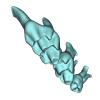
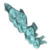
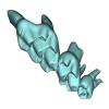
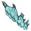
The present 3D dataset contains the 3D models analyzed in the publication: Rosa, R. M., Salvador, R. B., & Cavallari, D. C. (2025). The disappearing act of the magician tree snail: anatomy, distribution, and phylogenetic relationships of Drymaeus magus (Gastropoda: Bulimulidae), a long-lost species hidden in plain sight. Zoological Journal of the Linnean Society.
Drymaeus magus CMRP 1049 View specimen
|
M3#1597Internal organs of Drymaeus magus Type: "3D_surfaces"doi: 10.18563/m3.sf.1597 state:published |
Download 3D surface file |
|
M3#1598External surface of Drymaeus magus Type: "3D_surfaces"doi: 10.18563/m3.sf.1598 state:published |
Download 3D surface file |
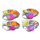
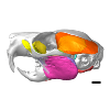
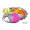
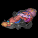
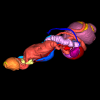
The present 3D Dataset contains the 3D models of extant Chiropteran endocranial casts, documenting 16 of the 19 extant bat families. They are used by Maugoust & Orliac (2023) to assess the correspondences between the brain and brain-surrounding tissues (i.e., neural tissues, blood vessels, meninges) and their imprint on the braincase, allowing for eventually proposing a Chiroptera-scale nomenclature of the endocast.
Balantiopteryx plicata UMMZ 102659 View specimen
|
M3#1132Endocranial cast of the corresponding cranium of Balantiopteryx plicata Type: "3D_surfaces"doi: 10.18563/m3.sf.1132 state:published |
Download 3D surface file |
Idiurus macrotis AMNH M-187705 View specimen
|
M3#1133Endocranial cast of the corresponding cranium of Nycteris macrotis Type: "3D_surfaces"doi: 10.18563/m3.sf.1133 state:published |
Download 3D surface file |
Thyroptera tricolor UMMZ 53240 View specimen
|
M3#1134Endocranial cast of the corresponding cranium of Thyroptera tricolor Type: "3D_surfaces"doi: 10.18563/m3.sf.1134 state:published |
Download 3D surface file |
Noctilio albiventris UMMZ 105827 View specimen
|
M3#1135Endocranial cast of the corresponding cranium of Noctilio albiventris Type: "3D_surfaces"doi: 10.18563/m3.sf.1135 state:published |
Download 3D surface file |
Mormoops blainvillii AMNH M-271513 View specimen
|
M3#1136Endocranial cast of the corresponding cranium of Mormoops blainvillii Type: "3D_surfaces"doi: 10.18563/m3.sf.1136 state:published |
Download 3D surface file |
Macrotus waterhousii UMMZ 95718 View specimen
|
M3#1137Endocranial cast of the corresponding cranium of Macrotus waterhousii Type: "3D_surfaces"doi: 10.18563/m3.sf.1137 state:published |
Download 3D surface file |
Nyctiellus lepidus UMMZ 105767 View specimen
|
M3#1138Endocranial cast of the corresponding cranium of Nyctiellus lepidus Type: "3D_surfaces"doi: 10.18563/m3.sf.1138 state:published |
Download 3D surface file |
Cheiromeles torquatus AMNH M-247585 View specimen
|
M3#1139Endocranial cast of the corresponding cranium of Cheiromeles torquatus Type: "3D_surfaces"doi: 10.18563/m3.sf.1139 state:published |
Download 3D surface file |
Miniopterus schreibersii UMMZ 156998 View specimen
|
M3#1140Endocranial cast of the corresponding cranium of Miniopterus schreibersii Type: "3D_surfaces"doi: 10.18563/m3.sf.1140 state:published |
Download 3D surface file |
Kerivoula pellucida UMMZ 161396 View specimen
|
M3#1141Endocranial cast of the corresponding cranium of Kerivoula pellucida Type: "3D_surfaces"doi: 10.18563/m3.sf.1141 state:published |
Download 3D surface file |
Scotophilus kuhlii UMMZ 157013 View specimen
|
M3#1142Endocranial cast of the corresponding cranium of Scotophilus kuhlii Type: "3D_surfaces"doi: 10.18563/m3.sf.1142 state:published |
Download 3D surface file |
Rhinolophus luctus MNHN CG-2006-87 View specimen
|
M3#1143Endocranial cast of the corresponding cranium of Rhinolophus luctus Type: "3D_surfaces"doi: 10.18563/m3.sf.1143 state:published |
Download 3D surface file |
Triaenops persicus AM RG-38552 View specimen
|
M3#1144Endocranial cast of the corresponding cranium of Triaenops persicus Type: "3D_surfaces"doi: 10.18563/m3.sf.1144 state:published |
Download 3D surface file |
Hipposideros armiger UM ZOOL-762-V View specimen
|
M3#1145Endocranial cast of the corresponding cranium of Hipposideros armiger Type: "3D_surfaces"doi: 10.18563/m3.sf.1145 state:published |
Download 3D surface file |
Lavia frons AM RG-12268 View specimen
|
M3#1146Endocranial cast of the corresponding cranium of Lavia frons Type: "3D_surfaces"doi: 10.18563/m3.sf.1146 state:published |
Download 3D surface file |
Rhinopoma hardwickii AM RG-M31166 View specimen
|
M3#1147Endocranial cast of the corresponding cranium of Rhinopoma hardwickii Type: "3D_surfaces"doi: 10.18563/m3.sf.1147 state:published |
Download 3D surface file |
Sphaerias blanfordi AMNH M-274330 View specimen
|
M3#1148Endocranial cast of the corresponding cranium of Sphaerias blanfordi Type: "3D_surfaces"doi: 10.18563/m3.sf.1148 state:published |
Download 3D surface file |
Rousettus aegyptiacus UMMZ 161026 View specimen
|
M3#1149Endocranial cast of the corresponding cranium of Rousettus aegyptiacus Type: "3D_surfaces"doi: 10.18563/m3.sf.1149 state:published |
Download 3D surface file |
Pteropus pumilus UMMZ 162253 View specimen
|
M3#1150Endocranial cast of the corresponding cranium of Pteropus pumilus Type: "3D_surfaces"doi: 10.18563/m3.sf.1150 state:published |
Download 3D surface file |
This contribution contains the 3D model of an endocranial cast analyzed in “A 10 ka intentionally deformed human skull from Northeast Asia”. There are many studies on the morphological characteristics of intentional cranial deformation (ICD), but few related 3D models were published. Here, we present the surface model of an intentionally deformed 10 ka human cranium for further research on ICD practice. The 3D model of the endocranial cast of this ICD cranium was discovered near Harbin City, Province Heilongjiang, Northeast China. The fossil preserved only the frontal, parietal, and occipital bones. To complete the endocast model of the specimen, we printed a 3D model and used modeling clay to reconstruct the missing part based on the general form of the modern human endocast morphology.
Homo sapiens IVPP-PA1616 View specimen
|
M3#972The frontal region of the endocast is flattened, probably formed by the constant pressure on the frontal bone during growth. There is a well-developed frontal crest on the endocranial surface. The endocast widens posteriorly from the frontal lobe. The widest point of the endocast is at the lateral border of the parietal lobe. The lower parietal areas display a marked lateral expansion. The overall shape of the endocast is asymmetrical, with the left side of the parietal lobe being more laterally expanded than the right side. Like the frontal lobe, the occipital lobe is also anteroposteriorly flattened. Type: "3D_surfaces"doi: 10.18563/m3.sf.972 state:published |
Download 3D surface file |
|
M3#976The original endocranial cast model (with texture) of IVPP-PA1616. It shows the original structures of the specimen, and was not altered in any way. Type: "3D_surfaces"doi: 10.18563/m3.sf.976 state:published |
Download 3D surface file |
The present 3D Dataset contains the 3D models of the endocranial cast of two specimens of Indohyus indirae described in the article entitled “The endocranial cast of Indohyus (Artiodactyla, Raoellidae): the origin of the cetacean brain” (Orliac and Thewissen, 2021). They represent the cast of the main cavity of the braincase as well as associated intraosseous sinuses.
Indohyus indirae RR 207 View specimen
|
M3#710cast of the main endocranial cavity and associated intraosseous sinuses Type: "3D_surfaces"doi: 10.18563/m3.sf.710 state:published |
Download 3D surface file |
Indohyus indirae RR 601 View specimen
|
M3#711casts of the main endocranial cavity Type: "3D_surfaces"doi: 10.18563/m3.sf.711 state:published |
Download 3D surface file |
This contribution contains the 3D model(s) described and figured in the following publication: The present 3D Dataset contains the 3D models and CT-Scan slices of the lower jaws and teeth analyzed in “A new prozostrodontian cynodont (Eucynodontia, Probainognathia) from the Upper Triassic of southern Brazil”. https://doi.org/10.1080/02724634.2020.1782415
Agudotherium gassenae CAPPA/UFSM 0262 View specimen
|
M3#546Left lower jaw and cheek teeth Type: "3D_surfaces"doi: 10.18563/m3.sf.546 state:published |
Download 3D surface file |
|
M3#5471578 slices Type: "3D_CT"doi: 10.18563/m3.sf.547 state:published |
Download CT data |
Agudotherium gassenae CAPPA/UFSM 0208 View specimen
|
M3#548right lower jaw Type: "3D_surfaces"doi: 10.18563/m3.sf.548 state:published |
Download 3D surface file |
|
M3#549CT data of CAPPA_UFSM_0208 Type: "3D_CT"doi: 10.18563/m3.sf.549 state:published |
Download CT data |


