3D models of Cainotheriids Ossicular chain
Explodable 3D Dog Skull for Veterinary Education
3D models of Kalakocetus, the earliest Cetacea
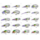
3D GM dataset of bird skeletal variation
Skeletal embryonic development in the catshark
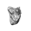
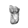
Bony connexions of the petrosal bone of extant hippos
bony labyrinth (14) , inner ear (11) , Eocene (11) , geometric morphometrics (10) , CT-scan (10) , Oligocene (9) , Micro-CT (9)
Lionel Hautier (25) , Maëva Judith Orliac (24) , Laurent Marivaux (18) , Renaud Lebrun (15) , Rodolphe Tabuce (15) , Pierre-Olivier Antoine (13) , Bastien Mennecart (13)
|
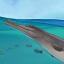
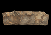
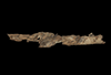
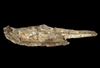
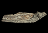
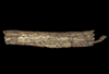
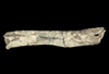
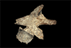
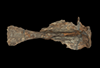
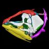
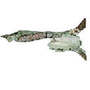
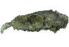
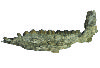
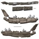
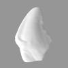
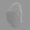
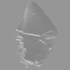
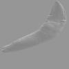
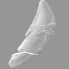
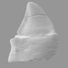
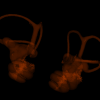
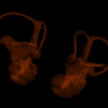
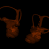
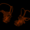
Virtual reconstruction of a Late Jurassic metriorhynchid skull from Switzerland and its use for scientific illustration and paleoartSophie De Sousa Oliveira, Léa Girard
Published online: 19/07/2023 |
|
M3#1037Left dentary (3 meshes) Type: "3D_surfaces"doi: 10.18563/m3.sf.1037 state:published |
Download 3D surface file |
|
M3#1038Right dentary (3 meshes) Type: "3D_surfaces"doi: 10.18563/m3.sf.1038 state:published |
Download 3D surface file |
|
M3#1039Left ramus (2 meshes) Type: "3D_surfaces"doi: 10.18563/m3.sf.1039 state:published |
Download 3D surface file |
|
M3#1040Right ramus (3 meshes) Type: "3D_surfaces"doi: 10.18563/m3.sf.1040 state:published |
Download 3D surface file |
|
M3#1041Left splenial (2 meshes) Type: "3D_surfaces"doi: 10.18563/m3.sf.1041 state:published |
Download 3D surface file |
|
M3#1042Right splenial (2 meshes) Type: "3D_surfaces"doi: 10.18563/m3.sf.1042 state:published |
Download 3D surface file |
|
M3#1043Frontal and left prefrontal Type: "3D_surfaces"doi: 10.18563/m3.sf.1043 state:published |
Download 3D surface file |
|
M3#1044Left maxilla (4 meshes) Type: "3D_surfaces"doi: 10.18563/m3.sf.1044 state:published |
Download 3D surface file |
|
M3#1045Right maxilla Type: "3D_surfaces"doi: 10.18563/m3.sf.1045 state:published |
Download 3D surface file |
|
M3#1046Left nasal Type: "3D_surfaces"doi: 10.18563/m3.sf.1046 state:published |
Download 3D surface file |
|
M3#1047Right nasal Type: "3D_surfaces"doi: 10.18563/m3.sf.1047 state:published |
Download 3D surface file |
|
M3#1048Parietal Type: "3D_surfaces"doi: 10.18563/m3.sf.1048 state:published |
Download 3D surface file |
|
M3#1049Right postorbital Type: "3D_surfaces"doi: 10.18563/m3.sf.1049 state:published |
Download 3D surface file |
|
M3#1050Right prefrontal Type: "3D_surfaces"doi: 10.18563/m3.sf.1050 state:published |
Download 3D surface file |
|
M3#1051Right premaxilla Type: "3D_surfaces"doi: 10.18563/m3.sf.1051 state:published |
Download 3D surface file |
|
M3#1052Left squamosal Type: "3D_surfaces"doi: 10.18563/m3.sf.1052 state:published |
Download 3D surface file |

|
M3#1053Right squamosal Type: "3D_surfaces"doi: 10.18563/m3.sf.1053 state:published |
Download 3D surface file |
|
M3#1054Reconstruction of the mandible Type: "3D_surfaces"doi: 10.18563/m3.sf.1054 state:published |
Download 3D surface file |

|
M3#1055Reconstruction of the cranium Type: "3D_surfaces"doi: 10.18563/m3.sf.1055 state:published |
Download 3D surface file |

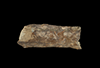
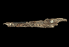
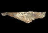
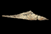
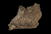
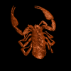
In this contribution a third new species of the rare genus Burmesescorpiops Lourenço, 2016 is described. The discovery of this new element belonging to the family Palaeoeuscorpiidae Lourenço, 2003 and to the subfamily Archaeoscorpiopinae Lourenço, 2015 brings further elements to support the validity of the genus Burmesescorpiops. This generic group remains however, poorly speciose. This is the latest discovery of Burmesescorpiops wunpawng, the name is derived from the Kachin Hilltribe peoples who are indigenous to the area. The data provided here is a 3D surface.
Burmesescorpiops wunpawng ps-gyi-01-25 View specimen
|
M3#18463d Surface Volume Type: "3D_surfaces"doi: 10.18563/m3.sf.1846 state:published |
Download 3D surface file |

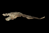
We provide a 3D reconstruction of the skull of Latimeria chalumnae that can be easily accessed and visualized for a better understanding of its cranial anatomy. Different skeletal elements are saved as separate PLY files that can be combined to visualize the entire skull or isolated to virtually dissect the skull. We included some guidelines for a fast and easy visualization of the 3D skull.
Latimeria chalumnae MHNG 1080.070 View specimen
|
M3#1254the skeletal elements of the skull of Latimeria chalumnae included in 26 different PLY files Type: "3D_surfaces"doi: 10.18563/m3.sf.1254 state:published |
Download 3D surface file |
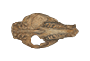
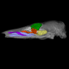
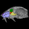
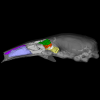
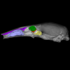
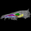
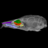
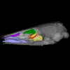
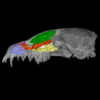
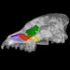
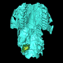
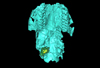
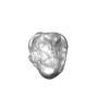
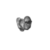
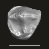
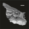
The present 3D dataset contains 3D models of the endocranial cast of the raoellid Khirtharia inflata retrieved from the middle Eocene of the Upper Subathu Formation in the Kalakot area (India). Raoellidae are closely related to stem cetaceans and bring crucial information to understand the earliest phase of land to water transition in Cetacea.
Khirtharia inflata GU/RJ/197 View specimen
|
M3#1608labeled cast of the endocranial cavity Type: "3D_surfaces"doi: 10.18563/m3.sf.1608 state:published |
Download 3D surface file |
|
M3#1609endocast and associated sinuses Type: "3D_surfaces"doi: 10.18563/m3.sf.1609 state:published |
Download 3D surface file |
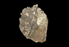
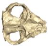
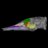
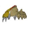
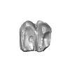
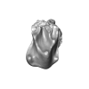
The present 3D Dataset contains the 3D model of the skull of the raoellid Indohyus indirae described in Patel et al. 2024.
Indohyus indirae RR 207 View specimen
|
M3#1259dorsoventrally crushed skull Type: "3D_surfaces"doi: 10.18563/m3.sf.1259 state:published |
Download 3D surface file |
Indohyus indirae RR 601 View specimen
|
M3#1268dorsoventrally crushed skull Type: "3D_surfaces"doi: 10.18563/m3.sf.1268 state:published |
Download 3D surface file |
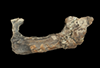
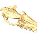
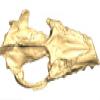
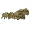
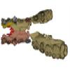
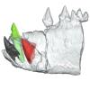
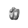
The present 3D Dataset contains the 3D model of a specimen of Metamynodon planifrons (UNISTRA.2015.0.1106) described and figured in: Veine-Tonizzo, L., Tissier, J., Bukhsianidze, M., Vasilyan, D., Becker, D., 2023, Cranial morphology and phylogenetic relationships of Amynodontidae Scott & Osborn, 1883 (Perissodactyla, Rhinocerotoidea).
Metamynodon planifrons UNISTRA.2015.0.1106 View specimen
|
M3#716Textured 3D surface model of the skull of the specimen UNISTRA.2015.0.1106 with right C1 and both rows of P2-M3. Type: "3D_surfaces"doi: 10.18563/m3.sf.716 state:published |
Download 3D surface file |
This contribution contains the 3D models described and figured in: New remains of Neotropical bunodont litopterns and the systematics of Megadolodinae (Mammalia: Litopterna). Geodiversitas.
Megadolodus molariformes VPPLT 974 View specimen

|
M3#1020Partial mandible with the symphysis and left body, bearing the alveoli of ?i2, right and left ?i3, alveolus of right c and p1, roots of left p1, and the left p2–m3 of Megadolodus molariformes (Litopterna, Mammalia) Type: "3D_surfaces"doi: 10.18563/m3.sf.1020 state:published |
Download 3D surface file |
Neodolodus colombianus VPPLT 1696 View specimen
|
M3#1021Almost complete skull with left and right ?I2 and P1–M3 Type: "3D_surfaces"doi: 10.18563/m3.sf.1021 state:published |
Download 3D surface file |
|
M3#1022Partial mandible with complete right and left dentition except for left ?i2 Type: "3D_surfaces"doi: 10.18563/m3.sf.1022 state:published |
Download 3D surface file |
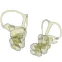
This contribution contains the three-dimensional models of the turbinal complex of 10 myrmecophagous and 10 non-myrmecophagous placental species. These specimens were analyzed and discussed in: Wright et. al (2024), Sniffing out morphological convergence in the turbinal complex of myrmecophagous placentals. https://doi.org/10.1002/ar.25603
Priodontes maximus NHMUK 732-a View specimen
|
M3#1536Turbinals of Priodontes maximus Type: "3D_surfaces"doi: 10.18563/m3.sf.1536 state:published |
Download 3D surface file |
Dasypus pilosus NHMUK 94-10-1-13 View specimen
|
M3#1537Turbinals of Dasypus pilosus Type: "3D_surfaces"doi: 10.18563/m3.sf.1537 state:published |
Download 3D surface file |
Dasypus novemcinctus AMNH 263287 View specimen
|
M3#1538Turbinals of Dasypus novemcinctus Type: "3D_surfaces"doi: 10.18563/m3.sf.1538 state:published |
Download 3D surface file |
Bradypus tridactylus UM 789N View specimen
|
M3#1539Turbinals of Bradypus tridactylus Type: "3D_surfaces"doi: 10.18563/m3.sf.1539 state:published |
Download 3D surface file |
Choloepus didactylus UM 767V View specimen
|
M3#1540Turbinals of Choloepus didactylus Type: "3D_surfaces"doi: 10.18563/m3.sf.1540 state:published |
Download 3D surface file |
Cyclopes didactylus NHMUK 88-8-8-14 View specimen
|
M3#1541Turbinals of Cyclopes didactylus Type: "3D_surfaces"doi: 10.18563/m3.sf.1541 state:published |
Download 3D surface file |
Myrmecophaga tridactyla UM 065V View specimen
|
M3#1542Turbinals of Myrmecophaga tridactyla Type: "3D_surfaces"doi: 10.18563/m3.sf.1542 state:published |
Download 3D surface file |
Tamandua tetradactyla NHMUK 3-7-7-135 View specimen
|
M3#1543Turbinals of Tamandua tetradactyla Type: "3D_surfaces"doi: 10.18563/m3.sf.1543 state:published |
Download 3D surface file |
Tamandua mexicana NHMUK 79-1-6-1 View specimen
|
M3#1544Turbinals of Tamandua mexicana Type: "3D_surfaces"doi: 10.18563/m3.sf.1544 state:published |
Download 3D surface file |
Orycteropus afer NHMUK 2-9-9-58 View specimen
|
M3#1545Turbinals of Orycteropus afer Type: "3D_surfaces"doi: 10.18563/m3.sf.1545 state:published |
Download 3D surface file |
Tenrec eucaudatus UM N439 View specimen
|
M3#1546Turbinals of Tenrec eucaudatus Type: "3D_surfaces"doi: 10.18563/m3.sf.1546 state:published |
Download 3D surface file |
Elephantulus rozeti UM N227 View specimen
|
M3#1547Turbinals of Elephantulus rozeti Type: "3D_surfaces"doi: 10.18563/m3.sf.1547 state:published |
Download 3D surface file |
Phataginus tetradactyla NHMUK 1-11-21-35 View specimen
|
M3#1548Turbinals of Phataginus tetradactyla Type: "3D_surfaces"doi: 10.18563/m3.sf.1548 state:published |
Download 3D surface file |
Smutsia gigantea KMMA 25479 View specimen
|
M3#1549Turbinals of Smutsia gigantea Type: "3D_surfaces"doi: 10.18563/m3.sf.1549 state:published |
Download 3D surface file |
Manis culionensis MNHN ZM-MO 1884-1822 View specimen
|
M3#1550Turbinals of Manis culionensis Type: "3D_surfaces"doi: 10.18563/m3.sf.1550 state:published |
Download 3D surface file |
Vulpes vulpes UM N140 View specimen
|
M3#1551Turbinals of Vulpes vulpes Type: "3D_surfaces"doi: 10.18563/m3.sf.1551 state:published |
Download 3D surface file |
Alopex lagopus UM N110 View specimen
|
M3#1552Turbinals of Alopex lagopus Type: "3D_surfaces"doi: 10.18563/m3.sf.1552 state:published |
Download 3D surface file |
Felis silvestris UM N149 View specimen
|
M3#1553Turbinals of Felis sylvestris Type: "3D_surfaces"doi: 10.18563/m3.sf.1553 state:published |
Download 3D surface file |
Hyaena hyaena UM N109 View specimen
|
M3#1554Turbinals of Hyaena hyaena Type: "3D_surfaces"doi: 10.18563/m3.sf.1554 state:published |
Download 3D surface file |
Proteles cristata NHMUK 4-3-1-58 View specimen
|
M3#1555Turbinals of Proteles cristata Type: "3D_surfaces"doi: 10.18563/m3.sf.1555 state:published |
Download 3D surface file |
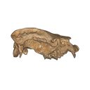
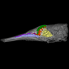
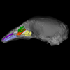
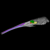
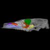
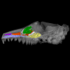
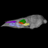
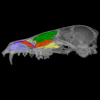
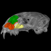
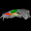
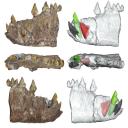
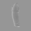
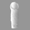
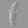
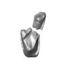
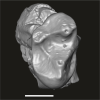
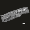
In this work, we digitally restore the snout of the raoellide Khirtharia inflata from the Kalakot area (Rajouri District, Jammu & Kashmir, India). Raoellids are small, semiaquatic ungulates closely related to cetaceans. The specimen is fairly complete and preserves left and right maxillaries, left premaxillary, and part of the anterior and jugal dentition. The digital restoration of this quite complete but deformed specimen of Khirtharia inflata is a welcome addition to the data available for raoellids and will be used to further the understanding of the origins of cetaceans.
Khirtharia inflata GU/RJ/157 View specimen
|
M3#1454deformed partial skull Type: "3D_surfaces"doi: 10.18563/m3.sf.1454 state:published |
Download 3D surface file |
|
M3#1455reconstruction of half snout Type: "3D_surfaces"doi: 10.18563/m3.sf.1455 state:published |
Download 3D surface file |
|
M3#1456reconstruction of complete snout Type: "3D_surfaces"doi: 10.18563/m3.sf.1456 state:published |
Download 3D surface file |
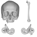
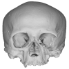
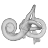
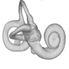
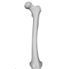
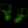
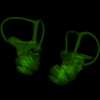
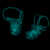
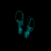
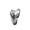
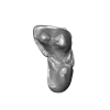
The present 3D Dataset contains the models analyzed in the publication: Menéndez L, Rios C, Acosta Morano C, Novellino P, Schmelzle T, Aguirre-Fernández G, Breidenstein A, Barquera R, Schuenemann VJ, Stafford TW, Sánchez-Villagra M, Barbieri C. (2025). A human skeleton from Última Esperanza, South-West Patagonia, Chile: Osteobiography, morphometric, and genetic analysis. The models include the skull, femur, and the segmented left and right inner ears of a late Holocene human skeleton from southern Patagonia. In the associated paper, we present the radiocarbon dating, an osteobiography profile evaluating some aspects of the life history of this individual, as well as genetic and morphometric analysis assessing biological relatedness to other individuals and populations.
Homo sapiens PIMUZ A/V 4612 View specimen
|
M3#1650Homo sapiens skull Type: "3D_surfaces"doi: 10.18563/m3.sf.1650 state:published |
Download 3D surface file |
|
M3#1652Homo sapiens left inner ear Type: "3D_surfaces"doi: 10.18563/m3.sf.1652 state:published |
Download 3D surface file |
|
M3#1653Homo sapiens right inner ear Type: "3D_surfaces"doi: 10.18563/m3.sf.1653 state:published |
Download 3D surface file |
Homo sapiens PIMUZ A/V 4613 View specimen
|
M3#1651Homo sapiens femur Type: "3D_surfaces"doi: 10.18563/m3.sf.1651 state:published |
Download 3D surface file |
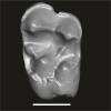
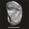
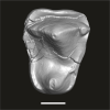
This contribution contains the three-dimensional models of the most informative fossil material attributed to both Peratherium musivum Gernelle, 2024, and Peratherium maximum (Crochet, 1979), respectively from early and middle early Eocene French localities. These specimens, which document the emergence of the relatively large peratheriines, were analyzed and discussed in: Gernelle et al. (2024), Dental morphology evolution in early peratheriines, including a new morphologically cryptic species and findings on the largest early Eocene European metatherian. https://doi.org/10.1080/08912963.2024.2403602
Peratherium musivum MNHN.F.SN122 View specimen
|
M3#16403D surface model of MNHN.F.SN122, right M3 Type: "3D_surfaces"doi: 10.18563/m3.sf.1640 state:published |
Download 3D surface file |
Peratherium musivum MNHN.F.RI220 View specimen
|
M3#16413D surface model of MNHN.F.RI220, left M2 (partial) Type: "3D_surfaces"doi: 10.18563/m3.sf.1641 state:published |
Download 3D surface file |
Peratherium musivum MNHN.F.RI296 View specimen
|
M3#16423D surface model of MNHN.F.RI296, right M1 (partial) Type: "3D_surfaces"doi: 10.18563/m3.sf.1642 state:published |
Download 3D surface file |
Peratherium musivum MNHN.F.RI368 View specimen
|
M3#16433D surface model of MNHN.F.RI368, right m2 Type: "3D_surfaces"doi: 10.18563/m3.sf.1643 state:published |
Download 3D surface file |
Peratherium musivum MNHN.F.RI385 View specimen
|
M3#16443D surface model of MNHN.F.RI385, left m1 Type: "3D_surfaces"doi: 10.18563/m3.sf.1644 state:published |
Download 3D surface file |
Peratherium maximum UM-BRI-17 View specimen
|
M3#16453D surface model of UM-BRI-17, right hemi-mandible with p1-p3, m1-m3 alveoli, and m4 Type: "3D_surfaces"doi: 10.18563/m3.sf.1645 state:published |
Download 3D surface file |
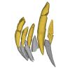
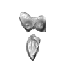
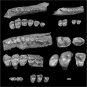
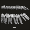
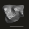
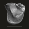
The present 3D dataset contains 3D models of new material from the middle Eocene of the Upper Subathu Formation in the Kalakot area (India), documenting the anterior dentition of the raoellid Indohyus indirae. Raoellidae are closely related to stem cetaceans and bring crucial information to understand the earliest phase of land to water transition in Cetacea.
Indohyus indirae GU/RJ/31 View specimen
|
M3#1505Right i1 Type: "3D_surfaces"doi: 10.18563/m3.sf.1505 state:published |
Download 3D surface file |
Indohyus indirae GU/RJ/32 View specimen
|
M3#1506Right i1 Type: "3D_surfaces"doi: 10.18563/m3.sf.1506 state:published |
Download 3D surface file |
Indohyus indirae GU/RJ/16 View specimen
|
M3#1507Left I3 Type: "3D_surfaces"doi: 10.18563/m3.sf.1507 state:published |
Download 3D surface file |
Indohyus indirae GU/RJ/23 View specimen
|
M3#1508Left I2 Type: "3D_surfaces"doi: 10.18563/m3.sf.1508 state:published |
Download 3D surface file |
Indohyus indirae GU/RJ/25 View specimen
|
M3#1509Left I1 Type: "3D_surfaces"doi: 10.18563/m3.sf.1509 state:published |
Download 3D surface file |
Indohyus indirae GU/RJ/26 View specimen
|
M3#1510Right I3 Type: "3D_surfaces"doi: 10.18563/m3.sf.1510 state:published |
Download 3D surface file |
Indohyus indirae GU/RJ/57 View specimen
|
M3#1511Left I1 Type: "3D_surfaces"doi: 10.18563/m3.sf.1511 state:published |
Download 3D surface file |
Indohyus indirae GU/RJ/61 View specimen
|
M3#1512Right upper canine Type: "3D_surfaces"doi: 10.18563/m3.sf.1512 state:published |
Download 3D surface file |
Indohyus indirae GU/RJ/63 View specimen
|
M3#1513Left upper canine Type: "3D_surfaces"doi: 10.18563/m3.sf.1513 state:published |
Download 3D surface file |
Indohyus indirae GU/RJ/74 View specimen
|
M3#1514Left upper canine Type: "3D_surfaces"doi: 10.18563/m3.sf.1514 state:published |
Download 3D surface file |
Indohyus indirae GU/RJ/439 View specimen
|
M3#1515Left upper canine Type: "3D_surfaces"doi: 10.18563/m3.sf.1515 state:published |
Download 3D surface file |
Indohyus indirae GU/RJ/457 View specimen
|
M3#1516Left upper canine Type: "3D_surfaces"doi: 10.18563/m3.sf.1516 state:published |
Download 3D surface file |
Indohyus indirae GU/RJ/846 View specimen
|
M3#1517Left upper canine Type: "3D_surfaces"doi: 10.18563/m3.sf.1517 state:published |
Download 3D surface file |
Indohyus indirae GU/RJ/822 View specimen
|
M3#1518Right fragmentary maxillary with decidual canine and I3 Type: "3D_surfaces"doi: 10.18563/m3.sf.1518 state:published |
Download 3D surface file |
Indohyus indirae GU/RJ/824 View specimen
|
M3#1519Right fragmentary mandible with lower canine and small part of the i3 Type: "3D_surfaces"doi: 10.18563/m3.sf.1519 state:published |
Download 3D surface file |
Indohyus indirae GU/RJ/838 View specimen
|
M3#1520Right fragmentary mandible with permanent i2, i3 and canine and small part of the root of the decidual i3 Type: "3D_surfaces"doi: 10.18563/m3.sf.1520 state:published |
Download 3D surface file |
Indohyus indirae GU/RJ/842 View specimen
|
M3#1521Left fragmentary mandible with decidual and permanent canine Type: "3D_surfaces"doi: 10.18563/m3.sf.1521 state:published |
Download 3D surface file |
Indohyus indirae GU/RJ/26,32,56,57,457,822,838,842 : Composite Anterior dentition View specimen
|
M3#15293D composite reconstruction of the anterior dentition of Indohyus indirae with GU/RJ/57 (I1), 56 (I2), 26 (I3), 457 (Upper canine), 822 (P1), 32 (i1), 838 (i2, i3 and lower canine) and 842 (p1) Type: "3D_surfaces"doi: 10.18563/m3.sf.1529 state:published |
Download 3D surface file |
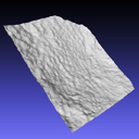
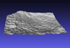
The present 3D Dataset contains the 3D model of the skin of Allosaurus described in Hendrickx, C. et al. in press. Morphology and distribution of scales, dermal ossifications, and other non-feather integumentary structures in non-avialan theropod dinosaurs. Biological Reviews.
Allosaurus jimmadseni UMNH VP C481 View specimen
|
M3#902The material consists of a 3D reconstruction of the counterpart of a 30 cm2 patch of skin impression associated with the anterior dorsal ribs/pectoral region of the specimen of Allosaurus jimmadseni UMNH VP C481. The skin shows a semi-uniform basement of 1-2 mm diameter pebbles with a smaller number of slightly larger (up to 3 mm) ovoid scales. The irregular shape, distribution, and overall small size of these larger scales suggest that they are not classifiable as feature scales but rather as variations in the basement scales. Type: "3D_surfaces"doi: 10.18563/m3.sf.902 state:published |
Download 3D surface file |
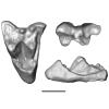
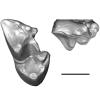
The present 3D Dataset contains the 3D models of the holotype (NMB Sth. 833) of the new species Micromeryx? eiselei analysed in the article Aiglstorfer, M., Costeur, L., Mennecart, B., Heizmann, E.P.J.. 2017. Micromeryx? eiselei - a new moschid species from Steinheim am Albuch, Germany, and the first comprehensive description of moschid cranial material from the Miocene of Central Europe. PlosOne https://doi.org/10.1371/journal.pone.0185679
Micromeryx? eiselei NMB Sth. 833 View specimen
|
M3#284The 3 D surfaces comprises the skull, petrosal, and bony labyrinth of NMB Sth.833, the holotype of Micromeryx? eiselei Type: "3D_surfaces"doi: 10.18563/m3.sf.284 state:published |
Download 3D surface file |
This contribution contains 3D models of left and right house mouse (Mus musculus domesticus) inner ears analyzed in Renaud et al. (2024). The studied mice belong to four groups: wild-trapped mice, wild-derived lab offspring, a typical laboratory strain (Swiss) and hybrids between wild-derived and Swiss mice. They have been analyzed to assess the impact of mobility reduction on inner ear morphology, including patterns of divergence, levels of inter-individual variance (disparity) and intra-individual variance (fluctuating asymmetry)
Mus musculus Tourch_7819 View specimen
|
M3#1366Two bony labyrinths Type: "3D_surfaces"doi: 10.18563/m3.sf.1366 state:published |
Download 3D surface file |
Mus musculus Tourch_7821 View specimen
|
M3#1367Two bony labyrinths Type: "3D_surfaces"doi: 10.18563/m3.sf.1367 state:published |
Download 3D surface file |
Mus musculus Tourch_7839 View specimen
|
M3#1368Two bony labyrinths Type: "3D_surfaces"doi: 10.18563/m3.sf.1368 state:published |
Download 3D surface file |
Mus musculus Tourch_7873 View specimen
|
M3#1369Two bony labyrinths Type: "3D_surfaces"doi: 10.18563/m3.sf.1369 state:published |
Download 3D surface file |
Mus musculus Tourch_7877 View specimen
|
M3#1370Two bony labyrinths Type: "3D_surfaces"doi: 10.18563/m3.sf.1370 state:published |
Download 3D surface file |
Mus musculus Tourch_7922 View specimen
|
M3#1371Two bony labyrinths Type: "3D_surfaces"doi: 10.18563/m3.sf.1371 state:published |
Download 3D surface file |
Mus musculus Tourch_7923 View specimen
|
M3#1372Two bony labyrinths Type: "3D_surfaces"doi: 10.18563/m3.sf.1372 state:published |
Download 3D surface file |
Mus musculus Tourch_7925 View specimen
|
M3#1373Two bony labyrinths Type: "3D_surfaces"doi: 10.18563/m3.sf.1373 state:published |
Download 3D surface file |
Mus musculus Tourch_7927 View specimen
|
M3#1374Two bony labyrinths Type: "3D_surfaces"doi: 10.18563/m3.sf.1374 state:published |
Download 3D surface file |
Mus musculus Tourch_7932 View specimen
|
M3#1375Two bony labyrinths Type: "3D_surfaces"doi: 10.18563/m3.sf.1375 state:published |
Download 3D surface file |
Mus musculus Bal02 View specimen
|
M3#1320Two bony labyrinths Type: "3D_surfaces"doi: 10.18563/m3.sf.1320 state:published |
Download 3D surface file |
Mus musculus Bal04 View specimen
|
M3#1321Two bony labyrinths Type: "3D_surfaces"doi: 10.18563/m3.sf.1321 state:published |
Download 3D surface file |
Mus musculus Bal06 View specimen
|
M3#1322Two bony labyrinths Type: "3D_surfaces"doi: 10.18563/m3.sf.1322 state:published |
Download 3D surface file |
Mus musculus Bal08 View specimen
|
M3#1323Two bony labyrinths Type: "3D_surfaces"doi: 10.18563/m3.sf.1323 state:published |
Download 3D surface file |
Mus musculus Bal11 View specimen
|
M3#1324Two bony labyrinths Type: "3D_surfaces"doi: 10.18563/m3.sf.1324 state:published |
Download 3D surface file |
Mus musculus Bal12 View specimen
|
M3#1325Two bony labyrinths Type: "3D_surfaces"doi: 10.18563/m3.sf.1325 state:published |
Download 3D surface file |
Mus musculus Bal15 View specimen
|
M3#1326Two bony labyrinths Type: "3D_surfaces"doi: 10.18563/m3.sf.1326 state:published |
Download 3D surface file |
Mus musculus Bal16 View specimen
|
M3#1327Two bony labyrinths Type: "3D_surfaces"doi: 10.18563/m3.sf.1327 state:published |
Download 3D surface file |
Mus musculus Bal17 View specimen
|
M3#1328Two bony labyrinths Type: "3D_surfaces"doi: 10.18563/m3.sf.1328 state:published |
Download 3D surface file |
Mus musculus Bal18 View specimen
|
M3#1329Two bony labyrinths Type: "3D_surfaces"doi: 10.18563/m3.sf.1329 state:published |
Download 3D surface file |
Mus musculus Bal19 View specimen
|
M3#1330Two bony labyrinths Type: "3D_surfaces"doi: 10.18563/m3.sf.1330 state:published |
Download 3D surface file |
Mus musculus Bal20 View specimen
|
M3#1331Two bony labyrinths Type: "3D_surfaces"doi: 10.18563/m3.sf.1331 state:published |
Download 3D surface file |
Mus musculus Bal21 View specimen
|
M3#1332Two bony labyrinths Type: "3D_surfaces"doi: 10.18563/m3.sf.1332 state:published |
Download 3D surface file |
Mus musculus Bal22 View specimen

|
M3#1333Two bony labyrinths Type: "3D_surfaces"doi: 10.18563/m3.sf.1333 state:published |
Download 3D surface file |
Mus musculus Bal23 View specimen
|
M3#1334Two bony labyrinths Type: "3D_surfaces"doi: 10.18563/m3.sf.1334 state:published |
Download 3D surface file |
Mus musculus Bal24 View specimen
|
M3#1335Two bony labyrinths Type: "3D_surfaces"doi: 10.18563/m3.sf.1335 state:published |
Download 3D surface file |
Mus musculus Bal25 View specimen
|
M3#1336Two bony labyrinths Type: "3D_surfaces"doi: 10.18563/m3.sf.1336 state:published |
Download 3D surface file |
Mus musculus Balan_LAB_035 View specimen
|
M3#1337Two bony labyrinths Type: "3D_surfaces"doi: 10.18563/m3.sf.1337 state:published |
Download 3D surface file |
Mus musculus Balan_LAB_046 View specimen
|
M3#1338Two bony labyrinths Type: "3D_surfaces"doi: 10.18563/m3.sf.1338 state:published |
Download 3D surface file |
Mus musculus Balan_LAB_054 View specimen
|
M3#1339Two bony labyrinths Type: "3D_surfaces"doi: 10.18563/m3.sf.1339 state:published |
Download 3D surface file |
Mus musculus Balan_LAB_056 View specimen
|
M3#1340Two bony labyrinths Type: "3D_surfaces"doi: 10.18563/m3.sf.1340 state:published |
Download 3D surface file |
Mus musculus Balan_LAB_082 View specimen
|
M3#1341Two bony labyrinths Type: "3D_surfaces"doi: 10.18563/m3.sf.1341 state:published |
Download 3D surface file |
Mus musculus Balan_LAB_086 View specimen

|
M3#1342Two bony labyrinths Type: "3D_surfaces"doi: 10.18563/m3.sf.1342 state:published |
Download 3D surface file |
Mus musculus Balan_LAB_092 View specimen
|
M3#1343Two bony labyrinths Type: "3D_surfaces"doi: 10.18563/m3.sf.1343 state:published |
Download 3D surface file |
Mus musculus Balan_LAB_319 View specimen
|
M3#1344Two bony labyrinths Type: "3D_surfaces"doi: 10.18563/m3.sf.1344 state:published |
Download 3D surface file |
Mus musculus Balan_LAB_325 View specimen
|
M3#1345Two bony labyrinths Type: "3D_surfaces"doi: 10.18563/m3.sf.1345 state:published |
Download 3D surface file |
Mus musculus Balan_LAB_329 View specimen
|
M3#1346Two bony labyrinths Type: "3D_surfaces"doi: 10.18563/m3.sf.1346 state:published |
Download 3D surface file |
Mus musculus Balan_LAB_330 View specimen
|
M3#1347Two bony labyrinths Type: "3D_surfaces"doi: 10.18563/m3.sf.1347 state:published |
Download 3D surface file |
Mus musculus Balan_LAB_F2b View specimen
|
M3#1348Two bony labyrinths Type: "3D_surfaces"doi: 10.18563/m3.sf.1348 state:published |
Download 3D surface file |
Mus musculus Balan_LAB_BB3weeks View specimen
|
M3#1349Two bony labyrinths Type: "3D_surfaces"doi: 10.18563/m3.sf.1349 state:published |
Download 3D surface file |
Mus musculus SW0ter View specimen
|
M3#1376Two bony labyrinths Type: "3D_surfaces"doi: 10.18563/m3.sf.1376 state:published |
Download 3D surface file |
Mus musculus SW343 View specimen
|
M3#1377Two bony labyrinths Type: "3D_surfaces"doi: 10.18563/m3.sf.1377 state:published |
Download 3D surface file |
Mus musculus SW1 View specimen
|
M3#1378Two bony labyrinths Type: "3D_surfaces"doi: 10.18563/m3.sf.1378 state:published |
Download 3D surface file |
Mus musculus SW2 View specimen
|
M3#1379Two bony labyrinths Type: "3D_surfaces"doi: 10.18563/m3.sf.1379 state:published |
Download 3D surface file |
Mus musculus BAL_F1_30x17_27j View specimen
|
M3#1350Two bony labyrinths Type: "3D_surfaces"doi: 10.18563/m3.sf.1350 state:published |
Download 3D surface file |
Mus musculus BAL_F1_167_48j View specimen
|
M3#1351Two bony labyrinths Type: "3D_surfaces"doi: 10.18563/m3.sf.1351 state:published |
Download 3D surface file |
Mus musculus BAL_F1_188_32j View specimen
|
M3#1352Two bony labyrinths Type: "3D_surfaces"doi: 10.18563/m3.sf.1352 state:published |
Download 3D surface file |
Mus musculus BAL_F1_192_28j View specimen
|
M3#1353Two bony labyrinths Type: "3D_surfaces"doi: 10.18563/m3.sf.1353 state:published |
Download 3D surface file |
Mus musculus BAL_F1_194_46j View specimen
|
M3#1354Two bony labyrinths Type: "3D_surfaces"doi: 10.18563/m3.sf.1354 state:published |
Download 3D surface file |
Mus musculus BAL_F1_196_44j View specimen
|
M3#1355Two bony labyrinths Type: "3D_surfaces"doi: 10.18563/m3.sf.1355 state:published |
Download 3D surface file |
Mus musculus BAL_F2_40x56_24j View specimen

|
M3#1356Two bony labyrinths Type: "3D_surfaces"doi: 10.18563/m3.sf.1356 state:published |
Download 3D surface file |
Mus musculus BAL_F2_47x61_22j View specimen
|
M3#1357Two bony labyrinths Type: "3D_surfaces"doi: 10.18563/m3.sf.1357 state:published |
Download 3D surface file |
Mus musculus Gardouch_3419 View specimen
|
M3#1358Two bony labyrinths Type: "3D_surfaces"doi: 10.18563/m3.sf.1358 state:published |
Download 3D surface file |
Mus musculus Gardouch_3432 View specimen
|
M3#1359Two bony labyrinths Type: "3D_surfaces"doi: 10.18563/m3.sf.1359 state:published |
Download 3D surface file |
Mus musculus Gardouch_3437 View specimen
|
M3#1360Two bony labyrinths Type: "3D_surfaces"doi: 10.18563/m3.sf.1360 state:published |
Download 3D surface file |
Mus musculus Gardouch_3439 View specimen
|
M3#1361Two bony labyrinths Type: "3D_surfaces"doi: 10.18563/m3.sf.1361 state:published |
Download 3D surface file |
Mus musculus Gardouch_3450 View specimen
|
M3#1362Two bony labyrinths Type: "3D_surfaces"doi: 10.18563/m3.sf.1362 state:published |
Download 3D surface file |
Mus musculus Gardouch_3453 View specimen
|
M3#1363Two bony labyrinths Type: "3D_surfaces"doi: 10.18563/m3.sf.1363 state:published |
Download 3D surface file |
Mus musculus Gardouch_3459 View specimen
|
M3#1364Two bony labyrinths Type: "3D_surfaces"doi: 10.18563/m3.sf.1364 state:published |
Download 3D surface file |
Mus musculus Gardouch_3462 View specimen
|
M3#1365Two bony labyrinths Type: "3D_surfaces"doi: 10.18563/m3.sf.1365 state:published |
Download 3D surface file |
Mus musculus SW5 View specimen
|
M3#1380Two bony labyrinths Type: "3D_surfaces"doi: 10.18563/m3.sf.1380 state:published |
Download 3D surface file |
Mus musculus SWF3 View specimen
|
M3#1381Two bony labyrinths Type: "3D_surfaces"doi: 10.18563/m3.sf.1381 state:published |
Download 3D surface file |
Mus musculus SW342 View specimen
|
M3#1382Two bony labyrinths Type: "3D_surfaces"doi: 10.18563/m3.sf.1382 state:published |
Download 3D surface file |
Mus musculus SW341 View specimen
|
M3#1383Two bony labyrinths Type: "3D_surfaces"doi: 10.18563/m3.sf.1383 state:published |
Download 3D surface file |
Mus musculus SW339 View specimen
|
M3#1384Two bony labyrinths Type: "3D_surfaces"doi: 10.18563/m3.sf.1384 state:published |
Download 3D surface file |
Mus musculus SWF4 View specimen
|
M3#1385Two bony labyrinths Type: "3D_surfaces"doi: 10.18563/m3.sf.1385 state:published |
Download 3D surface file |
Mus musculus SW0bis_350 View specimen
|
M3#1386Two bony labyrinths Type: "3D_surfaces"doi: 10.18563/m3.sf.1386 state:published |
Download 3D surface file |
Mus musculus SW0_348 View specimen
|
M3#1387Two bony labyrinths Type: "3D_surfaces"doi: 10.18563/m3.sf.1387 state:published |
Download 3D surface file |
Mus musculus SW347 View specimen
|
M3#1388Two bony labyrinths Type: "3D_surfaces"doi: 10.18563/m3.sf.1388 state:published |
Download 3D surface file |
Mus musculus SW345 View specimen
|
M3#1389Two bony labyrinths Type: "3D_surfaces"doi: 10.18563/m3.sf.1389 state:published |
Download 3D surface file |
Mus musculus hyb_125xSW_01 View specimen
|
M3#1390Two bony labyrinths Type: "3D_surfaces"doi: 10.18563/m3.sf.1390 state:published |
Download 3D surface file |
Mus musculus hyb_125xSW_02 View specimen
|
M3#1391Two bony labyrinths Type: "3D_surfaces"doi: 10.18563/m3.sf.1391 state:published |
Download 3D surface file |
Mus musculus hyb_SWx126_01 View specimen
|
M3#1392Two bony labyrinths Type: "3D_surfaces"doi: 10.18563/m3.sf.1392 state:published |
Download 3D surface file |
Mus musculus hyb_SWx126_02 View specimen
|
M3#1393Two bony labyrinths Type: "3D_surfaces"doi: 10.18563/m3.sf.1393 state:published |
Download 3D surface file |

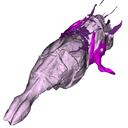
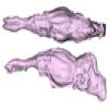
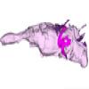
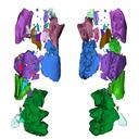
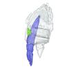
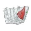
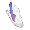
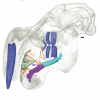
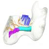
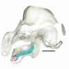
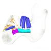
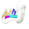
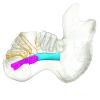
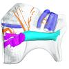
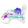
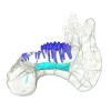
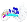
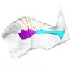
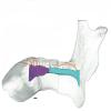
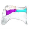
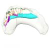
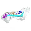
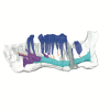
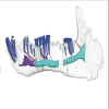
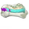
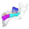
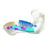
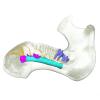
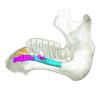
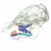
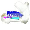
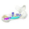
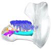
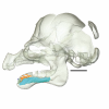
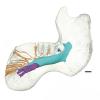
This contribution contains the 3D models described and figured in the following publication: Hautier L, Gomes Rodrigues H, Ferreira-Cardoso S, Emerling CA, Porcher M-L, Asher R, Portela Miguez R, Delsuc F. 2023. From teeth to pad: tooth loss and development of keratinous structures in sirenians. Proceedings of the Royal Society B. https://doi.org/10.1098/rspb.2023.1932
Dugong dugon 2005.51 View specimen
|
M3#1275Internal mandibular morphology. Orange = dorsal canaliculi; purple = mental branches; cyan = mandibular canal; dark blue = teeth; green = tooth alveoli. Type: "3D_surfaces"doi: 10.18563/m3.sf.1275 state:published |
Download 3D surface file |
Dugong dugon 2023.66 View specimen
|
M3#1274Internal mandibular morphology. Orange = dorsal canaliculi; purple = mental branches; cyan = mandibular canal; dark blue = teeth; green = tooth alveoli. Type: "3D_surfaces"doi: 10.18563/m3.sf.1274 state:published |
Download 3D surface file |
Dugong dugon 5386 View specimen
|
M3#1276Internal mandibular morphology. Orange = dorsal canaliculi; purple = mental branches; cyan = mandibular canal; dark blue = teeth; green = tooth alveoli. Type: "3D_surfaces"doi: 10.18563/m3.sf.1276 state:published |
Download 3D surface file |
Dugong dugon 1848.8.29.7/GERM 1027g View specimen
|
M3#1277Internal mandibular morphology. Orange = dorsal canaliculi; purple = mental branches; cyan = mandibular canal; dark blue = teeth. Type: "3D_surfaces"doi: 10.18563/m3.sf.1277 state:published |
Download 3D surface file |
Dugong dugon 1991.413 View specimen
|
M3#1278Internal mandibular morphology. Orange = dorsal canaliculi; purple = mental branches; cyan = mandibular canal; dark blue = teeth. Type: "3D_surfaces"doi: 10.18563/m3.sf.1278 state:published |
Download 3D surface file |
Dugong dugon 1991.427 View specimen
|
M3#1279Internal mandibular morphology. Orange = dorsal canaliculi; purple = mental branches; cyan = mandibular canal; dark blue = teeth. Type: "3D_surfaces"doi: 10.18563/m3.sf.1279 state:published |
Download 3D surface file |
Dugong dugon 2017-3-9 View specimen
|
M3#1280Internal mandibular morphology. Orange = dorsal canaliculi; purple = mental branches; cyan = mandibular canal; dark blue = teeth. Type: "3D_surfaces"doi: 10.18563/m3.sf.1280 state:published |
Download 3D surface file |
Eosiren lybica 1913-22 View specimen
|
M3#1281Internal mandibular morphology. Orange = dorsal canaliculi; purple = mental branches; cyan = mandibular canal; dark blue = teeth; green = tooth alveoli. Type: "3D_surfaces"doi: 10.18563/m3.sf.1281 state:published |
Download 3D surface file |
Halitherium taulannense RGHP C001 View specimen
|
M3#1282Internal mandibular morphology. Cyan = mandibular canal; dark blue = teeth. Type: "3D_surfaces"doi: 10.18563/m3.sf.1282 state:published |
Download 3D surface file |
Halitherium taulannense RGHP C009 View specimen
|
M3#1283Internal mandibular morphology. Orange = dorsal canaliculi; purple = mental branches; cyan = mandibular canal; dark blue = teeth; green = tooth alveoli. Type: "3D_surfaces"doi: 10.18563/m3.sf.1283 state:published |
Download 3D surface file |
Hydrodamalis gigas 1947.10.21.1 View specimen
|
M3#1284Internal mandibular morphology. Orange = dorsal canaliculi; purple = mental branches; cyan = mandibular canal Type: "3D_surfaces"doi: 10.18563/m3.sf.1284 state:published |
Download 3D surface file |
Hydrodamalis gigas C1021 View specimen

|
M3#1285Anterior part of the mandible Type: "3D_surfaces"doi: 10.18563/m3.sf.1285 state:published |
Download 3D surface file |
|
M3#1286Posterior part of the mandible Type: "3D_surfaces"doi: 10.18563/m3.sf.1286 state:published |
Download 3D surface file |
Hydrodamalis gigas 2023.67 View specimen
|
M3#1287Internal mandibular morphology. Orange = dorsal canaliculi; purple = mental branches; cyan = mandibular canal Type: "3D_surfaces"doi: 10.18563/m3.sf.1287 state:published |
Download 3D surface file |
Libysiren sickenbergi M.82429 View specimen
|
M3#1288Internal mandibular morphology. Orange = dorsal canaliculi; purple = mental branches; cyan = mandibular canal; dark blue = teeth; green = tooth alveoli. Type: "3D_surfaces"doi: 10.18563/m3.sf.1288 state:published |
Download 3D surface file |
Libysiren sickenbergi M.45675 View specimen
|
M3#1289Internal mandibular morphology. Orange = dorsal canaliculi; purple = mental branches; cyan = mandibular canal; dark blue = teeth; green = tooth alveoli. Type: "3D_surfaces"doi: 10.18563/m3.sf.1289 state:published |
Download 3D surface file |
Prorastomus sirenoides OR.448976 View specimen
|
M3#1290Internal morphology of the left mandible. Orange = dorsal canaliculi; purple = mental branches; cyan = mandibular canal; dark blue = teeth; green = tooth alveoli. Type: "3D_surfaces"doi: 10.18563/m3.sf.1290 state:published |
Download 3D surface file |
|
M3#1304Internal morphology of the right mandible. Orange = dorsal canaliculi; purple = mental branches; cyan = mandibular canal; dark blue = teeth. Type: "3D_surfaces"doi: 10.18563/m3.sf.1304 state:published |
Download 3D surface file |
Ribodon limbatus M.7073 View specimen
|
M3#1292Internal mandibular morphology. Orange = dorsal canaliculi; purple = mental branches; cyan = mandibular canal; dark blue = teeth; green = tooth alveoli. Type: "3D_surfaces"doi: 10.18563/m3.sf.1292 state:published |
Download 3D surface file |
Rytiodus capgrandi PAL2017-8-1 View specimen
|
M3#1293Internal mandibular morphology. Orange = dorsal canaliculi; purple = mental branches; cyan = mandibular canal; dark blue = teeth; green = tooth alveoli. Type: "3D_surfaces"doi: 10.18563/m3.sf.1293 state:published |
Download 3D surface file |
Trichechus inunguis 1868.12.19.2 View specimen
|
M3#1294Internal mandibular morphology. Orange = dorsal canaliculi; purple = mental branches; cyan = mandibular canal; dark blue = teeth; green = tooth alveoli. Type: "3D_surfaces"doi: 10.18563/m3.sf.1294 state:published |
Download 3D surface file |
Trichechus manatus 1843.3.10.12 View specimen
|
M3#1295Internal mandibular morphology. Orange = dorsal canaliculi; purple = mental branches; cyan = mandibular canal; dark blue = teeth. Type: "3D_surfaces"doi: 10.18563/m3.sf.1295 state:published |
Download 3D surface file |
Trichechus manatus 1864.6.5.1 View specimen
|
M3#1296Internal mandibular morphology. Orange = dorsal canaliculi; purple = mental branches; cyan = mandibular canal; dark blue = teeth; green = tooth alveoli. Type: "3D_surfaces"doi: 10.18563/m3.sf.1296 state:published |
Download 3D surface file |
Trichechus manatus 1950.1.23.1 View specimen
|
M3#1297Internal mandibular morphology. Orange = dorsal canaliculi; purple = mental branches; cyan = mandibular canal; dark blue = teeth; green = tooth alveoli. Type: "3D_surfaces"doi: 10.18563/m3.sf.1297 state:published |
Download 3D surface file |
Trichechus senegalensis 1885.6.30.2 View specimen
|
M3#1298Internal mandibular morphology. Orange = dorsal canaliculi; purple = mental branches; cyan = mandibular canal; dark blue = teeth; green = tooth alveoli. Type: "3D_surfaces"doi: 10.18563/m3.sf.1298 state:published |
Download 3D surface file |
Trichechus senegalensis 1894.7.25.8 View specimen
|
M3#1299Internal mandibular morphology. Orange = dorsal canaliculi; purple = mental branches; cyan = mandibular canal; dark blue = teeth. Type: "3D_surfaces"doi: 10.18563/m3.sf.1299 state:published |
Download 3D surface file |
Trichechus senegalensis V97 View specimen
|
M3#1302Mandibular internal morphology. Orange = dorsal canaliculi; purple = mental branches; cyan = mandibular canal; dark blue = teeth. Type: "3D_surfaces"doi: 10.18563/m3.sf.1302 state:published |
Download 3D surface file |
Trichechus sp. 65.4.28.9 View specimen
|
M3#1300Internal mandibular morphology. Orange = dorsal canaliculi; purple = mental branches; cyan = mandibular canal; dark blue = teeth; green = tooth alveoli. Type: "3D_surfaces"doi: 10.18563/m3.sf.1300 state:published |
Download 3D surface file |
Dugong dugon 1946.8.6.2 View specimen
|
M3#1301Mandibular internal morphology. Orange = dorsal canaliculi; purple = mental branches; cyan = mandibular canal; dark blue = teeth. Type: "3D_surfaces"doi: 10.18563/m3.sf.1301 state:published |
Download 3D surface file |
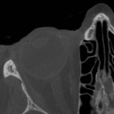
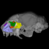
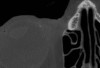
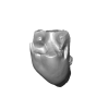
The present Dataset contains the micro-CT scan of the head of an anonymous 54 year old female donor, at a voxel resolution of 145µm. The skin of the face has been masked in order to avoid the donor to be recognized.
Homo sapiens UM_HS_2018_09_13 View specimen
|
M3#1152Micro-ct data set Type: "3D_CT"doi: 10.18563/m3.sf.1152 state:published |
Download CT data |
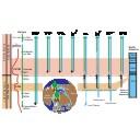
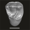
This contribution contains the 3D models described and figured in the following publication: Tabuce R., Marandat B., Adnet S., Gernelle K., Girard F., Marivaux L., Solé F., Schnyder J., Steurbaut E., Storme J.-Y., Vianey-Liaud M., Yans J. (2025). European mammal turnover driven by a global rapid warming event preceding the Paleocene-Eocene Thermal Maximum. PNAS. https://doi.org/10.1073/pnas.2505795122
Acritoparamys aff. atavus UM-ALB-41 View specimen
|
M3#17653D digital model Type: "3D_surfaces"doi: 10.18563/m3.sf.1765 state:published |
Download 3D surface file |
Acritoparamys aff. atavus UM-ALB-42 View specimen
|
M3#1766m1 (right) Type: "3D_surfaces"doi: 10.18563/m3.sf.1766 state:published |
Download 3D surface file |
Acritoparamys aff. atavus UM-ALB-43 View specimen
|
M3#1767M3 (right) Type: "3D_surfaces"doi: 10.18563/m3.sf.1767 state:published |
Download 3D surface file |
indet. indet. UM-ALB-7 View specimen
|
M3#1768M1or2 (left) Type: "3D_surfaces"doi: 10.18563/m3.sf.1768 state:published |
Download 3D surface file |
Arcius cf. rougieri UM-ALB-3 View specimen
|
M3#1769m2 (left) Type: "3D_surfaces"doi: 10.18563/m3.sf.1769 state:published |
Download 3D surface file |
Arfia sp. UM-ALB-2 View specimen
|
M3#1770M1or2 (right) Type: "3D_surfaces"doi: 10.18563/m3.sf.1770 state:published |
Download 3D surface file |
Bustylus sp. UM-ALB-37 View specimen
|
M3#1771M1 (left) Type: "3D_surfaces"doi: 10.18563/m3.sf.1771 state:published |
Download 3D surface file |
?Corbarimys sp. UM-ALB-44 View specimen

|
M3#1772M1or2 (left) Type: "3D_surfaces"doi: 10.18563/m3.sf.1772 state:published |
Download 3D surface file |
indet. indet. UM-ALB-26 View specimen
|
M3#1773upper molar (right) Type: "3D_surfaces"doi: 10.18563/m3.sf.1773 state:published |
Download 3D surface file |
indet. indet. UM-ALB-39 View specimen
|
M3#1774m1or2 (left) Type: "3D_surfaces"doi: 10.18563/m3.sf.1774 state:published |
Download 3D surface file |
Paschatherium marianae UM-ALB-4 View specimen
|
M3#1775P4 (right) Type: "3D_surfaces"doi: 10.18563/m3.sf.1775 state:published |
Download 3D surface file |
Paschatherium marianae UM-ALB-5 View specimen
|
M3#1776DP4 (right) Type: "3D_surfaces"doi: 10.18563/m3.sf.1776 state:published |
Download 3D surface file |
Paschatherium marianae UM-ALB-8 View specimen
|
M3#1777mandible with m2 and talonid of m1 (left) Type: "3D_surfaces"doi: 10.18563/m3.sf.1777 state:published |
Download 3D surface file |
Paschatherium marianae UM-ALB-10 View specimen
|
M3#1778M3 (righ Type: "3D_surfaces"doi: 10.18563/m3.sf.1778 state:published |
Download 3D surface file |
Paschatherium marianae UM-ALB-22 View specimen
|
M3#1779m3 (right) Type: "3D_surfaces"doi: 10.18563/m3.sf.1779 state:published |
Download 3D surface file |
Paschatherium marianae UM-ALB-33 View specimen
|
M3#1780M2 (right) Type: "3D_surfaces"doi: 10.18563/m3.sf.1780 state:published |
Download 3D surface file |
Peratherium sp. UM-ALB-12 View specimen
|
M3#1781?m2 (left) Type: "3D_surfaces"doi: 10.18563/m3.sf.1781 state:published |
Download 3D surface file |
Peratherium sp. UM-ALB-23 View specimen
|
M3#1782?M2 (right) Type: "3D_surfaces"doi: 10.18563/m3.sf.1782 state:published |
Download 3D surface file |
Peratherium sp. UM-ALB-25 View specimen
|
M3#1783?M3 (left) Type: "3D_surfaces"doi: 10.18563/m3.sf.1783 state:published |
Download 3D surface file |
Plagioctenodon cf. dormaalensis UM-ALB-16 View specimen
|
M3#1784M1or2 (right) Type: "3D_surfaces"doi: 10.18563/m3.sf.1784 state:published |
Download 3D surface file |
Plagioctenodon cf. dormaalensis UM-ALB-18 View specimen
|
M3#1785P4 (right) Type: "3D_surfaces"doi: 10.18563/m3.sf.1785 state:published |
Download 3D surface file |
gen. nov. sp. nov. UM-ALB-27 View specimen
|
M3#1786M1or2 (left) Type: "3D_surfaces"doi: 10.18563/m3.sf.1786 state:published |
Download 3D surface file |
Teilhardimys cf. reisi UM-ALB-36a View specimen
|
M3#1787M2 (right) Type: "3D_surfaces"doi: 10.18563/m3.sf.1787 state:published |
Download 3D surface file |
Teilhardimys cf. reisi UM-ALB-36b View specimen
|
M3#1788M1 (right) Type: "3D_surfaces"doi: 10.18563/m3.sf.1788 state:published |
Download 3D surface file |
Wyonycteris sp. UM-ALB-19 View specimen
|
M3#1789M1or2 (right) Type: "3D_surfaces"doi: 10.18563/m3.sf.1789 state:published |
Download 3D surface file |
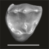
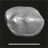
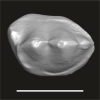
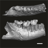
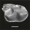
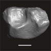
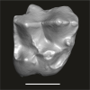
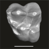
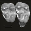
This contribution contains the three-dimensional digital models of the dental fossil material of strepsirrhine primates (Azibiidae and ?Djebelemuridae) from the late early to early middle Eocene of the Gour Lazib Complex in western Algeria and of Djebel Chambi in central-western Tunisia. These fossils were described, figured and discussed in the following publication: Marivaux et al. (2025), New insights into the diversity of strepsirrhine primates from the late early – early middle Eocene of North Africa (Algeria and Tunisia). Journal of Human Evolution, 103729. https://doi.org/10.1016/j.jhevol.2025.103729
Algeripithecus minimissimus ONM-CBI-1-38 View specimen
|
M3#1715Isolated right P3 Type: "3D_surfaces"doi: 10.18563/m3.sf.1715 state:published |
Download 3D surface file |
Algeripithecus minimissimus ONM-CBI-1-37 View specimen
|
M3#1716Isolated right P4 Type: "3D_surfaces"doi: 10.18563/m3.sf.1716 state:published |
Download 3D surface file |
Algeripithecus minimissimus ONM-CBI-1-1206 View specimen
|
M3#1717Isolated right p4 Type: "3D_surfaces"doi: 10.18563/m3.sf.1717 state:published |
Download 3D surface file |
Algeripithecus minimissimus ONM-CBI-1-1207 View specimen
|
M3#1718Isolated right p4 Type: "3D_surfaces"doi: 10.18563/m3.sf.1718 state:published |
Download 3D surface file |
Algeripithecus minimissimus ONM-CBI-1-1205 View specimen
|
M3#1719Fragment of right mandible bearing m1-3 (Holotype) Type: "3D_surfaces"doi: 10.18563/m3.sf.1719 state:published |
Download 3D surface file |
Algeripithecus minimissimus ONM-CBI-1-1209 View specimen
|
M3#1720Isolated left m2 Type: "3D_surfaces"doi: 10.18563/m3.sf.1720 state:published |
Download 3D surface file |
Algeripithecus minimissimus ONM-CBI-1-1208 View specimen
|
M3#1721Isolated right m2 Type: "3D_surfaces"doi: 10.18563/m3.sf.1721 state:published |
Download 3D surface file |
Algeripithecus minutus UM-HGL50-294 View specimen
|
M3#1722Left DP4 Type: "3D_surfaces"doi: 10.18563/m3.sf.1722 state:published |
Download 3D surface file |
Algeripithecus minutus UM-HGL50-297 View specimen
|
M3#1723Isolated right P2 Type: "3D_surfaces"doi: 10.18563/m3.sf.1723 state:published |
Download 3D surface file |
Algeripithecus minutus UM-HGL50-298 View specimen
|
M3#1724Isolated right P3 Type: "3D_surfaces"doi: 10.18563/m3.sf.1724 state:published |
Download 3D surface file |
Algeripithecus minutus UM-HGL50-299 View specimen
|
M3#1725Isolated right P4 Type: "3D_surfaces"doi: 10.18563/m3.sf.1725 state:published |
Download 3D surface file |
Algeripithecus minutus UM-HGL50-303 View specimen
|
M3#1726Isolated left P4 Type: "3D_surfaces"doi: 10.18563/m3.sf.1726 state:published |
Download 3D surface file |
Algeripithecus minutus UM-GZC-7 View specimen
|
M3#1727Isolated left M1 (lingually broken) Type: "3D_surfaces"doi: 10.18563/m3.sf.1727 state:published |
Download 3D surface file |
Algeripithecus minutus UM-GZC-1 View specimen
|
M3#1728Isolated left M2 (Holotype) Type: "3D_surfaces"doi: 10.18563/m3.sf.1728 state:published |
Download 3D surface file |
Algeripithecus minutus UM-HGL50-319 View specimen
|
M3#1729Isolated left M3 Type: "3D_surfaces"doi: 10.18563/m3.sf.1729 state:published |
Download 3D surface file |
Algeripithecus minutus UM-HGL50-397 View specimen
|
M3#1730Fragment of left mandible bearing p3-m3 Type: "3D_surfaces"doi: 10.18563/m3.sf.1730 state:published |
Download 3D surface file |
Azibius magnus UM-HGL50-258 View specimen
|
M3#1731Isolated right P3 or P4 Type: "3D_surfaces"doi: 10.18563/m3.sf.1731 state:published |
Download 3D surface file |
Azibius magnus UM-HGL50-260 View specimen
|
M3#1732Isolated right M2 Type: "3D_surfaces"doi: 10.18563/m3.sf.1732 state:published |
Download 3D surface file |
Azibius magnus UM-HGL50-261 View specimen
|
M3#1733Isolated left M3 Type: "3D_surfaces"doi: 10.18563/m3.sf.1733 state:published |
Download 3D surface file |
Azibius magnus UM-HGL50-263 View specimen
|
M3#1734Isolated left p3 Type: "3D_surfaces"doi: 10.18563/m3.sf.1734 state:published |
Download 3D surface file |
Azibius magnus UM-HGL50-264 View specimen
|
M3#1735Isolated right m1 (Holotype) Type: "3D_surfaces"doi: 10.18563/m3.sf.1735 state:published |
Download 3D surface file |
Azibius magnus UM-HGL50-265 View specimen
|
M3#1736Isolated right m1 (lingually broken) Type: "3D_surfaces"doi: 10.18563/m3.sf.1736 state:published |
Download 3D surface file |
Azibius magnus UM-HGL50-266 View specimen
|
M3#1738Isolated right m2 (corroded) Type: "3D_surfaces"doi: 10.18563/m3.sf.1738 state:published |
Download 3D surface file |
Azibius trerki UM-HGL50-166 View specimen
|
M3#1739Isolated right DP4 Type: "3D_surfaces"doi: 10.18563/m3.sf.1739 state:published |
Download 3D surface file |
Azibius trerki UM-HGL50-295 View specimen
|
M3#1740Isolated left DP4 Type: "3D_surfaces"doi: 10.18563/m3.sf.1740 state:published |
Download 3D surface file |
Azibius trerki UM-HGL51-46 View specimen
|
M3#1741Fragment of right maxillary bearing P3-4 Type: "3D_surfaces"doi: 10.18563/m3.sf.1741 state:published |
Download 3D surface file |
|
M3#1742Fragment of right maxillary bearing M3 Type: "3D_surfaces"doi: 10.18563/m3.sf.1742 state:published |
Download 3D surface file |
Azibius trerki UM-GZC-41 View specimen
|
M3#1743Isolated left P4 Type: "3D_surfaces"doi: 10.18563/m3.sf.1743 state:published |
Download 3D surface file |
Azibius trerki UM-HGL50-396 View specimen
|
M3#1744Boneless fragment of a left maxillary bearing M1-2 Type: "3D_surfaces"doi: 10.18563/m3.sf.1744 state:published |
Download 3D surface file |
Azibius trerki UM-HGL50-270 View specimen
|
M3#1745Fragment (talonid) of an isolated right dp4 Type: "3D_surfaces"doi: 10.18563/m3.sf.1745 state:published |
Download 3D surface file |
Azibius trerki UM-HGL50-248 View specimen
|
M3#1746Isolated left m1 Type: "3D_surfaces"doi: 10.18563/m3.sf.1746 state:published |
Download 3D surface file |
Azibius trerki UM-HGL50-256 View specimen
|
M3#1753Fragment of left mandible bearing p4-m3 Type: "3D_surfaces"doi: 10.18563/m3.sf.1753 state:published |
Download 3D surface file |
Lazibadapis anchomomyinopsis UM-HGL50-326 View specimen
|
M3#1747Isolated right M1 (buccally broken) Type: "3D_surfaces"doi: 10.18563/m3.sf.1747 state:published |
Download 3D surface file |
Lazibadapis anchomomyinopsis UM-HGL50-169 View specimen
|
M3#1748Isolated right M2 (corroded) Type: "3D_surfaces"doi: 10.18563/m3.sf.1748 state:published |
Download 3D surface file |
Lazibadapis anchomomyinopsis UM-HGL50-170 View specimen
|
M3#1749Isolated right M2 or M3 Type: "3D_surfaces"doi: 10.18563/m3.sf.1749 state:published |
Download 3D surface file |
Lazibadapis anchomomyinopsis UM-HGL50-325 View specimen
|
M3#1750Boneless fragment of left mandible preserving m2-3 (Holotype) -> m2 Type: "3D_surfaces"doi: 10.18563/m3.sf.1750 state:published |
Download 3D surface file |

|
M3#1751Boneless fragment of left mandible preserving m2-3 (Holotype) -> m3 Type: "3D_surfaces"doi: 10.18563/m3.sf.1751 state:published |
Download 3D surface file |
Lazibadapis anchomomyinopsis UM-HGL50-290 View specimen
|
M3#1752Isolated left m3 Type: "3D_surfaces"doi: 10.18563/m3.sf.1752 state:published |
Download 3D surface file |
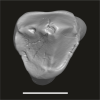
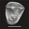
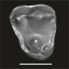
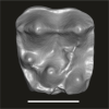
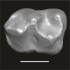
This contribution contains the 3D models described and figured in the following publication: Pujos F., Hautier L., Antoine P-O., Boivin M., Moison B, Salas-Gismondi R, Tejada J.V. , Varas-Malca R.M., Yans J., Marivaux L. (2025). Unexpected pampatheriid from the early Oligocene of Peruvian Amazonia: insights into the tropical differentiation of cingulate xenarthrans. Historical Biology.
Bradypus tridactylus UM-ZOOL-V69 View specimen
|
M3#1600Molariform and associated dentinal microstructure Type: "3D_surfaces"doi: 10.18563/m3.sf.1600 state:published |
Download 3D surface file |
Choloepus didactylus UM-ZOOL-V12 View specimen
|
M3#1601Molariform and associated dentinal microstructure Type: "3D_surfaces"doi: 10.18563/m3.sf.1601 state:published |
Download 3D surface file |
Dasypus mexicanus UM-ZOOL-2787 View specimen

|
M3#1602Molariform and associated dentinal microstructure Type: "3D_surfaces"doi: 10.18563/m3.sf.1602 state:published |
Download 3D surface file |
Tolypeutes matacus UM-ZOOL-2789 View specimen
|
M3#1603Molariform and associated dentinal microstructure Type: "3D_surfaces"doi: 10.18563/m3.sf.1603 state:published |
Download 3D surface file |
Euphractus sexcinctus UM-ZOOL-2790 View specimen
|
M3#1604Molariform and associated dentinal microstructure Type: "3D_surfaces"doi: 10.18563/m3.sf.1604 state:published |
Download 3D surface file |
Holmesina septrionalis UM-FLD-1 View specimen
|
M3#1605Molariform and associated dentinal microstructure Type: "3D_surfaces"doi: 10.18563/m3.sf.1605 state:published |
Download 3D surface file |
Megatherium sp. UM-TAR-1 View specimen
|
M3#1607Molariform and associated dentinal microstructure Type: "3D_surfaces"doi: 10.18563/m3.sf.1607 state:published |
Download 3D surface file |
Indet indet MUSM-3965 View specimen
|
M3#1606Molariform and associated dentinal microstructure Type: "3D_surfaces"doi: 10.18563/m3.sf.1606 state:published |
Download 3D surface file |


