3D models of Cainotheriids Ossicular chain
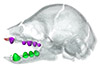
Explodable 3D Dog Skull for Veterinary Education
3D models of Kalakocetus, the earliest Cetacea
3D GM dataset of bird skeletal variation
Skeletal embryonic development in the catshark
Bony connexions of the petrosal bone of extant hippos
bony labyrinth (14) , inner ear (11) , Eocene (11) , geometric morphometrics (10) , CT-scan (10) , Oligocene (9) , Micro-CT (9)
Lionel Hautier (25) , Maëva Judith Orliac (24) , Laurent Marivaux (18) , Renaud Lebrun (15) , Rodolphe Tabuce (15) , Pierre-Olivier Antoine (13) , Bastien Mennecart (13)
|
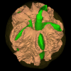
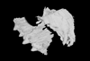
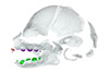
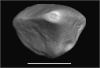
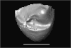
3D models related to the publication: Ontogenetic development of the otic region in the new model organism, Leucoraja erinacea (Chondrichthyes; Rajidae).
|
|
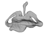
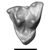
M3#3673D model of the right skeletal labyrinth of the adult specimen of Leucoraja erincea. T Type: "3D_surfaces"doi: 10.18563/m3.sf.367 state:published |
Download 3D surface file |
Leucoraja erinacea 2018.9.25.2 View specimen
|
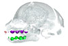
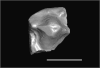
M3#3683D model of the right skeletal labyrinth of the stage 34 specimen of Leucoraja erincea. Type: "3D_surfaces"doi: 10.18563/m3.sf.368 state:published |
Download 3D surface file |
Leucoraja erinacea 2018.9.25.3 View specimen
|
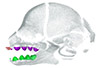
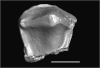
M3#3693D model of the right skeletal labyrinth of the stage 32 specimen of Leucoraja erinacea. Type: "3D_surfaces"doi: 10.18563/m3.sf.369 state:published |
Download 3D surface file |
|
M3#3723D model of the right membranous system of stage 32 of Leucoraja erincea. Type: "3D_surfaces"doi: 10.18563/m3.sf.372 state:published |
Download 3D surface file |
Leucoraja erinacea 2018.9.25.4 View specimen
|
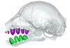
M3#3703D model of the right skeletal labyrinth of the stage 31 specimen of Leucoraja erinacea. Type: "3D_surfaces"doi: 10.18563/m3.sf.370 state:published |
Download 3D surface file |
Leucoraja erinacea 2018.9.26.5 View specimen

|
M3#3763D model of the right skeletal labyrinth of the stage 29 specimen of Leucoraja erinacea. Type: "3D_surfaces"doi: 10.18563/m3.sf.376 state:published |
Download 3D surface file |
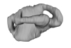
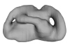
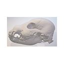
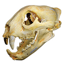
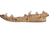
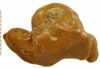
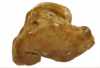
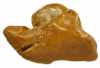
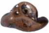
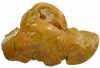
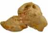
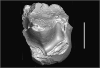
The present 3D Dataset contains the 3D model analyzed in the following publication: Solé et al. (2018), Niche partitioning of the European carnivorous mammals during the paleogene. Palaios. https://doi.org/10.2110/palo.2018.022
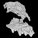
Hyaenodon leptorhynchus FSL848325 View specimen
|
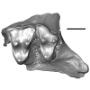
M3#336The specimen FSL848325 is separated in two fragments: the anterior part bears the incisors, the deciduous and permanent canines, while the posterior part bears the right P3, P4, M1 and M2. The P2 is isolated. When combined, the cranium length is approximatively 10.5 cm long. The anterior part is 6.9 cm long and 2.15 cm wide (taken at the level of the P1). The posterior part is 4.8 cm long. The anterior part of the cranium is very narrow. Type: "3D_surfaces"doi: 10.18563/m3.sf.336 state:published |
Download 3D surface file |
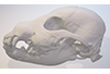
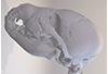
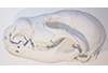
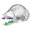
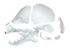
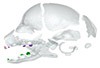
This contribution comprises the 3D models of three wolf pup skulls, which were used for the publication by Geiger et al. 2017 on Neomorphosis and heterochrony of skull shape in dog domestication.
Canis lupus CLL2 View specimen
|
M3#3123d model of a wolf pup skull Type: "3D_surfaces"doi: 10.18563/m3.sf.312 state:published |
Download 3D surface file |
Canis lupus CLL4 View specimen
|
M3#3133d model of a wolf pup skull Type: "3D_surfaces"doi: 10.18563/m3.sf.313 state:published |
Download 3D surface file |
Canis lupus CLL5 View specimen
|
M3#3143d model of a wolf pup skull Type: "3D_surfaces"doi: 10.18563/m3.sf.314 state:published |
Download 3D surface file |
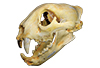
The present 3D Dataset contains the 3D model of a skull analyzed in “A Puma concolor (Carnivora: Felidae) in the Middle-Late Holocene landscapes of the Brazilian Northeast (Bahia): submerged cave deposits and stable isotopes”. The 3D model was generated by photogrammetry.
Puma concolor MN 57461 View specimen
|
M3#843Cranium Type: "3D_surfaces"doi: 10.18563/m3.sf.843 state:published |
Download 3D surface file |
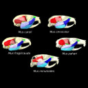
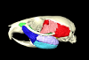
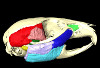
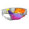
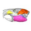
The present 3D Dataset contains the 3D models analyzed in the article entitled "One skull to rule them all? Descriptive and comparative anatomy of the masticatory apparatus in five mice species based on traditional and digital dissections" (Ginot et al. 2018, Journal of Morphology, https://doi.org/10.1002/jmor.20845).
Mus cervicolor R7314 View specimen
|
M3#343.ply surfaces of the skull and masticatory muscles of Mus cervicolor. Created with MorphoDig, .pos and .ntw files also included. Scans were obtained thanks to the Institut des Sciences de l'Evolution de Montpellier MRI platform. Type: "3D_surfaces"doi: 10.18563/m3.sf.343 state:published |
Download 3D surface file |
Mus caroli R7264 View specimen
|
M3#344.ply surfaces of the skull and masticatory muscles of Mus caroli. Created with MorphoDig, .pos and .ntw files also included. Scans were obtained thanks to the Institut des Sciences de l'Evolution de Montpellier MRI platform. Type: "3D_surfaces"doi: 10.18563/m3.sf.344 state:published |
Download 3D surface file |
Mus fragilicauda R7260 View specimen
|
M3#345.ply surfaces of the skull and masticatory muscles of Mus fragilicauda. Created with MorphoDig, .pos and .ntw files also included. Scans were obtained thanks to the Institut des Sciences de l'Evolution de Montpellier MRI platform. Type: "3D_surfaces"doi: 10.18563/m3.sf.345 state:published |
Download 3D surface file |
Mus pahari R7226 View specimen
|
M3#346.ply surfaces of the skull and masticatory muscles of Mus pahari. Created with MorphoDig, .pos and .ntw files also included. Scans were obtained thanks to the Institut des Sciences de l'Evolution de Montpellier MRI platform. Type: "3D_surfaces"doi: 10.18563/m3.sf.346 state:published |
Download 3D surface file |
Mus minutoides minutoides-1 View specimen

|
M3#347.ply surfaces of the skull and masticatory muscles of Mus minutoides. Created with MorphoDig, .pos and .ntw files also included. Scans were obtained thanks to the Institut des Sciences de l'Evolution de Montpellier MRI platform. Type: "3D_surfaces"doi: 10.18563/m3.sf.347 state:published |
Download 3D surface file |
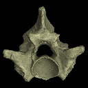
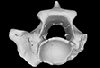
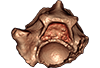
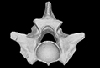
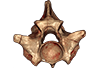
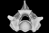
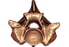
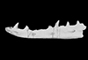
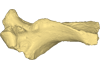
The present 3D Dataset contains the 3D models analyzed in the following publication: Georgalis, G. L., and T. M. Scheyer. A new species of Palaeopython (Serpentes) and other extinct squamates from the Eocene of Dielsdorf (Zurich, Switzerland). Swiss Journal of Geosciences (in press). https://doi.org/10.1007/s00015-019-00341-6
Palaeopython helveticus PIMUZ A/III 631 View specimen
|
M3#399ZIP file containing .ply of vertebra PIMUZ A/III 631 from Palaeopython helveticus n. sp. Type: "3D_surfaces"doi: 10.18563/m3.sf.399 state:published |
Download 3D surface file |
|
M3#403dataset of snake vertebra PIMUZ A/III 631 Type: "3D_CT"doi: 10.18563/m3.sf.403 state:published |
Download CT data |
Palaeopython helveticus PIMUZ A/III 634 View specimen
|
M3#400ZIP file containing .ply of vertebra PIMUZ A/III 634 from Palaeopython helveticus n. sp. (holotype) Type: "3D_surfaces"doi: 10.18563/m3.sf.400 state:published |
Download 3D surface file |
|
M3#404dataset of snake vertbra PIMUZ A/III 634 (holotype) Type: "3D_CT"doi: 10.18563/m3.sf.404 state:published |
Download CT data |
Palaeopython helveticus PIMUZ A/III 636 View specimen
|
M3#401ZIP file containing .ply of vertebra PIMUZ A/III 636 from Palaeopython helveticus n. sp. Type: "3D_surfaces"doi: 10.18563/m3.sf.401 state:published |
Download 3D surface file |
|
M3#406dataset of snake vertebra PIMUZ A/III 636 Type: "3D_CT"doi: 10.18563/m3.sf.406 state:published |
Download CT data |
Palaeovaranus sp. PIMUZ A/III 234 View specimen
|
M3#402ZIP file containing .ply of dentary PIMUZ A/III 234 of Palaeovaranus sp. Type: "3D_surfaces"doi: 10.18563/m3.sf.402 state:published |
Download 3D surface file |
|
M3#405dataset of dentary of Palaeovaranus sp. (PIMUZ A/III 234) Type: "3D_CT"doi: 10.18563/m3.sf.405 state:published |
Download CT data |
This contribution contains the 3D models described and figured in the following publication: Hautier L., Gomes Rodrigues H., Billet G., Asher R.J., 2016. The hidden teeth of sloths: evolutionary vestiges and the development of a simplified dentition. Scientific Reports. doi: 10.1038/srep27763
Bradypus variegatus ZMB 33812 View specimen
|
M3#110Three-dimensional reconstruction of the teeth, mandibles, maxillary and premaxillary bones Type: "3D_surfaces"doi: 10.18563/m3.sf.110 state:published |
Download 3D surface file |
Bradypus variegatus ZMB 41122 View specimen
|
M3#109Three-dimensional reconstruction of the teeth, mandibles, maxillary and premaxillary bones Type: "3D_surfaces"doi: 10.18563/m3.sf.109 state:published |
Download 3D surface file |
Bradypus variegatus MNHN-ZM-MO-1995-326A View specimen
|
M3#111Three-dimensional reconstruction of the teeth, mandibles, maxillary and premaxillary bones Type: "3D_surfaces"doi: 10.18563/m3.sf.111 state:published |
Download 3D surface file |
Bradypus variegatus MNHN-ZM-MO-1995-326B View specimen
|
M3#112Three-dimensional reconstruction of the teeth, mandibles, maxillary and premaxillary bones Type: "3D_surfaces"doi: 10.18563/m3.sf.112 state:published |
Download 3D surface file |
Bradypus sp. MNHN-ZM-MO-1902-325 View specimen
|
M3#113Three-dimensional reconstruction of the teeth, mandibles, maxillary, and premaxillary bones Type: "3D_surfaces"doi: 10.18563/m3.sf.113 state:published |
Download 3D surface file |
Bradypus sp. MNHN-ZM-MO-1995-327 View specimen
|
M3#114Three-dimensional reconstruction of the teeth, mandibles, maxillary and premaxillary bones Type: "3D_surfaces"doi: 10.18563/m3.sf.114 state:published |
Download 3D surface file |
Choloepus didactylus MNHN-ZM-MO-1882-625 View specimen
|
M3#115Three-dimensional reconstruction of the teeth, mandibles, maxillary and premaxillary bones Type: "3D_surfaces"doi: 10.18563/m3.sf.115 state:published |
Download 3D surface file |
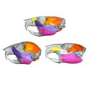
This contribution contains the 3D models described and figured in the following publication: Aguirre-Fernández G, Jost J, and Hilfiker S. 2022. First records of extinct kentriodontid and squalodelphinid dolphins from the Upper Marine Molasse (Burdigalian age) of Switzerland and a reappraisal of the Swiss cetacean fauna.
Kentriodon sp. NMBE 5023944 View specimen
|
M3#8583D models of left periotic and bony labyrinth of NMBE 5023944 (Kentriodon sp.) Type: "3D_surfaces"doi: 10.18563/m3.sf.858 state:published |
Download 3D surface file |
Kentriodon sp. NMBE 5023945 View specimen
|
M3#8593D models of right periotic and bony labyrinth of NMBE 5023945 (Kentriodontidae indet.) Type: "3D_surfaces"doi: 10.18563/m3.sf.859 state:published |
Download 3D surface file |
Kentriodon sp. NMBE 5023946 View specimen
|
M3#8603D models of left periotic and bony labyrinth of NMBE 5023946 (Kentriodon sp.) Type: "3D_surfaces"doi: 10.18563/m3.sf.860 state:published |
Download 3D surface file |
Kentriodon sp. NMBE 5036436 View specimen
|
M3#8613D models of right periotic and bony labyrinth of NMBE 5036436 (Kentriodontidae indet.) Type: "3D_surfaces"doi: 10.18563/m3.sf.861 state:published |
Download 3D surface file |
indet. indet. NMBE 5023942 View specimen
|
M3#8623D models of right periotic and bony labyrinth of NMBE 5023942 (Squalodelphinidae indet.) Type: "3D_surfaces"doi: 10.18563/m3.sf.862 state:published |
Download 3D surface file |
indet. indet. NMBE 5023943 View specimen
|
M3#8633D models of left periotic and bony labyrinth of NMBE 5023943 (Squalodelphinidae indet.) Type: "3D_surfaces"doi: 10.18563/m3.sf.863 state:published |
Download 3D surface file |
indet. indet. NMBE 5036437 View specimen
|
M3#8643D models of left periotic and bony labyrinth of NMBE 5036437 (Physeteridae indet.) Type: "3D_surfaces"doi: 10.18563/m3.sf.864 state:published |
Download 3D surface file |
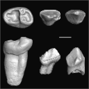
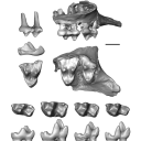
This contribution contains the 3D models of the fossil teeth of a small-bodied platyrrhine primate, Neosaimiri cf. fieldsi (Cebinae, Cebidae, Platyrrhini) discovered from Laventan deposits (late Middle Miocene) of Peruvian Amazonia, San Martín Department (TAR-31: Tarapoto/Juan Guerra vertebrate fossil-bearing locus n°31). These fossils were described and figured in the following publication: Marivaux et al. (2020), New record of Neosaimiri (Cebidae, Platyrrhini) from the late Middle Miocene of Peruvian Amazonia. Journal of Human Evolution. https://doi.org/10.1016/j.jhevol.2020.102835
Neosaimiri cf. fieldsi MUSM-3888 View specimen
|
M3#538MUSM-3888, right m3 of Neosaimiri cf. fieldsi. Type: "3D_surfaces"doi: 10.18563/m3.sf.538 state:published |
Download 3D surface file |
Neosaimiri cf. fieldsi MUSM-3890 View specimen
|
M3#540MUSM-3890, left dp2 of Neosaimiri cf. fieldsi. Type: "3D_surfaces"doi: 10.18563/m3.sf.540 state:published |
Download 3D surface file |
Neosaimiri cf. fieldsi MUSM-3895 View specimen
|
M3#541MUSM-3895, right DC1 of Neosaimiri cf. fieldsi. Type: "3D_surfaces"doi: 10.18563/m3.sf.541 state:published |
Download 3D surface file |
Neosaimiri cf. fieldsi MUSM-3891 View specimen
|
M3#542MUSM-3891, lingual part of a fragmentary right M1 or M2 of Neosaimiri cf. fieldsi. Type: "3D_surfaces"doi: 10.18563/m3.sf.542 state:published |
Download 3D surface file |
Neosaimiri cf. fieldsi MUSM-3892 View specimen
|
M3#543MUSM-3892, distobuccal part of a fragmentary right upper molar (metacone region) of Neosaimiri cf. fieldsi. Type: "3D_surfaces"doi: 10.18563/m3.sf.543 state:published |
Download 3D surface file |
Neosaimiri cf. fieldsi MUSM-3893 View specimen
|
M3#544MUSM-3893, buccal part of a fragmentary right P3 or P4 of Neosaimiri cf. fieldsi. Type: "3D_surfaces"doi: 10.18563/m3.sf.544 state:published |
Download 3D surface file |
Neosaimiri cf. fieldsi MUSM-3894 View specimen
|
M3#545MUSM-3894, lingual part of a fragmentary left P3 or P4 of Neosaimiri cf. fieldsi. Type: "3D_surfaces"doi: 10.18563/m3.sf.545 state:published |
Download 3D surface file |
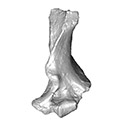
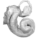
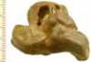
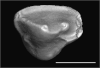
This contribution contains the 3D model described and figured in the following publication: Crochet, J.-Y., Hautier, L., Lehmann, T., 2015. A pangolin (Manidae, Pholidota, Mammalia) from the French Quercy phosphorites (Pech du Fraysse, Saint-Projet, Tarn-et-Garonne, late Oligocene, MP 28). Palaeovertebrata 39(2)-e4. doi: 10.18563/pv.39.2.e4
Necromanis franconica UM PFY 4051 View specimen
|
M3#12A partial left humerus from Pech du Fraysse (Saint-Projet, Tarn-et-Garonne, France), MP 28 (late Oligocene) Type: "3D_surfaces"doi: 10.18563/m3.sf12 state:published |
Download 3D surface file |
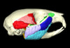
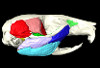
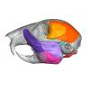
This contribution contains the 3D models described and figured in the following publication: Hautier L, Da Cunha L, Alves Filho M, Moison B, Besson L, Fabre P-H. 20XX. A 3D atlas of the trigeminal nerve and its relevance for comparative studies of the masticatory apparatus in rodents. Journal of Anatomy
Rattus norvegicus UM-ZOOL-3105 View specimen
|
M3#19363D reconstruction of the masticatory musculature, trigeminal nerve and skull of the brown rat Rattus norvegicus Type: "3D_surfaces"doi: 10.18563/m3.sf.1936 state:in_press |
Download 3D surface file |
Cavia porcellus UM-ZOOL-499V View specimen
|
M3#19353D reconstruction of the masticatory musculature, trigeminal nerve and skull of the Guinea pig Cavia porcellus Type: "3D_surfaces"doi: 10.18563/m3.sf.1935 state:in_press |
Download 3D surface file |
Sciurus vulgaris BOUM-2022.2.12 View specimen
|
M3#19373D reconstruction of the masticatory musculature, trigeminal nerve and skull of the red squirrel Sciurus vulgaris Type: "3D_surfaces"doi: 10.18563/m3.sf.1937 state:in_press |
Download 3D surface file |
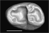
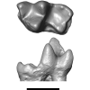
This contribution contains three-dimensional models of the holotypes of Europeradectes marivauxi Gernelle, 2026, and E. williamsoni Gernelle, 2026, as well as other fossil material referred to these species from their type locality, Prémontré (Paris Basin; late early Eocene, 50.4-50.3 Ma). These specimens, among the youngest known for Europeradectes Gernelle, 2026, were analyzed and discussed in: Gernelle et al. (2026), A comprehensive evolutionary history of Peradectidae (Mammalia: Metatheria) revealed by a new dental morphology-based phylogenetic framework, with insights from late early Eocene French specimens.
Europeradectes williamsoni MNHN.F.PRE26 View specimen
|
M3#1889right m3 (holotype) Type: "3D_surfaces"doi: 10.18563/m3.sf.1889 state:in_press |
Download 3D surface file |
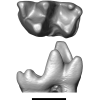
Europeradectes williamsoni MNHN.F.PRE643 View specimen
|
M3#1890left m2 Type: "3D_surfaces"doi: 10.18563/m3.sf.1890 state:in_press |
Download 3D surface file |
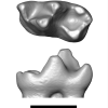
Europeradectes williamsoni MNHN.F.PRE272 View specimen
|
M3#1891right m1 Type: "3D_surfaces"doi: 10.18563/m3.sf.1891 state:in_press |
Download 3D surface file |
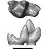
Europeradectes williamsoni MNHN.F.PMT167 View specimen
|
M3#1892left m4 Type: "3D_surfaces"doi: 10.18563/m3.sf.1892 state:in_press |
Download 3D surface file |
Europeradectes marivauxi MNHN.F.PMT169 View specimen
|
M3#1893left M2 Type: "3D_surfaces"doi: 10.18563/m3.sf.1893 state:in_press |
Download 3D surface file |
Europeradectes marivauxi MNHN.F.PMT166 View specimen
|
M3#1894partial right maxilla with M3-M4 (holotype) Type: "3D_surfaces"doi: 10.18563/m3.sf.1894 state:in_press |
Download 3D surface file |