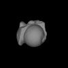
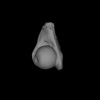
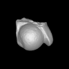
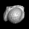
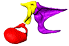
3D models of Cainotheriids Ossicular chain
Explodable 3D Dog Skull for Veterinary Education
3D models of Kalakocetus, the earliest Cetacea
3D GM dataset of bird skeletal variation
Skeletal embryonic development in the catshark
Bony connexions of the petrosal bone of extant hippos
bony labyrinth (14) , inner ear (11) , Eocene (11) , geometric morphometrics (10) , CT-scan (10) , Oligocene (9) , Micro-CT (9)
Lionel Hautier (25) , Maëva Judith Orliac (24) , Laurent Marivaux (18) , Renaud Lebrun (15) , Rodolphe Tabuce (15) , Pierre-Olivier Antoine (13) , Bastien Mennecart (13)
|
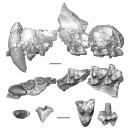
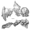
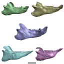
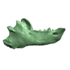
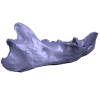
3D model related to the publication: A new fossil of Tayassuidae (Mammalia: Cetartiodactyla) from the Pleistocene of northern BrazilPaula Lopes Copetti
Published online: 29/01/2021 |
|
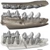
M3#498Left dentary with m1-m3 Type: "3D_surfaces"doi: 10.18563/m3.sf.498 state:published |
Download 3D surface file |

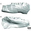
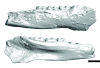
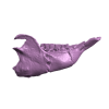
This project presents a µCT dataset and an associated 3D surface model of the holotype of Donrussellia magna (UM PAT 17; Primates, Adapiformes). UM PAT17 is the only known specimen for the species and consists of a well-preserved left lower jaw with p4-m3. It documents one of the oldest European primates, eventually dated near the Paleocene Eocene Thermal Maximum.
Donrussellia magna UM PAT 17 View specimen

|
M3#173D surface file model of UM PAT 17 (type specimen of Donrussellia magna), which is a well preserved left lower jaw with p4-m3. The teeth (and roots) were manually segmented. Type: "3D_surfaces"doi: 10.18563/m3.sf17 state:published |
Download 3D surface file |
|
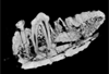
M3#18CT Scan Data of Donrussellia magna UM PAT 17. Voxel size (in µm): 36µm (isotropic voxels). Dimensions in x,y,z : 594 pixels, 294 pixels, 1038 pixels. Image type : 8-bit voxels. Image format : raw data format (no header). Type: "3D_CT"doi: 10.18563/m3.sf18 state:published |
Download CT data |
This contribution contains the three-dimensional models of the most complete and/or informative fossil materials attributed to Peradectes crocheti Gernelle, 2024, the earliest peradectid metatherian species of Europe, from its type locality (Palette, Provence, ~55 Ma). These specimens were analyzed and discussed in: Gernelle et al. (2024), Taxonomy and evolutionary history of peradectids (Metatheria): new data from the early Eocene of France. https://doi.org/10.1007/s10914-024-09724-5
Peradectes crocheti MHN.AIX.PV.2018.26.14 View specimen
|
M3#14993D surface model of MHN.AIX.PV.2018.26.14, fragmentary left maxilla with C-P1, anterior root of P2, and M1-M3 Type: "3D_surfaces"doi: 10.18563/m3.sf.1499 state:published |
Download 3D surface file |
Peradectes crocheti MHN.AIX.PV.2017.6.6 View specimen
|
M3#15003D surface model of MHN.AIX.PV.2017.6.6, left P2 Type: "3D_surfaces"doi: 10.18563/m3.sf.1500 state:published |
Download 3D surface file |
Peradectes crocheti MHN.AIX.PV.2017.6.7 View specimen
|
M3#15013D surface model of MHN.AIX.PV.2017.6.7, left M3 Type: "3D_surfaces"doi: 10.18563/m3.sf.1501 state:published |
Download 3D surface file |
Peradectes crocheti MHN.AIX.PV.2017.6.8 View specimen
|
M3#15023D surface model of MHN.AIX.PV.2017.6.8, right hemi-mandible fragment with canine alveolus, posterior root of p1, partial p2, p3, partial m1, and m2-m3 Type: "3D_surfaces"doi: 10.18563/m3.sf.1502 state:published |
Download 3D surface file |
Peradectes crocheti MHN.AIX.PV.2017.6.9 View specimen
|
M3#15033D surface model of MHN.AIX.PV.2017.6.9, leftm1-m4 row with fragments of dentary Type: "3D_surfaces"doi: 10.18563/m3.sf.1503 state:published |
Download 3D surface file |
Peradectes crocheti MHN.AIX.PV.2017.6.14 View specimen
|
M3#15043D surface model of MHN.AIX.PV.2017.6.14, right astragalus Type: "3D_surfaces"doi: 10.18563/m3.sf.1504 state:published |
Download 3D surface file |

This contribution contains 3D models of the cranial endoskeleton of three specimens of the Permian ‘acanthodian’ stem-group chondrichthyan (cartilaginous fish) Acanthodes confusus, obtained using computed tomography. These datasets were described and analyzed in Dearden et al. (2024) “3D models related to the publication: The pharynx of the iconic stem-group chondrichthyan Acanthodes Agassiz, 1833 revisited with micro computed tomography.” Zoological Journal of the Linnean Society
Acanthodes confusus MNHN-F-SAA20 View specimen
|
M3#14703D surfaces representing the three-dimensionally fossilised head of Acanthodes confusus Type: "3D_surfaces"doi: 10.18563/m3.sf.1470 state:published |
Download 3D surface file |
Acanthodes confusus MNHN-F-SAA21 View specimen
|
M3#14713D surfaces representing the three-dimensionally fossilised head of Acanthodes confusus Type: "3D_surfaces"doi: 10.18563/m3.sf.1471 state:published |
Download 3D surface file |
Acanthodes confusus MNHN-F-SAA24 View specimen
|
M3#14723D surfaces representing the three-dimensionally fossilised head of Acanthodes confusus Type: "3D_surfaces"doi: 10.18563/m3.sf.1472 state:published |
Download 3D surface file |
The present contribution contains the 3D models of fossil humeri and ilia of anurans from various Eocene-Miocene deposits of Peruvian Amazonia. These fossils were described and figured in the following publication: Jansen et al. (2023), First Eocene–Miocene anuran fossils from Peruvian Amazonia: insights into Neotropical frog evolution and diversity. Papers in Palaeontology, The Palaeontological Association.
Indet. indet. MUSM 4746 View specimen
|
M3#1231Humeral fragment (distal end) Type: "3D_surfaces"doi: 10.18563/m3.sf.1231 state:published |
Download 3D surface file |
Indet. indet. MUSM 4747 View specimen
|
M3#1232Humeral fragment (distal end) Type: "3D_surfaces"doi: 10.18563/m3.sf.1232 state:published |
Download 3D surface file |
Indet. indet. MUSM 4748 View specimen
|
M3#1233Humeral fragment (distal end) Type: "3D_surfaces"doi: 10.18563/m3.sf.1233 state:published |
Download 3D surface file |
Indet. indet. MUSM 4755 View specimen
|
M3#1234Humeral fragment (distal end) Type: "3D_surfaces"doi: 10.18563/m3.sf.1234 state:published |
Download 3D surface file |
Indet. indet. MUSM 4756 View specimen
|
M3#1235Humeral fragment (distal end) Type: "3D_surfaces"doi: 10.18563/m3.sf.1235 state:published |
Download 3D surface file |
Indet. indet. MUSM 4757 View specimen
|
M3#1236Humeral fragment (distal end) Type: "3D_surfaces"doi: 10.18563/m3.sf.1236 state:published |
Download 3D surface file |
Indet. indet. MUSM 4761 View specimen
|
M3#1237Humeral fragment (distal end) Type: "3D_surfaces"doi: 10.18563/m3.sf.1237 state:published |
Download 3D surface file |
Indet. indet. MUSM 4763 View specimen
|
M3#1238Humeral fragment (distal end) Type: "3D_surfaces"doi: 10.18563/m3.sf.1238 state:published |
Download 3D surface file |
Indet. indet. MUSM 4765 View specimen
|
M3#1239Humeral fragment (distal end) Type: "3D_surfaces"doi: 10.18563/m3.sf.1239 state:published |
Download 3D surface file |
Indet. indet. MUSM 4766 View specimen
|
M3#1240Humeral fragment (distal end) Type: "3D_surfaces"doi: 10.18563/m3.sf.1240 state:published |
Download 3D surface file |
Indet. indet. MUSM 4775 View specimen
|
M3#1241Humeral fragment (distal end) Type: "3D_surfaces"doi: 10.18563/m3.sf.1241 state:published |
Download 3D surface file |
cf. Pipa sp. MUSM 4776 View specimen
|
M3#1242Humeral fragment (distal end) Type: "3D_surfaces"doi: 10.18563/m3.sf.1242 state:published |
Download 3D surface file |
Indet. indet. MUSM 4788 View specimen
|
M3#1243Ilial fragment Type: "3D_surfaces"doi: 10.18563/m3.sf.1243 state:published |
Download 3D surface file |
Indet. indet. MUSM 4789 View specimen
|
M3#1244Ilial fragment Type: "3D_surfaces"doi: 10.18563/m3.sf.1244 state:published |
Download 3D surface file |
Indet. indet. MUSM 4790 View specimen
|
M3#1245Ilial fragment Type: "3D_surfaces"doi: 10.18563/m3.sf.1245 state:published |
Download 3D surface file |
Indet. indet. MUSM 4792 View specimen
|
M3#1246Ilial fragment Type: "3D_surfaces"doi: 10.18563/m3.sf.1246 state:published |
Download 3D surface file |
Indet. indet. MUSM 4793 View specimen
|
M3#1247Ilial fragment Type: "3D_surfaces"doi: 10.18563/m3.sf.1247 state:published |
Download 3D surface file |
Indet. indet. MUSM 4794 View specimen
|
M3#1249Ilial fragment Type: "3D_surfaces"doi: 10.18563/m3.sf.1249 state:published |
Download 3D surface file |
Indet. indet. MUSM 4795 View specimen
|
M3#1250Ilial fragment Type: "3D_surfaces"doi: 10.18563/m3.sf.1250 state:published |
Download 3D surface file |
cf. Pipa sp. MUSM 4796 View specimen
|
M3#1251Ilial fragment Type: "3D_surfaces"doi: 10.18563/m3.sf.1251 state:published |
Download 3D surface file |
cf. Pipa sp. MUSM 4797 View specimen
|
M3#1252Ilial fragment Type: "3D_surfaces"doi: 10.18563/m3.sf.1252 state:published |
Download 3D surface file |
The present 3D Dataset contains the 3D models analyzed in 3D Finite Element Analysis and Geometric Morphometrics of Sloths (Xenarthra, Folivora) Mandibles Show Insights on the Dietary Specializations of Fossil Taxa. Journal of South American Earth Sciences. https://doi.org/10.1016/j.jsames.2023.104445
Mylodon darwinii CAV 379 View specimen
|
M3#1159Right hemimandible Type: "3D_surfaces"doi: 10.18563/m3.sf.1159 state:published |
Download 3D surface file |
Scelidotherium leptocephalum MNHN-M 137,722 View specimen
|
M3#1160Mandible Type: "3D_surfaces"doi: 10.18563/m3.sf.1160 state:published |
Download 3D surface file |
Glossotherium robustum MNHN-M 914 View specimen
|
M3#1161Mandible Type: "3D_surfaces"doi: 10.18563/m3.sf.1161 state:published |
Download 3D surface file |
Lestodon armatus MPAC 899 View specimen
|
M3#1162Mandible Type: "3D_surfaces"doi: 10.18563/m3.sf.1162 state:published |
Download 3D surface file |
Valgipes bucklandi NHMD.Z.M.K. 1/1845:3540 View specimen
|
M3#1163Mandible Type: "3D_surfaces"doi: 10.18563/m3.sf.1163 state:published |
Download 3D surface file |
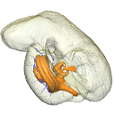
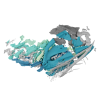
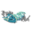
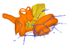
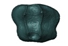
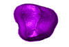
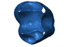
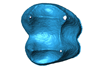
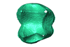
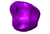
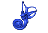
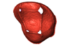
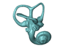
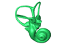
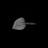
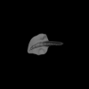
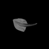
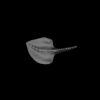
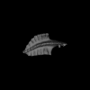
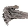
In this contribution, we describe the external and internal morphology of a delphinid petrosal bone collected from Ahu Tahai, a burial site located on the Southwestern coast of Easter Island, at Hangaroa. We discuss the taxonomic attribution of this archaeological item and describe its internal structures based on µCT data, including the bony labyrinth and the nerve and vein patterns. Identification of the nerves exists lead us to relocate the identification of the foramen singulare in delphinid petrosals.
indet. indet. AT1 View specimen
|
M3#420Stapes Type: "3D_surfaces"doi: 10.18563/m3.sf.420 state:published |
Download 3D surface file |
|
M3#421petrosal bone Type: "3D_surfaces"doi: 10.18563/m3.sf.421 state:published |
Download 3D surface file |
|
M3#422in situ bony labyrinth Type: "3D_surfaces"doi: 10.18563/m3.sf.422 state:published |
Download 3D surface file |
|
M3#423bony labyrinth and associated nerves and blood vessels Type: "3D_surfaces"doi: 10.18563/m3.sf.423 state:published |
Download 3D surface file |
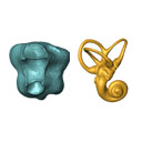
The present 3D Dataset contains the 3D models of the enamel-dentine junctions of upper third molars and of the bony labyrinths of the extant cercopithecoid specimens analyzed in the following publication: Beaudet, A., Dumoncel, J., Thackeray, J.F., Bruxelles, L., Duployer, B., Tenailleau, C., Bam, L., Hoffman, J., de Beer, F., Braga, J.: Upper third molar internal structural organization and semicircular canal morphology in Plio-Pleistocene South African cercopithecoids. Journal of Human Evolution 95, 104-120. https://doi.org/10.1016/j.jhevol.2016.04.004
Cercocebus atys 81.007-M-0041 View specimen
|
M3#4453D model of the enamel-dentine junction of the right upper third molar. Type: "3D_surfaces"doi: 10.18563/m3.sf.445 state:published |
Download 3D surface file |
Cercocebus torquatus 73.018-M-0359 View specimen
|
M3#4463D model of the enamel-dentine junction of the right upper third molar. Type: "3D_surfaces"doi: 10.18563/m3.sf.446 state:published |
Download 3D surface file |

|
M3#4963D model of the left bony labyrinth. Type: "3D_surfaces"doi: 10.18563/m3.sf.496 state:published |
Download 3D surface file |
Mandrillus leucophaeus 73.029-M-0106 View specimen
|
M3#4473D model of the enamel-dentine junction of the right upper third molar. Type: "3D_surfaces"doi: 10.18563/m3.sf.447 state:published |
Download 3D surface file |

|
M3#4703D model of the right bony labyrinth. Type: "3D_surfaces"doi: 10.18563/m3.sf.470 state:published |
Download 3D surface file |
Lophocebus albigena 73.029-M-0109 View specimen
|
M3#4483D model of the enamel-dentine junction of the right upper third molar. Type: "3D_surfaces"doi: 10.18563/m3.sf.448 state:published |
Download 3D surface file |

|
M3#4713D model of the right bony labyrinth. Type: "3D_surfaces"doi: 10.18563/m3.sf.471 state:published |
Download 3D surface file |
Piliocolobus foai 91.060-M-0071 View specimen
|
M3#4493D model of the enamel-dentine junction of the right upper third molar. Type: "3D_surfaces"doi: 10.18563/m3.sf.449 state:published |
Download 3D surface file |

|
M3#4723D model of the right bony labyrinth. Type: "3D_surfaces"doi: 10.18563/m3.sf.472 state:published |
Download 3D surface file |
Colobus guereza 1215 View specimen
|
M3#4503D model of the enamel-dentine junction of the right upper third molar. Type: "3D_surfaces"doi: 10.18563/m3.sf.450 state:published |
Download 3D surface file |

|
M3#4733D model of the right bony labyrinth. Type: "3D_surfaces"doi: 10.18563/m3.sf.473 state:published |
Download 3D surface file |
Colobus guereza 2800 View specimen
|
M3#4513D model of the enamel-dentine junction of the right upper third molar. Type: "3D_surfaces"doi: 10.18563/m3.sf.451 state:published |
Download 3D surface file |

|
M3#4743D model of the right bony labyrinth. Type: "3D_surfaces"doi: 10.18563/m3.sf.474 state:published |
Download 3D surface file |
Papio cynocephalus kindae 3503 View specimen
|
M3#4523D model of the enamel-dentine junction of the right upper third molar. Type: "3D_surfaces"doi: 10.18563/m3.sf.452 state:published |
Download 3D surface file |

|
M3#4753D model of the right bony labyrinth. Type: "3D_surfaces"doi: 10.18563/m3.sf.475 state:published |
Download 3D surface file |
Erythrocebus patas 8452 View specimen
|
M3#4533D model of the enamel-dentine junction of the right upper third molar. Type: "3D_surfaces"doi: 10.18563/m3.sf.453 state:published |
Download 3D surface file |

|
M3#4763D model of the right bony labyrinth. Type: "3D_surfaces"doi: 10.18563/m3.sf.476 state:published |
Download 3D surface file |
Papio cynocephalus kindae 17979 View specimen
|
M3#4543D model of the enamel-dentine junction of the right upper third molar. Type: "3D_surfaces"doi: 10.18563/m3.sf.454 state:published |
Download 3D surface file |

|
M3#4773D model of the right bony labyrinth. Type: "3D_surfaces"doi: 10.18563/m3.sf.477 state:published |
Download 3D surface file |
Colobus angolensis 25456 View specimen
|
M3#4553D model of the enamel-dentine junction of the right upper third molar. Type: "3D_surfaces"doi: 10.18563/m3.sf.455 state:published |
Download 3D surface file |

|
M3#4783D model of the right bony labyrinth. Type: "3D_surfaces"doi: 10.18563/m3.sf.478 state:published |
Download 3D surface file |
Chlorocebus pygerythrus 37477 View specimen
|
M3#4563D model of the enamel-dentine junction of the right upper third molar. Type: "3D_surfaces"doi: 10.18563/m3.sf.456 state:published |
Download 3D surface file |
|
M3#4813D model of the right bony labyrinth. Type: "3D_surfaces"doi: 10.18563/m3.sf.481 state:published |
Download 3D surface file |
Chlorocebus pygerythrus 37478 View specimen
|
M3#4573D model of the enamel-dentine junction of the right upper third molar. Type: "3D_surfaces"doi: 10.18563/m3.sf.457 state:published |
Download 3D surface file |

|
M3#4823D model of the right bony labyrinth. Type: "3D_surfaces"doi: 10.18563/m3.sf.482 state:published |
Download 3D surface file |
Lophocebus albigena 37572 View specimen
|
M3#4583D model of the enamel-dentine junction of the right upper third molar. Type: "3D_surfaces"doi: 10.18563/m3.sf.458 state:published |
Download 3D surface file |

|
M3#4833D model of the right bony labyrinth. Type: "3D_surfaces"doi: 10.18563/m3.sf.483 state:published |
Download 3D surface file |
Lophocebus albigena 37579 View specimen
|
M3#4593D model of the enamel-dentine junction of the right upper third molar. Type: "3D_surfaces"doi: 10.18563/m3.sf.459 state:published |
Download 3D surface file |
Erythrocebus patas OST.2002-26 View specimen
|
M3#4603D model of the enamel-dentine junction of the right upper third molar. Type: "3D_surfaces"doi: 10.18563/m3.sf.460 state:published |
Download 3D surface file |
|
M3#4843D model of the right bony labyrinth. Type: "3D_surfaces"doi: 10.18563/m3.sf.484 state:published |
Download 3D surface file |
Mandrillus sphinx OST.AC.488 View specimen
|
M3#4613D model of the enamel-dentine junction of the right upper third molar. Type: "3D_surfaces"doi: 10.18563/m3.sf.461 state:published |
Download 3D surface file |
|
M3#4853D model of the left bony labyrinth. Type: "3D_surfaces"doi: 10.18563/m3.sf.485 state:published |
Download 3D surface file |
Macaca mulatta OST.AC.492 View specimen
|
M3#4623D model of the enamel-dentine junction of the right upper third molar. Type: "3D_surfaces"doi: 10.18563/m3.sf.462 state:published |
Download 3D surface file |

|
M3#4863D model of the right bony labyrinth. Type: "3D_surfaces"doi: 10.18563/m3.sf.486 state:published |
Download 3D surface file |
Chlorocebus aethiops OST.AC.523 View specimen

|
M3#4633D model of the enamel-dentine junction of the right upper third molar. Type: "3D_surfaces"doi: 10.18563/m3.sf.463 state:published |
Download 3D surface file |

|
M3#4913D model of the right bony labyrinth. Type: "3D_surfaces"doi: 10.18563/m3.sf.491 state:published |
Download 3D surface file |
Cercopithecus cephus OST.AC.533 View specimen
|
M3#4643D model of the enamel-dentine junction of the right upper third molar. Type: "3D_surfaces"doi: 10.18563/m3.sf.464 state:published |
Download 3D surface file |

|
M3#4933D model of the right bony labyrinth. Type: "3D_surfaces"doi: 10.18563/m3.sf.493 state:published |
Download 3D surface file |
Chlorocebus aethiops OST.AC.540 View specimen
|
M3#4653D model of the enamel-dentine junction of the right upper third molar. Type: "3D_surfaces"doi: 10.18563/m3.sf.465 state:published |
Download 3D surface file |

|
M3#4943D model of the right bony labyrinth. Type: "3D_surfaces"doi: 10.18563/m3.sf.494 state:published |
Download 3D surface file |
Mandrillus sphinx OST.AC.543 View specimen
|
M3#4663D model of the enamel-dentine junction of the right upper third molar. Type: "3D_surfaces"doi: 10.18563/m3.sf.466 state:published |
Download 3D surface file |
|
M3#4953D model of the right bony labyrinth. Type: "3D_surfaces"doi: 10.18563/m3.sf.495 state:published |
Download 3D surface file |
Cercocebus torquatus 73.018-M-389 View specimen

|
M3#4683D model of the right bony labyrinth. Type: "3D_surfaces"doi: 10.18563/m3.sf.468 state:published |
Download 3D surface file |
Mandrillus leucophaeus 73.029-M-0105 View specimen
|
M3#4693D model of the right bony labyrinth. Type: "3D_surfaces"doi: 10.18563/m3.sf.469 state:published |
Download 3D surface file |
Mandrillus leucophaeus 28425 View specimen

|
M3#4793D model of the right bony labyrinth. Type: "3D_surfaces"doi: 10.18563/m3.sf.479 state:published |
Download 3D surface file |
Cercocebus atys 28998 View specimen

|
M3#4803D model of the right bony labyrinth. Type: "3D_surfaces"doi: 10.18563/m3.sf.480 state:published |
Download 3D surface file |
Macaca sylvanus OST.AC.493 View specimen

|
M3#4873D model of the right bony labyrinth. Type: "3D_surfaces"doi: 10.18563/m3.sf.487 state:published |
Download 3D surface file |
Chlorocebus aethiops OST.AC.508 View specimen

|
M3#4883D model of the left bony labyrinth. Type: "3D_surfaces"doi: 10.18563/m3.sf.488 state:published |
Download 3D surface file |
Cercopithecus cephus OST.AC.515 View specimen

|
M3#4893D model of the right bony labyrinth. Type: "3D_surfaces"doi: 10.18563/m3.sf.489 state:published |
Download 3D surface file |
Colobus guereza OST.AC.519 View specimen

|
M3#4903D model of the right bony labyrinth. Type: "3D_surfaces"doi: 10.18563/m3.sf.490 state:published |
Download 3D surface file |
Macaca sp. OST.AC.532 View specimen

|
M3#4923D model of the left bony labyrinth. Type: "3D_surfaces"doi: 10.18563/m3.sf.492 state:published |
Download 3D surface file |
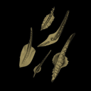
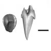
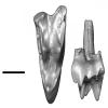
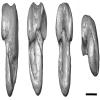
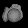
The present 3D Dataset contains the 3D models analyzed in Assemat et al. 2023: Shape diversity in conodont elements, a quantitative study using 3D topography. Marine Micropaleontology 184. https://doi.org/10.1016/j.marmicro.2023.102292
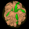
P1 elements represent dental components of the conodont apparatus that perform the final stage of food processing before ingestion. Consequently, quantifying the shape of P1 elements across the topographic indices of different conodont species becomes crucial for deciphering the diversity in feeding behavior within this group.
Bispathodus aculeatus UM CTB 082 View specimen
|
M3#1404P element Type: "3D_surfaces"doi: 10.18563/m3.sf.1404 state:published |
Download 3D surface file |
Bispathodus aculeatus UM CTB 083 View specimen

|
M3#1405P element Type: "3D_surfaces"doi: 10.18563/m3.sf.1405 state:published |
Download 3D surface file |
Bispathodus aculeatus UM CTB 086 View specimen
|
M3#1406P element Type: "3D_surfaces"doi: 10.18563/m3.sf.1406 state:published |
Download 3D surface file |
Bispathodus ultimus UM CTB 088 View specimen

|
M3#1407P element Type: "3D_surfaces"doi: 10.18563/m3.sf.1407 state:published |
Download 3D surface file |
Bispathodus aculeatus UM CTB 089 View specimen
|
M3#1408P element Type: "3D_surfaces"doi: 10.18563/m3.sf.1408 state:published |
Download 3D surface file |
Bispathodus costatus UM CTB 090 View specimen
|
M3#1409P element Type: "3D_surfaces"doi: 10.18563/m3.sf.1409 state:published |
Download 3D surface file |
Bispathodus ultimus UM CTB 092 View specimen
|
M3#1410P element Type: "3D_surfaces"doi: 10.18563/m3.sf.1410 state:published |
Download 3D surface file |
Bispathodus costatus UM CTB 093 View specimen
|
M3#1411P element Type: "3D_surfaces"doi: 10.18563/m3.sf.1411 state:published |
Download 3D surface file |
Bispathodus spinulicostatus UM CTB 094 View specimen
|
M3#1412P element Type: "3D_surfaces"doi: 10.18563/m3.sf.1412 state:published |
Download 3D surface file |
Bispathodus aculeatus UM CTB 096 View specimen
|
M3#1413P element Type: "3D_surfaces"doi: 10.18563/m3.sf.1413 state:published |
Download 3D surface file |
Bispathodus ultimus UM CTB 098 View specimen
|
M3#1414P element Type: "3D_surfaces"doi: 10.18563/m3.sf.1414 state:published |
Download 3D surface file |
Bispathodus costatus UM CTB 060 View specimen
|
M3#1415P element Type: "3D_surfaces"doi: 10.18563/m3.sf.1415 state:published |
Download 3D surface file |
Bispathodus spinulicostatus UM CTB 073 View specimen
|
M3#1416P element Type: "3D_surfaces"doi: 10.18563/m3.sf.1416 state:published |
Download 3D surface file |
Branmehla suprema UM CTB 049 View specimen

|
M3#1417P element Type: "3D_surfaces"doi: 10.18563/m3.sf.1417 state:published |
Download 3D surface file |
Branmehla inornata UM CTB 100 View specimen
|
M3#1418P element Type: "3D_surfaces"doi: 10.18563/m3.sf.1418 state:published |
Download 3D surface file |
Bispathodus stabilis (morphe 1) UM CTB 101 View specimen

|
M3#1419P element Type: "3D_surfaces"doi: 10.18563/m3.sf.1419 state:published |
Download 3D surface file |
Branmehla suprema UM CTB 102 View specimen

|
M3#1420P element Type: "3D_surfaces"doi: 10.18563/m3.sf.1420 state:published |
Download 3D surface file |
Branmehla suprema UM CTB 103 View specimen

|
M3#1421P element Type: "3D_surfaces"doi: 10.18563/m3.sf.1421 state:published |
Download 3D surface file |
Branmehla suprema UM CTB 104 View specimen
|
M3#1422P element Type: "3D_surfaces"doi: 10.18563/m3.sf.1422 state:published |
Download 3D surface file |
Branmehla suprema UM CTB 105 View specimen

|
M3#1423P element Type: "3D_surfaces"doi: 10.18563/m3.sf.1423 state:published |
Download 3D surface file |
Branmehla suprema UM CTB 106 View specimen
|
M3#1424P element Type: "3D_surfaces"doi: 10.18563/m3.sf.1424 state:published |
Download 3D surface file |
Branmehla suprema UM CTB 072 View specimen

|
M3#1425P element Type: "3D_surfaces"doi: 10.18563/m3.sf.1425 state:published |
Download 3D surface file |
Branmehla suprema UM CTB 107 View specimen
|
M3#1426P element Type: "3D_surfaces"doi: 10.18563/m3.sf.1426 state:published |
Download 3D surface file |
Branmehla suprema UM CTB 108 View specimen

|
M3#1427P element Type: "3D_surfaces"doi: 10.18563/m3.sf.1427 state:published |
Download 3D surface file |
Branmehla suprema UM CTB 109 View specimen
|
M3#1428P element Type: "3D_surfaces"doi: 10.18563/m3.sf.1428 state:published |
Download 3D surface file |
Bispathodus stabilis (morphe 1) UM CTB 110 View specimen

|
M3#1429P element Type: "3D_surfaces"doi: 10.18563/m3.sf.1429 state:published |
Download 3D surface file |
Palmatolepis gracilis UM CTB 112 View specimen

|
M3#1430P element Type: "3D_surfaces"doi: 10.18563/m3.sf.1430 state:published |
Download 3D surface file |
Palmatolepis gracilis UM CTB 061 View specimen

|
M3#1431P element Type: "3D_surfaces"doi: 10.18563/m3.sf.1431 state:published |
Download 3D surface file |
Palmatolepis gracilis UM CTB 115 View specimen
|
M3#1432P element Type: "3D_surfaces"doi: 10.18563/m3.sf.1432 state:published |
Download 3D surface file |
Palmatolepis gracilis UM CTB 116 View specimen

|
M3#1433P element Type: "3D_surfaces"doi: 10.18563/m3.sf.1433 state:published |
Download 3D surface file |
Palmatolepis gracilis UM CTB 117 View specimen
|
M3#1434P element Type: "3D_surfaces"doi: 10.18563/m3.sf.1434 state:published |
Download 3D surface file |
Palmatolepis gracilis UM CTB 062 View specimen
|
M3#1435P element Type: "3D_surfaces"doi: 10.18563/m3.sf.1435 state:published |
Download 3D surface file |
Palmatolepis gracilis UM CTB 118 View specimen
|
M3#1436P element Type: "3D_surfaces"doi: 10.18563/m3.sf.1436 state:published |
Download 3D surface file |
Palmatolepis gracilis UM CTB 119 View specimen

|
M3#1437P element Type: "3D_surfaces"doi: 10.18563/m3.sf.1437 state:published |
Download 3D surface file |
Palmatolepis gracilis UM CTB 120 View specimen
|
M3#1438P element Type: "3D_surfaces"doi: 10.18563/m3.sf.1438 state:published |
Download 3D surface file |
Polygnathus communis UM CTB 075 View specimen
|
M3#1439P element Type: "3D_surfaces"doi: 10.18563/m3.sf.1439 state:published |
Download 3D surface file |
Polygnathus communis UM CTB 121 View specimen
|
M3#1440P element Type: "3D_surfaces"doi: 10.18563/m3.sf.1440 state:published |
Download 3D surface file |
Polygnathus communis UM CTB 122 View specimen
|
M3#1441P element Type: "3D_surfaces"doi: 10.18563/m3.sf.1441 state:published |
Download 3D surface file |
Polygnathus communis UM CTB 123 View specimen
|
M3#1442P element Type: "3D_surfaces"doi: 10.18563/m3.sf.1442 state:published |
Download 3D surface file |
Polygnathus communis UM CTB 125 View specimen
|
M3#1443P element Type: "3D_surfaces"doi: 10.18563/m3.sf.1443 state:published |
Download 3D surface file |
Polygnathus communis UM CTB 126 View specimen
|
M3#1444P element Type: "3D_surfaces"doi: 10.18563/m3.sf.1444 state:published |
Download 3D surface file |
Polygnathus communis UM CTB 128 View specimen
|
M3#1445P element Type: "3D_surfaces"doi: 10.18563/m3.sf.1445 state:published |
Download 3D surface file |
Polygnathus communis UM CTB 130 View specimen
|
M3#1446P element Type: "3D_surfaces"doi: 10.18563/m3.sf.1446 state:published |
Download 3D surface file |
Polygnathus communis UM CTB 131 View specimen
|
M3#1447P element Type: "3D_surfaces"doi: 10.18563/m3.sf.1447 state:published |
Download 3D surface file |
Polygnathus communis UM CTB 132 View specimen
|
M3#1448P element Type: "3D_surfaces"doi: 10.18563/m3.sf.1448 state:published |
Download 3D surface file |
Polygnathus communis UM CTB 133 View specimen
|
M3#1449P element Type: "3D_surfaces"doi: 10.18563/m3.sf.1449 state:published |
Download 3D surface file |
Polygnathus symmetricus UM CTB 139 View specimen
|
M3#1450P element Type: "3D_surfaces"doi: 10.18563/m3.sf.1450 state:published |
Download 3D surface file |
Polygnathus symmetricus UM CTB 140 View specimen
|
M3#1451P element Type: "3D_surfaces"doi: 10.18563/m3.sf.1451 state:published |
Download 3D surface file |
Polygnathus symmetricus UM CTB 141 View specimen
|
M3#1452P element Type: "3D_surfaces"doi: 10.18563/m3.sf.1452 state:published |
Download 3D surface file |
Polygnathus symmetricus UM CTB 142 View specimen
|
M3#1453P element Type: "3D_surfaces"doi: 10.18563/m3.sf.1453 state:published |
Download 3D surface file |
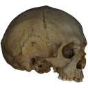
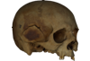
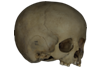
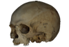
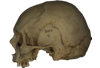
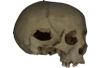
This contribution contains the 3D models described and figured in the following publications:
- Marini E., Lussu P., 2020. A virtual physical anthropology lab. Teaching in the time of coronavirus, in prep.;
- Lussu P., Bratzu D., Marini E., 2020. Cloud-based ultra close-range digital photogrammetry: validation of an approach for the effective virtual reconstruction of skeletal remains, in prep.
Homo sapiens MSAE 59 View specimen
|
M3#509MSAE 59 Type: "3D_surfaces"doi: 10.18563/m3.sf.509 state:published |
Download 3D surface file |
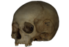
Homo sapiens MSAE 62 View specimen
|
M3#510MSAE 62 Type: "3D_surfaces"doi: 10.18563/m3.sf.510 state:published |
Download 3D surface file |
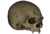
Homo sapiens MSAE 63 View specimen
|
M3#512MSAE 63 Type: "3D_surfaces"doi: 10.18563/m3.sf.512 state:published |
Download 3D surface file |
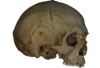
Homo sapiens MSAE 78 View specimen
|
M3#514MSAE 78 Type: "3D_surfaces"doi: 10.18563/m3.sf.514 state:published |
Download 3D surface file |
Homo sapiens MSAE 95 View specimen

|
M3#515MSAE 95 Type: "3D_surfaces"doi: 10.18563/m3.sf.515 state:published |
Download 3D surface file |
Homo sapiens MSAE 1852 View specimen
|
M3#516MSAE 1852 Type: "3D_surfaces"doi: 10.18563/m3.sf.516 state:published |
Download 3D surface file |
Homo sapiens MSAE 6426 View specimen
|
M3#517MSAE 6426 Type: "3D_surfaces"doi: 10.18563/m3.sf.517 state:published |
Download 3D surface file |
Homo sapiens MSAE 6428 View specimen
|
M3#518MSAE 6428 Type: "3D_surfaces"doi: 10.18563/m3.sf.518 state:published |
Download 3D surface file |
Homo sapiens MSAE 6992 View specimen
|
M3#519MSAE 6992 Type: "3D_surfaces"doi: 10.18563/m3.sf.519 state:published |
Download 3D surface file |
Homo sapiens MSAE 7688 View specimen

|
M3#520MSAE 7688 Type: "3D_surfaces"doi: 10.18563/m3.sf.520 state:published |
Download 3D surface file |
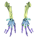
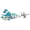
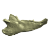
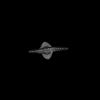
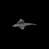
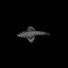
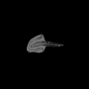
The present 3D Dataset contains the 3D model analyzed in Vautrin et al. (2019), Palaeontology, From limb to fin: an Eocene protocetid forelimb from Senegal sheds new light on the early locomotor evolution of early cetaceans.
?Carolinacetus indet. SNTB 2011-01 View specimen
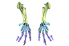
|
M3#3983D model of an articulated forelimb of a Carolinacetus-like protocetid from Senegal Type: "3D_surfaces"doi: 10.18563/m3.sf.398 state:published |
Download 3D surface file |
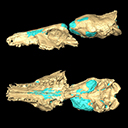
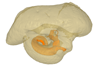
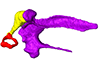
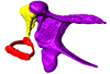
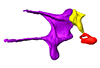
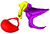
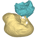
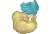
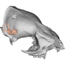
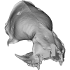
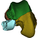
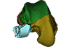
This contribution contains the 3D model described and figured in the following publication: Dubied, M., Mennecart, B. and Solé, F. 2019. The cranium of Proviverra typica (Mammalia, Hyaenodonta) and its impact on hyaenodont phylogeny and endocranial evolution. Palaeontology. https://doi.org/10.1111/pala.12437
Proviverra typica NMB Em18 View specimen
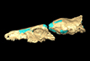
|
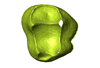
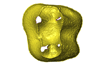
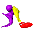
M3#355The file contain the cranium (yellow) and the endocast (blue) of the facial part and the brain case part of the type specimen of Proviverra typica (NMB Em18). Type: "3D_surfaces"doi: 10.18563/m3.sf.355 state:published |
Download 3D surface file |
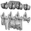
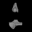
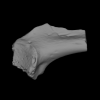
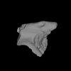
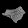
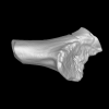
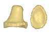
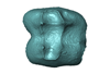
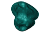
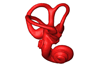
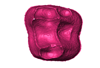
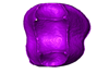
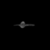
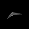
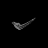
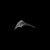
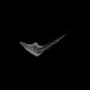
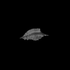
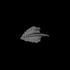
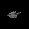
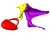
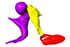
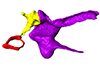
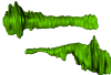
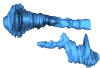
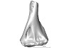
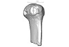
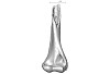
Considerable morphological variations are found in the middle ear among mammals. Here I present a three-dimensional atlas of the middle ear ossicles of eulipotyphlan mammals. This group has radiated into various environments as terrestrial, aquatic, and subterranean habitats independently in multiple lineages. Therefore, eulipotyphlans are an ideal group to explore the form-function relationship of the middle ear ossicles. This comparative atlas of hedgehogs, true shrews, water shrews, mole shrews, true moles, and shrew moles encourages future studies of the middle ear morphology of this diverse group.
Erinaceus europaeus DK2331 View specimen
|
M3#151Left middle ear ossicles Type: "3D_surfaces"doi: 10.18563/m3.sf.151 state:published |
Download 3D surface file |
Anourosorex yamashinai SIK_yamashinai View specimen
|
M3#152Left middle ear ossicles Type: "3D_surfaces"doi: 10.18563/m3.sf.152 state:published |
Download 3D surface file |
Blarina brevicauda M8003 View specimen
|
M3#153Right middle ear ossicles Type: "3D_surfaces"doi: 10.18563/m3.sf.153 state:published |
Download 3D surface file |
Chimarrogale platycephala DK5481 View specimen
|
M3#162Left middle ear ossicles Type: "3D_surfaces"doi: 10.18563/m3.sf.162 state:published |
Download 3D surface file |
Suncus murinus DK1227 View specimen
|
M3#155Left middle ear ossicles Type: "3D_surfaces"doi: 10.18563/m3.sf.155 state:published |
Download 3D surface file |
Condylura cristata SIK0050 View specimen
|
M3#156Right middle ear ossicles Type: "3D_surfaces"doi: 10.18563/m3.sf.156 state:published |
Download 3D surface file |
Euroscaptor klossi SIK0673 View specimen
|
M3#163Left middle ear ossicles Type: "3D_surfaces"doi: 10.18563/m3.sf.163 state:published |
Download 3D surface file |
Euroscaptor malayana SIK_malayana View specimen
|
M3#164Left middle ear ossicles Type: "3D_surfaces"doi: 10.18563/m3.sf.164 state:published |
Download 3D surface file |
Mogera wogura DK2551 View specimen
|
M3#159Left middle ear ossicles Type: "3D_surfaces"doi: 10.18563/m3.sf.159 state:published |
Download 3D surface file |
Talpa altaica SIK_altaica View specimen
|
M3#161Right middle ear ossicles Type: "3D_surfaces"doi: 10.18563/m3.sf.161 state:published |
Download 3D surface file |
Urotrichus talpoides DK0887 View specimen
|
M3#165Left middle ear ossicles Type: "3D_surfaces"doi: 10.18563/m3.sf.165 state:published |
Download 3D surface file |
Oreoscaptor mizura DK6545 View specimen
|
M3#166Left middle ear ossicles Type: "3D_surfaces"doi: 10.18563/m3.sf.166 state:published |
Download 3D surface file |
Scalopus aquaticus SIK_aquaticus View specimen
|
M3#167Left middle ear ossicles Type: "3D_surfaces"doi: 10.18563/m3.sf.167 state:published |
Download 3D surface file |
Scapanus orarius SIK_orarius View specimen

|
M3#168Left middle ear ossicles Type: "3D_surfaces"doi: 10.18563/m3.sf.168 state:published |
Download 3D surface file |
Neurotrichus gibbsii SIK_gibbsii View specimen
|
M3#169Left middle ear ossicles Type: "3D_surfaces"doi: 10.18563/m3.sf.169 state:published |
Download 3D surface file |
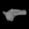
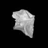
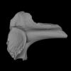
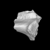
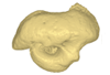
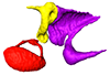
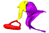
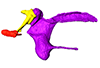
This contribution contains the 3D models described and figured in the following publication: Mourlam, M., Orliac, M. J. (2017), Protocetid (Cetacea, Artiodactyla) bullae and petrosals from the Middle Eocene locality of Kpogamé, Togo: new insights into the early history of cetacean hearing. Journal of Systematic Palaeontology https://doi.org/10.1080/14772019.2017.1328378
?Carolinacetus indet. UM KPG-M 164 View specimen
|
M3#132left petrosal of ?Carolinacetus sp. from the locality of Kpogamé, Togo Type: "3D_surfaces"doi: 10.18563/m3.sf.132 state:published |
Download 3D surface file |
indet. indet. UM KPG-M 73 View specimen

|
M3#133labelled surface of the left petrosal Type: "3D_surfaces"doi: 10.18563/m3.sf.133 state:published |
Download 3D surface file |
|
M3#134left bullaof Protocetidae indeterminate from Kpogamé, Togo Type: "3D_surfaces"doi: 10.18563/m3.sf.134 state:published |
Download 3D surface file |
|
M3#135petrotympanic complex of Protocetidae indeterminate from Kpogamé, Togo Type: "3D_surfaces"doi: 10.18563/m3.sf.135 state:published |
Download 3D surface file |
?Carolinacetus indet. UM KPG-M 33 View specimen
|
M3#136left auditory bulla of a juvenile specimen of ?Carolinacetus sp. from Kpogamé, Togo Type: "3D_surfaces"doi: 10.18563/m3.sf.136 state:published |
Download 3D surface file |
Togocetus traversei UM KPG-M 80 View specimen

|
M3#137fragmentary right auditory bulla of Togocetus traversei from Kpogamé, Togo Type: "3D_surfaces"doi: 10.18563/m3.sf.137 state:published |
Download 3D surface file |
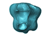
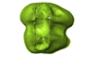
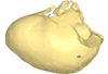
The present 3D Dataset contains 3D models of the cranium surface and of the bony labyrinth endocast of the stem bat Vielasia sigei. They are used by (Hand et al., 2023) to explore the phylogenetic position of this species, to infer its laryngeal echolocating capabilities, and to eventually discuss chiropteran evolution before the crown clade diversification.
Vielasia sigei UM VIE-250 View specimen
|
M3#1269External surface of the cranium Type: "3D_surfaces"doi: 10.18563/m3.sf.1269 state:published |
Download 3D surface file |
|
M3#1270Virtual endocast of the right bony labyrinth Type: "3D_surfaces"doi: 10.18563/m3.sf.1270 state:published |
Download 3D surface file |
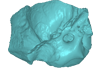
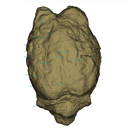
This contribution contains the 3D model(s) described and figured in the following publication: Carolina A. Hoffmann, P. G. Rodrigues, M. B. Soares & M. B. Andrade. 2021. Brain endocast of two non-mammaliaform cynodonts from southern Brazil: an ontogenetic and evolutionary approach, Historical Biology, 33:8, 1196-1207, https://doi.org/10.1080/08912963.2019.1685512
Probelesodon kitchingi MCP 1600 PV View specimen
|
M3#9783D model of the brain endocast of Probelesodon kitchingi. Type: "3D_surfaces"doi: 10.18563/m3.sf.978 state:published |
Download 3D surface file |
Massetognathus ochagaviae MCP 3871 PV View specimen
|
M3#9793D model of the brain endocast of Massetognathus ochagaviae. Type: "3D_surfaces"doi: 10.18563/m3.sf.979 state:published |
Download 3D surface file |
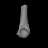
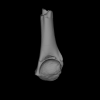
The present contribution contains the 3D virtual restoration of a Pliocene Lutrine right femur of Tobène, Senegal, described and figured in Lihoreau et al. (2021) : "A fossil terrestrial fauna from Tobène (Senegal) provides a unique early Pliocene window in Western Africa ". https://doi.org/10.1016/j.gr.2021.06.013
Indet indet SN-Tob-12-02 View specimen
|
M3#441Virtual restoration of SN-Tob-12-02 Type: "3D_surfaces"doi: 10.18563/m3.sf.441 state:published |
Download 3D surface file |
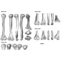
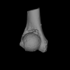
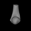
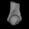
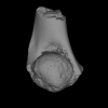
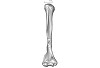
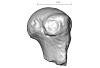
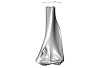
The present 3D Dataset contains the 3D models analyzed in the publication “Systematic and locomotor diversification of the Adapis group (Primates, Adapiformes) in the late Eocene of the Quercy (Southwest France), revealed by humeral remains”. In this paper, twenty humeral specimens from the old and new Quercy collections attributed to the fossil primates Adapis and Palaeolemur are described and analysed together. In this dataset only the scans of the fossils belonging to the collections of Université de Montpellier are provided.
In our paper (Marigó et al., 2019) we provide a qualitative and quantitative analysis of the different humeri, revealing that high variability is present within the “Adapis group” sample. Six different morphotypes are identified, confirming that what has often been called “Adapis parisiensis” is a mix of different species that present different locomotor adaptations.
Adapis sp. UM ROS 2-95 View specimen
|
M3#356Complete right humerus ROS 2-95 attributed to the Adapis group Type: "3D_surfaces"doi: 10.18563/m3.sf.356 state:published |
Download 3D surface file |
Adapis sp. UM ROS 2-536 View specimen
|
M3#357Proximal end of the right humerus ROS 2-536 attributed to the Adapis group Type: "3D_surfaces"doi: 10.18563/m3.sf.357 state:published |
Download 3D surface file |
Adapis sp. UM ROS 2-534 View specimen
|
M3#358Distal end of the left humerus ROS 2-534 attributed to the Adapis group Type: "3D_surfaces"doi: 10.18563/m3.sf.358 state:published |
Download 3D surface file |
Adapis sp. UM ROS 2-535 View specimen
|
M3#359Distal end of the left humerus ROS 2-535 attributed to the Adapis group Type: "3D_surfaces"doi: 10.18563/m3.sf.359 state:published |
Download 3D surface file |
Adapis sp. UM ROS 2-80 View specimen
|
M3#360Proximal end of the right humerus ROS 2-80 attributed to the Adapis group Type: "3D_surfaces"doi: 10.18563/m3.sf.360 state:published |
Download 3D surface file |
Adapis sp. UM ROS 2-79 View specimen
|
M3#361Distal end of the right humerus ROS 2-79 attributed to the Adapis group Type: "3D_surfaces"doi: 10.18563/m3.sf.361 state:published |
Download 3D surface file |
Adapis sp. UM ECA 1364 View specimen

|
M3#362Distal end of the left humerus ECA 1364 attributed to the Adapis group Type: "3D_surfaces"doi: 10.18563/m3.sf.362 state:published |
Download 3D surface file |
Adapis sp. UM ACQ-262 View specimen

|
M3#3733D model of ACQ 262. Humerus Type: "3D_surfaces"doi: 10.18563/m3.sf373 state:published |
Download 3D surface file |
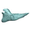
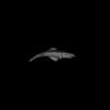
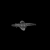
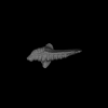
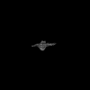
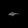
The present 3D Dataset contains the 3D model analyzed in The largest freshwater odontocete: a South Asian river dolphin relative from the Proto-Amazonia.
Pebanista yacuruna MUSM 4017 View specimen
|
M3#1394Holotype skull of Pebanista yacuruna MUSM 4017 Type: "3D_surfaces"doi: 10.18563/m3.sf.1394 state:published |
Download 3D surface file |
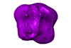
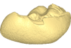
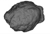
The present 3D Dataset contains the 3D model of the endocranial cast of Palaeolama sp. from the mid-Pleistocene (~1.2 Mya) of South America, analyzed in Balcarcel et al. 2023.
Palaeolama sp. PIMUZ A/V 4091 View specimen
|
M3#11283D model of a natural endocast Type: "3D_surfaces"doi: 10.18563/m3.sf.1128 state:published |
Download 3D surface file |